Step-by-Step: Robotic retroperitoneal partial nephrectomy
Robotic retroperitoneal partial nephrectomy: a step-by-step guide
Khurshid R. Ghani, James Porter*, Mani Menon and Craig Rogers
Vattikuti Urology Institute, Henry Ford Hospital, Detroit, MI, and *Department of Urology, Swedish Urology Group, Seattle, WA, USA
OBJECTIVE
To describe a step-by-step guide for successful implementation of the retroperitoneal approach to robotic partial nephrectomy (RPN)
PATIENTS AND METHODS
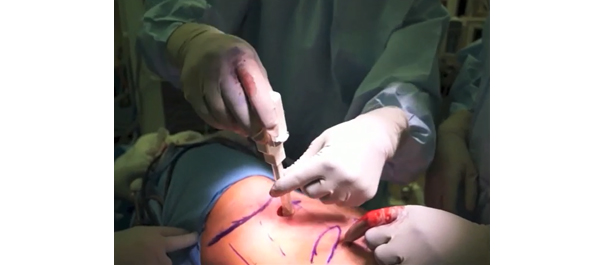
The patient is placed in the flank position and the table fully flexed to increase the space between the 12th rib and iliac crest. Access to the retroperitoneal space is obtained using a balloon-dilating device. Ports include a 12-mm camera port, two 8-mm robotic ports and a 12-mm assistant port placed in the anterior axillary line cephalad to the anterior superior iliac spine, and 7–8 cm caudal to the ipsilateral robotic port.
RESULTS
Positioning and port placement strategies for successful technique include: (i) Docking robot directly over the patient’s head parallel to the spine; (ii) incision for camera port ≈1.9 cm (1 fingerbreadth) above the iliac crest, lateral to the triangle of Petit; (iii) Seldinger technique insertion of kidney-shaped balloon dilator into retroperitoneal space; (iv) Maximising distance between all ports; (v) Ensuring camera arm is placed in the outer part of the ‘sweet spot’.
CONCLUSION
The retroperitoneal approach to RPN permits direct access to the renal hilum, no need for bowel mobilisation and excellent visualisation of posteriorly located tumours.


