Tag Archive for: women

Posts

Article of the Week: External urethral sphincter electromyography and the influence of the menstrual cycle

Every Week the Editor-in-Chief selects an Article of the Week from the current issue of BJUI. The abstract is reproduced below and you can click on the button to read the full article, which is freely available to all readers for at least 30 days from the time of this post.

In addition to the article itself, there is an accompanying editorial written by a prominent member of the urological community. This blog is intended to provoke comment and discussion and we invite you to use the comment tools at the bottom of each post to join the conversation.

Finally, the third post under the Article of the Month heading on the homepage will consist of additional material or media. This week we feature a video from Chris Betts and Kate Burnett discussing their paper. 

If you only have time to read one article this week, it should be this one.

External urethral sphincter electromyography in asymptomatic women and the influence of the menstrual cycle

Cecile Tawadros*, Katherine Burnett*, Laura F. Derbyshire*, Thomas Tawadros†, Noel W. Clarke*‡ and Christopher D. Betts*


*Department of Urology, Salford Royal NHS Foundation Trust, Salford, UK, Department of Urology, Centre Hospitalier Universitaire Vaudois, Lausanne, Switzerland, and Department of Urology, Christie Hospitals NHS Foundation Trust, Manchester, UK


OBJECTIVE

To investigate by electromyography (EMG), the presence of complex repetitive discharges (CRDs) and decelerating bursts (DBs) in the striated external urethral sphincter during the menstrual cycle in female volunteers with no urinary symptoms and complete bladder emptying.

SUBJECTS AND METHODS

Healthy female volunteers aged 20–40 years, with regular menstrual cycles and no urinary symptoms were recruited. Volunteers completed a menstruation chart, urinary symptom questionnaires, pregnancy test, urine dipstick, urinary free flow and post-void ultrasound bladder scan. Exclusion criteria included current pregnancy, use of hormonal medication or contraception, body mass index of >35 kg/m2, incomplete voiding and a history of pelvic surgery. Eligible participants underwent an external urethral sphincter EMG, using a needle electrode in the early follicular phase and the mid-luteal phase of their menstrual cycles. Serum oestradiol and progesterone were measured at each EMG test.

RESULTS

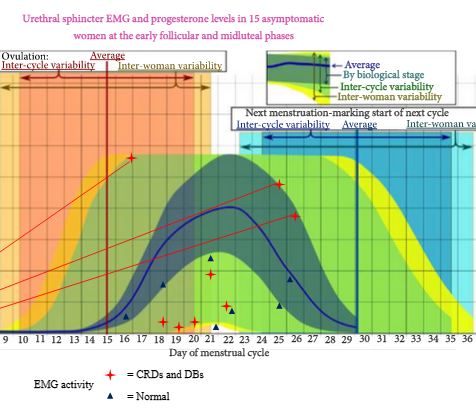
In all, 119 women enquired about the research and following screening, 18 were eligible to enter the study phase. Complete results were obtained in 15 women. In all, 30 EMG tests were undertaken in the 15 asymptomatic women. Sphincter EMG was positive for CRDs and DBs at one or both phases of the menstrual cycle in eight (53%) of the women. Three had CRDs and DBs in both early follicular and mid-luteal phases. Five had normal EMG activity in the early follicular phase and CRDs and DBs in the mid-luteal phase. No woman had abnormal EMG activity in the early follicular phase and normal activity in the luteal phase. There was no relationship between EMG activity and age, parity or serum levels of oestradiol and progesterone.

CONCLUSIONS

CRDs and DB activity in the external striated urethral sphincter is present in a high proportion of asymptomatic young women. This abnormal EMG activity has been shown for the first time to change during the menstrual cycle in individual women. CRDs and DBs are more commonly found in the luteal phase of the menstrual cycle. The importance of CRDs and DBs in the aetiology of urinary retention in young women remains uncertain. The distribution and or quantity of abnormal EMG activity in the external urethral sphincter may be important. In a woman with urinary retention the finding of CRDs and DBs by needle EMG does not automatically establish Fowler’s syndrome as the explanation for the bladder dysfunction. Urethral pressure profilometry may be helpful in establishing a diagnosis. Opiate use and psychological stress should be considered in young women with urinary retention.

Editorial: Do ‘whale noises’ help in the diagnosis of Fowler’s syndrome?

In 1985, Clare Fowler described the presence of abnormal electromyography (EMG) signals in the urethral sphincter of five women with unexplained urinary retention [1]. The presence of complex repetitive discharges (CRD) and decelerating bursts (DB) in women with urinary retention became an important diagnostic finding. Initially, it was described as an EMG finding resembling pseudomyotonia (delayed relaxation of striated muscle when deep tendon reflexes are elicited). Later, an association with polycystic ovary syndrome was hypothesized [2]. This hypothesis could not be proven, however, and the diagnostic criteria that were used at that time for diagnosing polycystic ovaries did not stand the test of time.

The main achievement of urethral sphincter EMG in women with urinary retention was the fact that, from that point onwards, women who were previously described and as being hysterical could now finally be assured that there was a measurable abnormality within their urethral sphincter. This was a major breakthrough in the management of women with urinary retention. Previously, most of these women ended up with permanent catheters or intermittent catheterization, while being stigmatized as psychiatric patients.

The presence of this EMG abnormality also seemed to correlate with the long-term outcome of sacral nerve stimulation [3]. Sacral nerve stimulation is now the standard of care for women with Fowler’s syndrome.

Many authors have questioned the value of these EMG findings. Ramm et al. [4] found the presence of CRD in 30% of healthy women and Tawadros et al. [5] also found this in 53% of healthy female volunteers. These authors showed that CRD and DB are mostly present during the luteal phase of the menstrual cycle. These results suggest at least some hormonal influence on the EMG findings. Fitzgerald et al. [6] showed that CRD correlated weakly with a history of strained voiding and urethral dilation, suggesting a higher prevalence in women with signs of obstructive voiding. Currently, however, the presence of CRD and DB is considered as non-specific.

While sphincter overactivity is generally accepted as the underlying aetiology in women with urinary retention, proving this overactivity remains a challenge. Urethral pressure profiles and urethral volume measurements by ultrasonography have a high interobserver variability and must be used with caution. Most publications originate from one centre and have not been reproduced by others [7]. MRI measurements of the urethral sphincter could possibly be helpful in the future.

While in healthy women CRD can be present, more research will be needed to establish the real value of sphincter EMG in women with pathological urinary retention. The design of good clinical trials will be difficult because of the limited number of patients with this condition. Currently, the diagnosis of Fowler syndrome remains a clinical one, based on a multimodal assessment of the patient.

Dirk De Ridder
Department of Urology, University Hospitals KU Leuven, Leuven, Belgium


References


2 Fowler CJ, Christmas TJ, Chapple CR, Parkhouse HF, Kirby RS, Jacobs HS. Abnormal electromyographic activity of the urethral sphincter, voiding dysfunction, and polycystic ovaries: a new syndrome? BMJ 1988; 297: 14368


4 Ramm O, Mueller ER, Brubaker L, Lowenstein L, Kenton K. Complex repetitive dischargesa feature of the urethral continence mechanism or pathological nding? J Urol 2012; 187: 21403


5 Tawadros C, Burnett K, Derbyshire LF, Tawadros T, Clarke NW, Betts CD. External urethral sphincter electromyography in asymptomatic women and the inuence of the menstrual cycle. BJU Int 2015; 42331


6 FitzGerald MP, Blazek B, Brubaker L. Complex repetitive discharges during urethral sphincter EMG: clinical correlates. Neurourol Urodyn 2000; 19: 57783


7 Wiseman OJ, Swinn MJ, Brady CM, Fowler CJ. Maximum urethral closure pressure and sphincter volume in women with urinary retention. J Urol 2002; 167: 134851; discussion 13512.


Video: External urethral sphincter electromyography and the influence of the menstrual cycle

External urethral sphincter electromyography in asymptomatic women and the influence of the menstrual cycle

Cecile Tawadros*, Katherine Burnett*, Laura F. Derbyshire*, Thomas Tawadros†, Noel W. Clarke*‡ and Christopher D. Betts*


*Department of Urology, Salford Royal NHS Foundation Trust, Salford, UK, Department of Urology, Centre Hospitalier Universitaire Vaudois, Lausanne, Switzerland, and Department of Urology, Christie Hospitals NHS Foundation Trust, Manchester, UK


OBJECTIVE

To investigate by electromyography (EMG), the presence of complex repetitive discharges (CRDs) and decelerating bursts (DBs) in the striated external urethral sphincter during the menstrual cycle in female volunteers with no urinary symptoms and complete bladder emptying.

SUBJECTS AND METHODS

Healthy female volunteers aged 20–40 years, with regular menstrual cycles and no urinary symptoms were recruited. Volunteers completed a menstruation chart, urinary symptom questionnaires, pregnancy test, urine dipstick, urinary free flow and post-void ultrasound bladder scan. Exclusion criteria included current pregnancy, use of hormonal medication or contraception, body mass index of >35 kg/m2, incomplete voiding and a history of pelvic surgery. Eligible participants underwent an external urethral sphincter EMG, using a needle electrode in the early follicular phase and the mid-luteal phase of their menstrual cycles. Serum oestradiol and progesterone were measured at each EMG test.

RESULTS

In all, 119 women enquired about the research and following screening, 18 were eligible to enter the study phase. Complete results were obtained in 15 women. In all, 30 EMG tests were undertaken in the 15 asymptomatic women. Sphincter EMG was positive for CRDs and DBs at one or both phases of the menstrual cycle in eight (53%) of the women. Three had CRDs and DBs in both early follicular and mid-luteal phases. Five had normal EMG activity in the early follicular phase and CRDs and DBs in the mid-luteal phase. No woman had abnormal EMG activity in the early follicular phase and normal activity in the luteal phase. There was no relationship between EMG activity and age, parity or serum levels of oestradiol and progesterone.

CONCLUSIONS

CRDs and DB activity in the external striated urethral sphincter is present in a high proportion of asymptomatic young women. This abnormal EMG activity has been shown for the first time to change during the menstrual cycle in individual women. CRDs and DBs are more commonly found in the luteal phase of the menstrual cycle. The importance of CRDs and DBs in the aetiology of urinary retention in young women remains uncertain. The distribution and or quantity of abnormal EMG activity in the external urethral sphincter may be important. In a woman with urinary retention the finding of CRDs and DBs by needle EMG does not automatically establish Fowler’s syndrome as the explanation for the bladder dysfunction. Urethral pressure profilometry may be helpful in establishing a diagnosis. Opiate use and psychological stress should be considered in young women with urinary retention.

© 2024 BJU International. All Rights Reserved.