Tag Archive for: Urine

Posts

Article of the week: A four‐group urine risk classifier for predicting outcomes in patients with prostate cancer

Every week, the Editor-in-Chief selects an Article of the Week from the current issue of BJUI. The abstract is reproduced below and you can click on the button to read the full article, which is freely available to all readers for at least 30 days from the time of this post.

In addition to the article itself, there is an editorial written by a prominent member of the urological community. These are intended to provoke comment and discussion and we invite you to use the comment tools at the bottom of each post to join the conversation. 

If you only have time to read one article this week, it should be this one.

A four‐group urine risk classifier for predicting outcomes in patients with prostate cancer

Shea P. Connell*, Marcelino Yazbek-Hanna*, Frank McCarthy, Rachel Hurst*, MartynWebb*, Helen Curley*, Helen Walker, Rob Mills, Richard Y. Ball, Martin G. Sanda§, Kathryn L. Pellegrini§, Dattatraya Patil§, Antoinette S. Perry, Jack Schalken**, Hardev Pandha††, Hayley Whitaker‡‡, Nening Dennis, Christine Stuttle, Ian G. Mills§§¶¶***, Ingrid Guldvik¶¶, Movember GAP1 Urine Biomarker Consortium1, Chris Parker†††, Daniel S. Brewer*‡‡‡, Colin S. Cooper* and Jeremy Clark*

*Norwich Medical School, University of East Anglia, Norwich, Institute of Cancer Research, Sutton, Norfolk and Norwich University Hospitals NHS Foundation Trust, Norwich, UK, §Department of Urology, Winship Cancer Institute, Emory University School of Medicine, Atlanta, GA, USA , School of Biology and Environmental Science, Science West, University College Dublin, Dublin 4, Ireland, **Nijmegen Medical Centre, Radboud University Medical Centre, Nijmegen, The Netherlands, ††Faculty of Health and Medical Sciences, The University of Surrey, Guildford, ‡‡Molecular Diagnostics and Therapeutics Group, University College London, London, §§School of Medicine, Dentistry and Biomedical Sciences, Institute for Health Sciences, Centre for Cancer Research and Cell Biology, Queen’s University Belfast, Belfast, UK, ¶¶Centre for Molecular Medicine, University of Oslo, Oslo, Norway, ***Nuffield Department of Surgical Sciences, University of Oxford, Oxford, †††Royal Marsden Hospital, Sutton and ‡‡‡Earlham Institute, Norwich, UK

Abstract

Objectives

To develop a risk classifier using urine‐derived extracellular vesicle (EV)‐RNA capable of providing diagnostic information on disease status prior to biopsy, and prognostic information for men on active surveillance (AS).

Patients and Methods

Post‐digital rectal examination urine‐derived EV‐RNA expression profiles (n = 535, multiple centres) were interrogated with a curated NanoString panel. A LASSO‐based continuation ratio model was built to generate four prostate urine risk (PUR) signatures for predicting the probability of normal tissue (PUR‐1), D’Amico low‐risk (PUR‐2), intermediate‐risk (PUR‐3), and high‐risk (PUR‐4) prostate cancer. This model was applied to a test cohort (n = 177) for diagnostic evaluation, and to an AS sub‐cohort (n = 87) for prognostic evaluation.

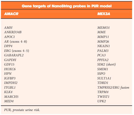
Table 2. NanoString gene probes incorporated by LASSO regularization in the final optimal model used to produce the prostate urine risk signatures

Results

Each PUR signature was significantly associated with its corresponding clinical category (P < 0.001). PUR‐4 status predicted the presence of clinically significant intermediate‐ or high‐risk disease (area under the curve = 0.77, 95% confidence interval [CI] 0.70–0.84). Application of PUR provided a net benefit over current clinical practice. In an AS sub‐cohort (n = 87), groups defined by PUR status and proportion of PUR‐4 had a significant association with time to progression (interquartile range hazard ratio [HR] 2.86, 95% CI 1.83–4.47; P < 0.001). PUR‐4, when used continuously, dichotomized patient groups with differential progression rates of 10% and 60% 5 years after urine collection (HR 8.23, 95% CI 3.26–20.81; P < 0.001).

Conclusion

Urine‐derived EV‐RNA can provide diagnostic information on aggressive prostate cancer prior to biopsy, and prognostic information for men on AS. PUR represents a new and versatile biomarker that could result in substantial alterations to current treatment of patients with prostate cancer.


Editorial: Do you need further assistance in diagnosing and risk stratifying prostate cancer?

I would hope the answer to the question posed in the title is a universal ‘yes’; at least that is my experience with this complex and common disease. The concept that in 2019, we have unmet needs in prostate cancer diagnostics is somewhat remarkable, given that we have access to: (i) one of the most widely used biomarkers in oncology (PSA), (ii) a readily accessible organ to examine (DRE), (iii) state of the art imaging (MRI, positron emission tomography), (iv) specialty biopsy systems (fusion/transperineal template), (v) enhanced risk stratification systems (National Comprehensive Cancer Network [NCCN], Cancer of the Prostate Risk Assessment [CAPRA], etc.), (vi) numerous nomograms, (vii) secondary urine/serum biomarkers (Prostate Health Index [PHI], prostate cancer antigen 3 [PCA3], SelectMDx, ExoDx, four‐kallikrein panel [4K]), and (viii) commercially available genomic platforms (Prolaris, OncotypeDx, Decipher).

The paper by Connell et al. [1] in this issue of BJUI asks you to consider adding another diagnostic test to your list. You might correctly assume from the title that the test is in discovery/validation stages, and lacks a fancy commercialised name. Many steps await any promising biomarker to make it to your clinic. So why pay attention to this one? Let me reiterate a few points made by the authors and suggest where new paradigms might emerge if the test delivers on its promises.

First, the test crosses over the current barriers between screening patients and active surveillance (AS). In both populations we care about Gleason Grade Group ≥2. Yet a SelectMDx or similar tests are validated for diagnosis but not for monitoring Grade Group 1 on AS. Genomic profiling tests have strong validation and prognostic value for AS, but require tissue and external laboratory work flows. This marker is being tested for both settings, with potentially meaningful distinctions for both patient groups.

Second, this test is in the urine and does not need imaging or needles to obtain samples. It may have serial use (if cost‐effective) for monitoring AS.

Third, for AS cohorts, the test seems to be able to identify progression well in advance. This would potentially allow for early intervention in the correct patients, and less intense monitoring in the remaining.

Fourth, the test metrics looked favourable in PSA screened and unscreened populations; will we ever see a novel biomarker bold enough to move to primary/independent screening status?

Fifth, some of the secondary biomarkers you may be using now are included in this model: PCA3, transmembrane protease serine 2:v‑ets erythroblastosis virus E26 oncogene homolog (TMPRSS2‐ERG), Homeobox C6 (HOXC6).

To be critical, this biomarker will need significant validation in other cohorts, and we can always hope for head‐to‐head data with existing strategies. I will remain optimistic these authors can move this biomarker strategy along and help bridge some of the gaps that remain in disease detection and risk stratification. I may even attempt to insert some of those lovely new equations in the methods section into future lectures.

Reference

  1. Connell SPYazbek‐Hanna MMcCarthy F et al. A four‐group urine risk classifier for predicting outcomes in patients with prostate cancer. BJU Int 2019124609– 20
Article of the week: Targeted deep sequencing of urothelial bladder cancers and associated urinary DNA: a 23‐gene panel with utility for non‐invasive diagnosis and risk stratification

Every week, the Editor-in-Chief selects an Article of the Week from the current issue of BJUI. The abstract is reproduced below and you can click on the button to read the full article, which is freely available to all readers for at least 30 days from the time of this post.

In addition to the article itself, there is an editorial written by a prominent member of the urological community and a video prepared by the authors. These are intended to provoke comment and discussion and we invite you to use the comment tools at the bottom of each post to join the conversation. 

If you only have time to read one article this week, it should be this one.

Targeted deep sequencing of urothelial bladder cancers and associated urinary DNA: a 23‐gene panel with utility for non‐invasive diagnosis and risk stratification

Douglas G. Ward*, Naheema S. Gordon*, Rebecca H. Boucher*, Sarah J. Pirrie*, Laura Baxter, Sascha Ott, Lee Silcock, Celina M. Whalley*, Joanne D. Stockton*, Andrew D. Beggs*, Mike Griffiths§, Ben Abbotts*, Hanieh Ijakipour*, Fathimath N.Latheef*, Robert A. Robinson*, Andrew J. White*, Nicholas D. James*, Maurice P.Zeegers, K. K. Cheng** and Richard T. Bryan*


*Institute of Cancer and Genomic Sciences, University of Birmingham, Birmingham, Department of Computer Science, University of Warwick, Coventry, Nonacus Limited, Birmingham Research Park, §West Midlands Regional Genetics Laboratory, Birmingham Women’s and Children’s NHS Foundation Trust, Birmingham, UK, NUTRIM School for Nutrition and Translational Research in Metabolism and CAPHRI Care and Public Health Research Institute, Maastricht University, Maastricht, The Netherlands and **Institute of Applied Health Research, University of Birmingham, Birmingham, UK

Read the full article

Abstract

Objectives

To develop a focused panel of somatic mutations (SMs) present in the majority of urothelial bladder cancers (UBCs), to investigate the diagnostic and prognostic utility of this panel, and to compare the identification of SMs in urinary cell‐pellet (cp) DNA and cell‐free (cf) DNA as part of the development of a non‐invasive clinical assay.

Patients and Methods

A panel of SMs was validated by targeted deep‐sequencing of tumour DNA from 956 patients with UBC. In addition, amplicon and capture‐based targeted sequencing measured mutant allele frequencies (MAFs) of SMs in 314 urine cpDNAs and 153 urine cfDNAs. The association of SMs with grade, stage and clinical outcomes was investigated by univariate and multivariate Cox models. Concordance between SMs detected in tumour tissue and cpDNA and cfDNA was assessed.


Results

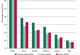
The panel comprised SMs in 23 genes: TERT (promoter), FGFR3, PIK3CA, TP53, ERCC2, RHOB, ERBB2, HRAS, RXRA, ELF3, CDKN1A, KRAS, KDM6A, AKT1, FBXW7, ERBB3, SF3B1, CTNNB1, BRAF, C3orf70, CREBBP, CDKN2A and NRAS; 93.5–98.3% of UBCs of all grades and stages harboured ≥1 SM (mean: 2.5 SMs/tumour). RAS mutations were associated with better overall survival (P = 0.04). Mutations in RXRA, RHOB and TERT (promoter) were associated with shorter time to recurrence (P < 0.05). MAFs in urinary cfDNA and cpDNA were highly correlated; using a capture‐based approach, >94% of tumour SMs were detected in both cpDNA and cfDNA.

Conclusions

SMs are reliably detected in urinary cpDNA and cfDNA. The technical capability to identify very low MAFs is essential to reliably detect UBC, regardless of the use of cpDNA or cfDNA. This 23‐gene panel shows promise for the non‐invasive diagnosis and risk stratification of UBC.

Read more Articles of the week


Editorial: Non‐invasive diagnosis and monitoring of urothelial bladder cancer: are we there yet?

In this issue of BJUI, Ward et al. [1] describe the development of DNA‐based urinary biomarkers for urothelial carcinoma (UC). The genomics of UC have been well characterized through interrogation of tumour issues in institutional series (e.g. the Memorial Sloan Kettering Cancer Center [MSKCC] experience), multi‐institutional collaborations (e.g. The Cancer Genome Atlas [TCGA]) and commercial platforms (e.g. the Foundation Medicine experience) [2]. Until recently, these have been largely academic pursuits, with possible impact on prognostication but limited clinical applicability and utility for therapy selection and monitoring of response; however, with the US Food and Drug Administration approval of erdafitinib several weeks ago, patients with advanced UC will routinely receive genomic assessment for FGFR2/3 mutation or fusion, the targets for this therapy [3]. In due time, it is anticipated that multiple other putative targets with associated therapies (e.g. ERBB2, CDKN2A), as well as potential predictive biomarkers, may also warrant testing.

The evolving landscape in advanced UC makes a non‐invasive biomarker particularly attractive. The authors of the present commentary have previously reported results from a series of 369 patients with advanced UC, demonstrating that genomic alterations in ctDNA could be identified in 91% of patients using a commercially available 73-gene panel [4]. More recently, Christensen et al. [5] assessed a cohort of 68 patients receiving neoadjuvant chemotherapy for muscle‐invasive disease, demonstrating 100% sensitivity and 98% specificity for the detection of relapsed disease with a patient‐specific ctDNA assessment (sequenced to a median target coverage of 105 000×) after cystectomy. Impressively, the data also showed that the dynamics of ctDNA appeared to be more useful than pathological downstaging in predicting relapse.

In contrast to these studies, Ward et al. have developed a 23‐gene panel based on frequently expressed genes in a cohort of 916 UC tissue specimens, largely derived from patients with non‐muscle‐invasive disease. Ultimately, with a cohort of 314 patients with DNA derived from a urinary cell pellet, sequencing identified 645 (71.4%) of 903 mutations detected in tumour. Using urinary supernatant, 353 (80.7%) of 437 mutations were detected. These relatively high sensitivities, if they can be interpreted as such, are promising but do not rise to the level of replacing existing strategies for UC detection, staging and monitoring. Notably, another study demonstrated that urinary ctDNA can be detected with high sensitivity and specificity in patients with localized early‐stage bladder cancer and for after‐treatment surveillance, providing the foundation for further studies evaluating the role of ctDNA in non‐invasive detection, genotyping and monitoring [6].

Beyond its use as a diagnostic tool, it is hoped that urinary ctDNA may also find applications in the selection of therapeutics. To this end, Ward et al. identified FGFR3, PIK3CA, ERCC2 and ERBB2 mutations in 45%, 32%, 14% and 7% of patients, respectively. The frequency of FGFR3 alteration decreased with increasing stage and grade, ranging from 72% in pTaG1 disease to just 13% in ≥pT2 disease, consistent with other reports [7]. These results may guide forthcoming studies evaluating FGFR inhibitors in non‐muscle‐invasive, muscle‐invasive and metastatic disease, where studies are ongoing. In reviewing the potential link between genomic alterations and clinical outcomes, perhaps the most curious finding is that between RAS mutations and improved overall survival (P = 0.04), the only such association found in multivariate analysis. These results stand in sharp contrast to reports in lung cancer, colorectal cancer and multiple other tumour types [8]. A closer look at the deleterious nature and functional impact of NRAS and KRAS mutations seen in this series is certainly warranted, along with further external validation in a more homogenous and larger patient population. There is also the potential application of monitoring treatment response by assessing eradication of urinary ctDNA, a hypothesis that is being evaluated in ongoing studies [9].

How will the results of this and other emerging urinary biomarker studies eventually make their way to the clinic? The answer is simple: incorporation of these biomarkers in prospective therapeutic trials. As the bladder cancer investigative community formulates novel trials for non‐muscle‐invasive and muscle‐invasive disease using targeted therapies, an excellent opportunity exists to correlate urinary, blood and tissue‐based biomarkers and to assess their relative predictive capabilities and clinical utility. Furthermore, with clinical surrogate endpoints likely to drive regulatory approval (e.g. landmark complete response rates for non‐muscle‐invasive disease, or pT0N0 rate for muscle‐invasive disease), a validated urinary biomarker could ultimately offer an alternative biological surrogate endpoint [10]. In an era of genomic revolution, prospective validation can help establish the potential clinical utility of promising biomarkers and help realize the dream of ‘precision oncology’.

by Rohit K. Jain, Petros Grivas and Sumanta K. Pal

References

  1. Ward DGGordon NSBoucher RH et al. Targeted deep sequencing of urothelial bladder cancers and associated urinary DNA: a 23‐gene panel with utility for non‐invasive diagnosis and risk stratification. BJU Int 2019
  2. Schiff JPBarata PCYu EYGrivas PPrecision therapy in advanced urothelial cancer. Expert Rev Precis Med Drug Dev 2019481– 93
  3. FDA grants accelerated approval to erdafitinib for metastatic urothelial carcinoma [press release] 2019.
  4. Agarwal NPal SKHahn AW et al. Characterization of metastatic urothelial carcinoma via comprehensive genomic profiling of circulating tumor DNA. Cancer 20181242115– 24
  5. Christensen EBirkenkamp‐Demtroder KSethi H et al. Early detection of metastatic relapse and monitoring of therapeutic efficacy by ultra‐deep sequencing of plasma cell‐free DNA in patients with urothelial bladder carcinoma. J Clin Oncol 2019371547– 57
  6. Dudley JCSchroers‐Martin JLazzareschi DV et al. Detection and surveillance of bladder cancer using urine tumor DNA. Cancer Discov 20199500– 9
  7. Tomlinson DCBaldo OHarnden PKnowles MAFGFR3 protein expression and its relationship to mutation status and prognostic variables in bladder cancer. J Pathol 200721391– 8
  8. Zhuang RLi SLi Q et al. The prognostic value of KRAS mutation by cell‐free DNA in cancer patients: a systematic review and meta‐analysis. PLoS One 201712e0182562
  9. Abbosh PHPlimack ERMolecular and clinical insights into the role and significance of mutated DNA repair genes in bladder cancer. Bladder Cancer 201849– 18
  10. Jarow JPLerner SPKluetz PG et al. Clinical trial design for the development of new therapies for nonmuscle‐invasive bladder cancer: report of a Food and Drug Administration and American Urological Association public workshop. Urology 201483262– 4


BJUI in the news: prostate urine risk

A recent BJUI article, A four‐group urine risk classifier for predicting outcomes in patients with prostate cancerby Shea Connell and coworkers from Norfolk and Norwich University Hospital (NNUH) has been featured on various news outlets including the BBC and ITV in the UK following its online publication.

The article describes a new urine test, the Prostate Urine Risk, for predicting potentially aggressive prostate cancer meaning many men may avoid needing invasive biopsies and unnecessary treatment. It is likely to be one of a range of tests including blood tests and MRI scans which will enter routine clinical practice for prostate cancer diagnosis.

The research team was led by Prof Colin Cooper, Dr Daniel Brewer and Dr Jeremy Clark, all from the University of East Anglia’s Norwich Medical School, with the support and expertise of Rob Mills, Marcel Hanna and Prof Richard Ball at the NNUH.

Read the full article

Article of the Week: Immunocytochemical expression of ERG in urine identifies prostate cancer

Every Week the Editor-in-Chief selects an Article of the Week from the current issue of BJUI. The abstract is reproduced below and you can click on the button to read the full article, which is freely available to all readers for at least 30 days from the time of this post.

In addition to the article itself, there is an accompanying editorial written by a prominent member of the urological community. This blog is intended to provoke comment and discussion and we invite you to use the comment tools at the bottom of each post to join the conversation.

If you only have time to read one article this week, it should be this one.

Immunocytochemical detection of ERG expression in exfoliated urinary cells identifies with high specificity patients with prostate cancer

Raj P. Pal*, Roger C. Kockelbergh, John Howard Pringl e*, Lara Cresswell, Roger
Hew§, John P. Dormer§, Colin Cooper, John Kilian Mellon, Julian G. Barwell** and Edward J. Hollox**


*Department of Cancer Studies and Molecular Medicine, University of Leicester, Department of Urology, Department of Cytogenetics, §Department of Cellular Pathology, University Hospitals of Leicester NHS Trust, Leicester, Department of Cancer Genetics, University of East Anglia, Norwich, and **Department of Genetics, University of Leicester, Leicester, UK


Read the full article

Objectives

To evaluate the immunocytochemical detection of ERG protein in exfoliated cells as a means of identifying patients with prostate cancer (PCa) before prostate biopsy.

Materials and Methods

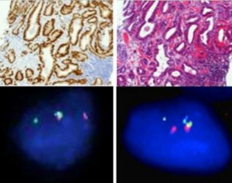
Urine samples (30 mL) were collected after digital rectal examination (DRE) from 159 patients with an elevated age-specific prostate-specific antigen (PSA) and/or an abnormal DRE who underwent prostate biopsy. In all cases, exfoliated urinary cells from half of the urine sample underwent immunocytochemical assessment for ERG protein expression. Exfoliated cells in the remaining half underwent assessment ofTMPRSS2:ERG status using either nested reverse-transcriptase (RT)-PCR (151 cases) or fluorescence in situ hybridization (FISH; eight cases). Corresponding tissue samples were evaluated using FISH to determine chromosomal gene fusion tissue status and immunohistochemistry (IHC) to determine ERG protein expression. Results were correlated with clinicopathological variables.

Results

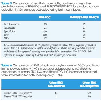
The sensitivity and specificity of urinary ERG immunocytochemistry (ICC) for PCa were 22.7 and 100%, respectively. ERG ICC results correlated with advanced tumour grade, stage and higher serum PSA. In comparison, urine TMPRSS2:ERG transcript analysis had 27% sensitivity and 98% specificity for PCa detection. On tissue IHC, ERG staining was highly specific for PCa. In all, 52% of cancers harboured foci of ERG staining; however, only 46% of cancers that were found to have ERG overexpression were positive on urine ICC. The ERG ICC results showed strong concordance with urinary RT-PCR and FISH, and tissue IHC and FISH.

ew

Conclusion

This is the first study to show that cytological gene fusion detection using ICC is feasible and identifies patients with adverse disease markers. ERG ICC was highly specific, but this technique was less sensitive than RT-PCR.

Read more articles of the week

Editorial: New possibilities for urinary molecular diagnostics

Fusion of androgen-driven serine protease transmembrane protease (TMPRSS) and oncogenic erythroblast transformation-specific-related gene (ERG) genes is a specific alteration in human prostate cancer and many studies have been performed in order to analyse its role as a biomarker or, even more interestingly, as a tumour promoter [1]. Recently, Nguyen et al. [1] demonstrated that ERG activates the Yes-associated protein 1 (YAP1) transcriptional programme thus inducing prostate carcinogenesis. YAP is a transcriptional coactivator involved in regulation of many biological processes. Thus, there is increasing evidence showing that ERG is causally involved in prostate cancer development. Since the original publication by Tomlins et al. [2], in which the gene fusion was discovered, researchers have addressed numerous scientific questions in order to reveal importance of the TMPRSS:ERG fusion in prostate cancer. This is particularly important because several other proteins have been proposed as prostate tumour markers; however, many of them cannot be used in clinical practice. The reasons for that are related to differences in sampling of tissues and methodologies. Thus, biomarker research will in future probably be restructured on the basis of standardisation of experimental procedures. Biomarker research work related to the TMPRSS:ERG fusion appears to be more advanced. In addition to tissue diagnostics, studies on detection of ERG may be performed in urine as evidenced in the paper by Pal et al. [3]. Moreover, the authors for the first time show that exfoliated urinary cells can be used to identify patients with prostate cancer by detection of positive ERG samples. The authors used nested PCR and urinary immunocytochemistry and fluorescence in situ hybridisation in this study. As the TMPRSS:ERG fusion is detectable in ≈50% of prostate cancer specimens, the data presented in the study published in this issue of BJUI are of considerable interest.

Combining the use of TMPRSS:ERG detection and other markers in urine should further improve diagnostics of prostate cancer. Another tumour marker, prostate cancer antigen 3 (PCA3), is frequently used in prostate cancer diagnostics. In urine, TMPRSS:ERG urinary transcript detection is superior to PSA and PCA3 for identifying patients with cancer [4]. The results support data recently published according to which the determination of urine TMPRSS2:ERG together with PCA3 may be used for a more individualised prostate cancer risk assessment [5].

As the authors state in the paper [3], differences between results of some marker studies on TMPRSS:ERG detection may be a consequence of the appearance of less common isoforms, which are not detectable by all assays.

In summary, Pal et al. [3] show for the first time that patients with prostate cancer could be identified by immunocytochemistry on the basis of detection of ERG in exfoliated urine cells. In addition, the authors [3] also show that the appearance of ERG in exfoliated urine cells is associated with a bad prognosis. Indirectly, the present paper [3], supports the concept that the presence of TMPRSS:ERG regulates various cellular events leading to increased migration and proliferation of prostate cancer cells [6]. It is expected that further improvements in urinary molecular diagnostics based on the presence of TMPRSS:ERG will be achieved in the near future. However, clinicians should also be aware that, despite its high specificity, there are limitations of methodology used in this paper, in particular a large proportion of tumours may remain undetected and not all cancers exfoliate into the urine.

Read the full article
Zoran Culig
Experimental Urology, Department of Urology, Medical University of Innsbruck, Anichstrasse 35, Innsbruck, A-6020, Austria


References


1 Nguyen LT, Tretiakova MS, Silvis MR et al. ERG activates the YAP1 transcriptional program and induces the development of age-related prostate tumors. Cancer Cell 2015; 27: 797808


2 Tomlins SA, Rhodes DR, Perner S et al. Recurrent fusion of TMPRSS2 and ETS transcription factor genes in prostate cancer. Science 2005; 310: 6448


5 Tomlins SA, Day JR, Lonigro RJ et al. Urine TMPRSS2:ERG plus PCA3 for individualized prostate cancer risk assessment. Eur Urol 2015; [Epub ahead of print] DOI: 10.1016/j.eururo.2015.04.039


Who let the Dogs Out?

Or

Let’s Paws for a Second!

Or

Lets paws for a second before we all start howling about nothing!


Recently while driving to a Day Surgery list at Heatherwood hospital in Ascot, I happened to listen to BBC Radio 4. There was a report regarding high accuracy for the detection of stomach cancer by a simple breath test. This reminded me of a study published some time ago in European Urology on the ability of a dog to detect prostate cancer by smelling a sample of urine. For a skeptic like me, reading that article in the platinum journal had indeed brought a sarcastic chuckle. Pondering over the paper and the report on the radio, it just dawned on to me as to why I would instantly believe a high-tech nanosensor detecting stomach cancer but not a mortal Belgian Malinois shepherd! This formed my basis of my blog.

It is well known that when someone is afflicted by a disease or cancer, there is a change that occurs in the internal milieu. In the vast majority, before this change can be manifested clinically, there is definite change seen biochemically.  There is emerging evidence that volatile organic compounds (VOC) that are exhaled either in the breath or in bodily fluids can indicate these changes reflecting the underlying pathology.  This is where the humble mongrel comes into play. Olfactory bulb in dogs is forty times bigger than of humans relative to total brain size. Having 125 to 220 million smell-sensitive receptors, their olfactory sense is up to one hundred thousand to one million times more sensitive than a human’s. So, there may be some sense and science in the paper that I had initially chuckled at. The earliest report is a letter to the Lancet reporting the diagnosis of melanoma made after the dog sniffed at a suspicious mole of its owner. Since then, there have been several reports on the ability for the trained dogs to detect various cancers and chronic illness, urological diseases. One of the earliest attempts to detect prostate cancer can be dated back to 2002. Further attempts were made to initiate trials in 2003 but no published results on Medline were found. Earliest published paper can be traced back to 2008, wherein the study did not support the concept of dogs being able to detect prostate cancer. This was recently challenged by the article by Cornu J et al that revealed a sensitivity and specificity of 91% for biopsy proven prostate cancer! In fact, one of the three patients who was wrongly classified as prostate cancer, was found to have cancer on a re-biopsy! The potential VOC that may be found in the urine of a patient with prostate cancer can be found in this letter to the editor. Indeed, to carry out more research, Medical Detection Dogs is aiming to recruit prostate cancer patients within the UK!

One would obviously think that if we can diagnose prostate cancer, why not bladder cancer? This is precisely what led to a “proof of principle” study headed by Carolyn Willis and findings were published in the BMJ. The dogs had a mean success rate of 41%, compared with 14% expected by chance alone. Multivariate analysis suggested that the dogs’ capacity to recognise a characteristic bladder cancer odour was independent of other chemical aspects of the urine detectable by urinalysis. What was astonishing about this study was on one occasion during training, all dogs unequivocally indicated as positive a sample from a participant recruited as a control on the basis of negative cystoscopy and ultrasonography. The consultant responsible for the patient was sufficiently concerned to bring forward further tests, and transitional cell carcinoma of the right kidney was discovered! The same group further reported specificity that ranged from 92% for urine samples obtained from healthy, young volunteers down to 56% for those taken from older patients with non-cancerous urological disease.

More trials are being carried out for detection of cancers affecting the lung, breast, ovary, bowel and others, a review of which can be found in this article. We may need to wait for a few more years to find out whether we are dealing with real science or we are going in circles like the dog chasing its own tail!

On the lighter note, if you hear an old male dog bark for no apparent reason, think prostate cancer!! Dog is the only other mammal that can be afflicted by prostate cancer and fortunately the doggie world will not be affected by the USPSTF recommendations, as canine prostate cancers do not secrete PSA!

Amrith Rao is a Consultant Urological Surgeon at Wexham Park Hospital, Wexham, UK

Tweet: @urorao


Comments on this blog are now closed.


© 2025 BJU International. All Rights Reserved.