Tag Archive for: TURBT

Posts

Editorial: Postoperative intravesical chemotherapy has an important role in reducing subsequent bladder tumours – why is it not routine?

Transurethral resection of bladder tumour (TURBT) is a frequent operation performed by urologists worldwide. Although on occasion the procedure can be quite challenging, the majority are relatively straightforward with little morbidity. In most cases, where the medical system allows, it is an outpatient procedure. Nonetheless, with the exception of small low‐grade tumours the patient is anaesthetised. It is costly, as the procedure requires medical clearance, an operating room team and equipment.

Most patients with bladder cancer have Ta or less frequently T1 tumours. Despite an initial TURBT, 30–80% of patients develop another tumour. Most are new tumours and some may be recurrences. The reasons for the high ‘recurrence’ rate are the continued impact of the carcinogen, e.g. cigarettes, incomplete resection, missed tumours, and tumour implantation on the altered urothelium. The urologist can help reduce these events by stressing the importance of limiting carcinogen exposure e.g. smoking cessation, striving to perform a complete TURBT, reviewing the entire bladder after the TURBT to avoid missing tumours (using narrow‐band imaging or fluorescent cystoscopy if available), and limiting implantation of tumour cells on the altered urothelial surface with the use of postoperative intravesical chemotherapy (POIVC).

There is a large body of evidence that POIVC reduces the chance of a subsequent tumour [1]. I became convinced that implantation occurs after animal studies demonstrated that bladder cancer cells placed into the bladder preferentially implant and grow only if the urothelial surface had been cauterised or otherwise damaged prior to exposure to the bladder cancer cells [2]. Prospective randomised trials eventually confirmed the benefit of POIVC [3]. The paper published in this issue of the BJUI by Bosschieter et al. [4] indicates that POIVC is equally effective if given the same day or the day after TURBT. Thus, if there are obstacles to instilling the medication on the day of the TURBT the drug can be administered the following day.

The evidence in favour of POIVC for bladder tumours is particularly impressive for Grade 1–2 Ta tumours. In my view, all patients with primary or ‘recurrent’ single or multiple papillary Grade 1–2 Ta tumours are the optimal candidates to receive POIVC [5]. POIVC is recommended by the European Association of Urology (EAU) and AUA/Society of Urologic Oncology (SUO) [6,7] yet, the adoption of this guideline is far from uniform. I queried my colleagues from the International Bladder Cancer group (IBCG), as they are conversant in the scientific basis for POIVC and represent several countries with different medical systems [8]. Their comments are pertinent and consistent with my understanding of the issues. Here are some of the common reasons for not following the guidelines: (i) Some urologists are not convinced that the reduction in the ‘recurrence’ rate is sufficient to use POIVC. (ii) The most common chemotherapeutic agent for POIVC in the USA is mitomycin C and it is expensive. The cost for 40 mg is ~$1000. It is approximately $500 in Europe. (iii) Hospitals have rules regarding the delivery of chemotherapy and the pharmacy and nursing departments may not make it easy to instil the drug in the postoperative setting. Some hospitals require notification a day before the surgery and the drug is wasted if the drug is not used. (iv) Urologists are concerned about extravasation and uncertainty of the tumour grade and stage. There may be other reasons but these help explain why POIVC is not routine.

On the other hand many patients with bladder cancer require frequent TURBTs. I am certain that following an uneventful TURBT or office cauterisation for Grade 1–2 Ta bladder cancer, they would choose to receive POIVC if properly informed. Urologists are proficient at judging whether a tumour fits the criteria for POIVC and if they underestimate the grade or stage the patient may still benefit. If urologists cannot instil the chemotherapy on the day of the TURBT, they can instil the drug the following day without compromising effectiveness. I believe it is our job to do what we can to help our patients and in this instance we should do our best to minimise subsequent tumour events, which includes the use of adjuvant chemotherapy.

Mark S. Soloway

Memorial Hospital Hollywood, Miami, FL, USA

References

  1. Perlis N, Zlotta AR, Beyene J, Finelli A, Fleshner NE, Kulkarni GS. Immediate post‐ transurethral resection of bladder tumor intravesical chemotherapy prevents non‐muscle invasive bladder tumor recurrence: an updated meta‐analysis on 2548 patients and quality –of‐evidence review. Eur Urol 2013; 64: 421–30
  2. Weldon TE, Soloway MS. Susceptibility of urothelium to neoplastic cellular implantation. Urology 1975; 5: 824–7
  3. Tolley DA, Hargreave TB, Smith PH et al. Effect of intravesical mitomycin C on recurrence of newly diagnosed superficial bladder cancer: interim report from the Medical Research Council Subgroup on Superficial Bladder cancer. Br Med J 1988; 296: 1259–61
  4. Bosschchieter J, von Moorselaar JA, Vis AN et al. The effect of timing of an immediate instillation of mitomycin C after transurethral resection in 941 patients with non‐muscle‐invasive bladder cancer. BJU Int 2018; 122: 571–5
  5. Klaassen Z, Soloway MS. European Association of Urology and American Urological Association/Society of Urologic Oncology guidelines on risk categories for non‐muscle‐invasive bladder cancer may lead to overtreatment for low‐grade Ta bladder tumors. Urology 2017; 105: 14–7
  6. Babjuk M, Böhle A, Burger M et al. EAU guidelines in non‐muscle invasive urothelial carcinoma of the bladder: update 2016. Eur Urol 2017; 71: 447–61
  7. Chang SS, Boorjian SA, Chou R et al. Diagnosis and treatment of non‐muscle invasive bladder cancer: AUA/SUO guideline. J Urol 2016; 196: 1021–9
  8. Brausi M, Witjes F, Lamm D et al. A review of current guidelines and best proactive recommendations for the management of nonmuscle invasive bladder cancer by the International Bladder Cancer Group. J Urol 2011; 186: 2158–67


Highlights from BAUS 2016

1.1

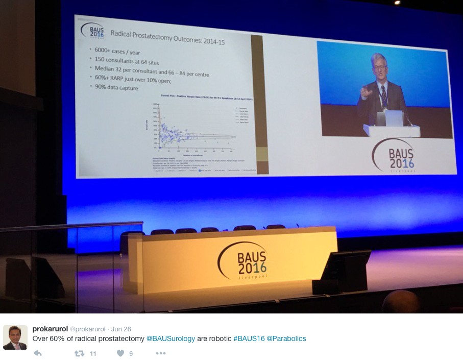
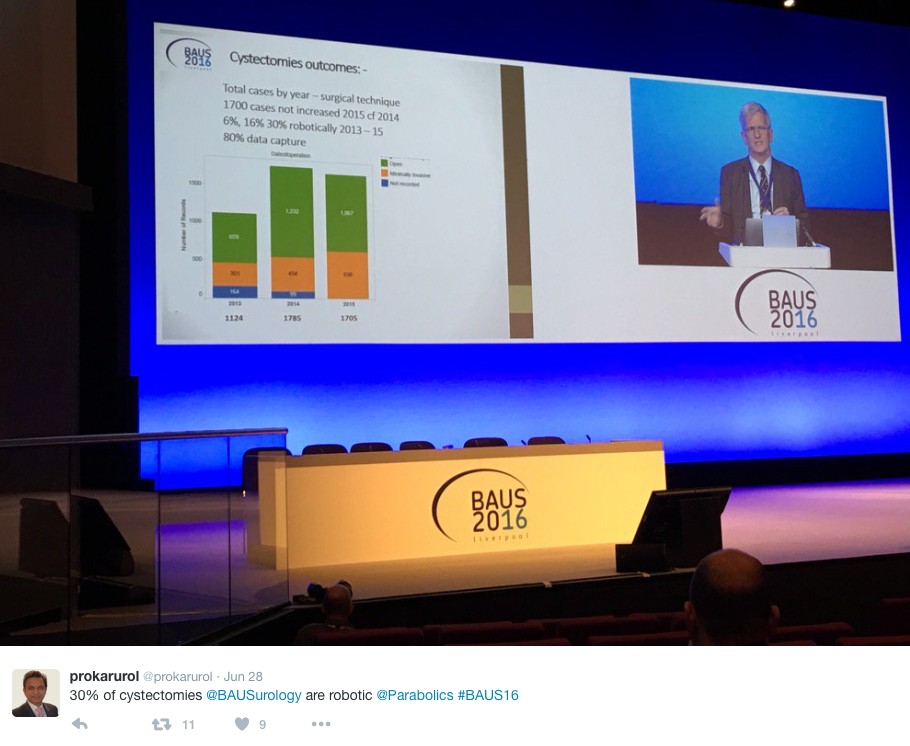
In the week following Britain’s exit from Europe after the BREXIT referendum, BAUS 2016 got underway in Liverpool’s BT convention Centre. This was the 72nd meeting of the British Association of Urological Surgeons and it was well attended with 1120 delegates (50% Consultant Member Urologists, 30% Trainees, 10% Non member Urologists/Other, 10% Nurses, HCP’S, Scientists).

1.2

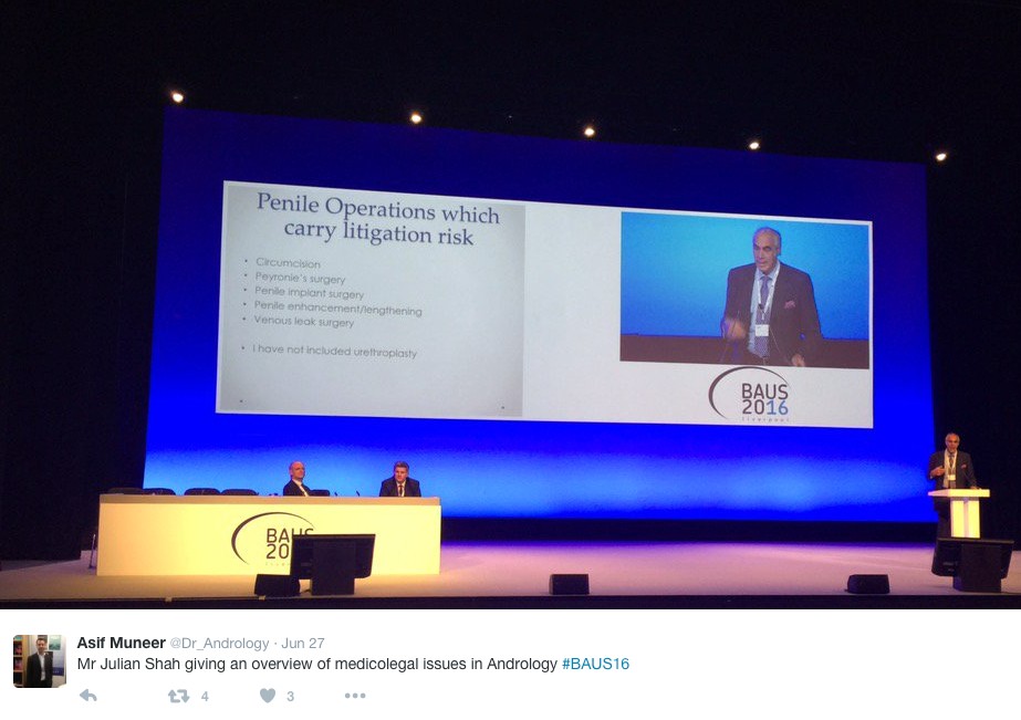
Monday saw a cautionary session on medicolegal aspects in Andrology, focusing on lawsuits over the last year. Mr Mark Speakman presented on the management issue of testicular torsion. This sparked further discussion on emergency cover for paediatrics with particular uncertainty noted at 4 and 5 year olds and great variation in approach dependent on local trust policy. Mr Julian Shah noted the most litigious areas of andrology, with focus on cosmesis following circumcisions. Therefore serving a reminder on the importance of good consent to manage patients’ expectations.

1.3

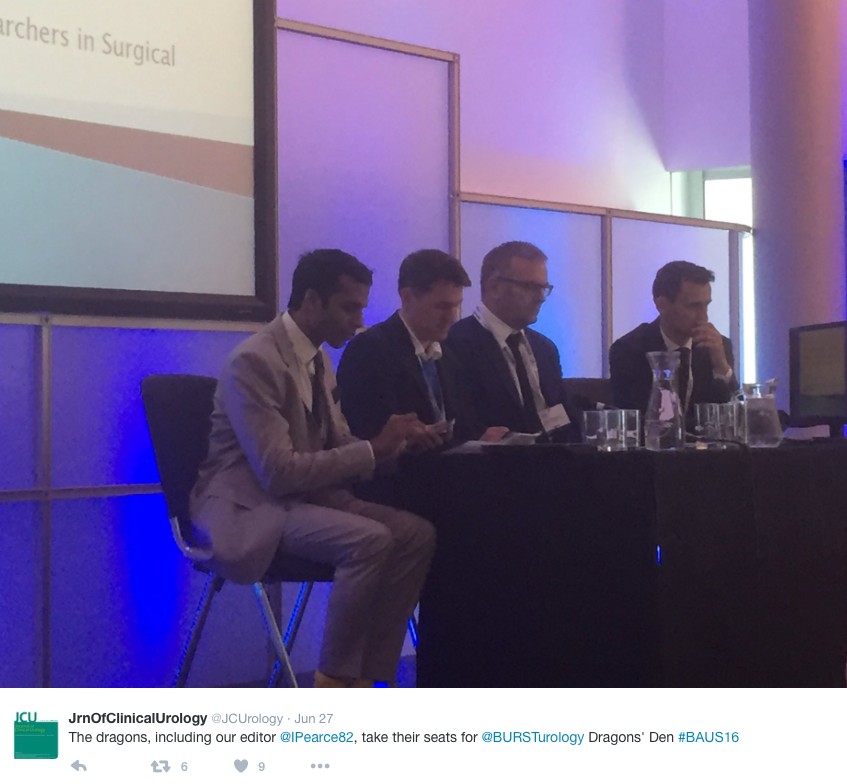
In the Dragons’ Den, like the TV show, junior urologists pitched their ideas for collaborative research projects, to an expert panel. This year’s panel was made up of – Mark Emberton, Ian Pearce, and Graeme MacLennan. The session was chaired by Veeru Kasivisvanathan, Chair of the BURST Research Collaborative.

1.4

Eventual winner Ben Lamb, a trainee from London, presented “Just add water”. The pitch was for an RCT to investigate the efficacy of water irrigation following TURBT against MMC in reducing tumour recurrence. Ben proposed that water, with its experimental tumouricidal properties, might provide a low risk, low cost alternative as an adjuvant agent following TURBT. Judges liked the scientific basis for this study and the initial planning for an RCT. The panel discussed the merits of non-inferiority vs. superiority methodology, and whether the team might compare MMC to MMC with the addition of water, or water instead of MMC. They Dragons’ suggested that an initial focus group to investigate patients’ views on chemotherapy might help to focus the investigation and give credence to the final research question, important when making the next pitch- to a funding body, or ethics committee.

Other proposals were from Ryad Chebbout, working with Marcus Cumberbatch, an academic trainee from Sheffield. Proposing to address the current controversy over the optimal surgical technique for orchidopexy following testicular torsion. His idea involved conducting a systematic review, a national survey of current practice followed by a Delphi consensus meeting to produce evidence based statement of best practice. The final presentation was from Sophia Cashman, East of England Trainee for an RCT to assess the optimal timing for a TWOC after urinary retention. The panel liked the idea of finally nailing down an answer to this age-old question.

1.5

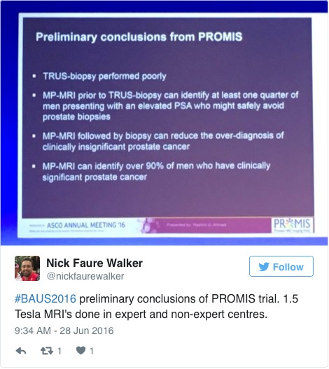
Waking up on Tuesday with England out of the European football cup as well as Europe the conference got underway with an update from the PROMIS trial (use of MRI to detect prostate cancer). Early data shows that multi-parametric MRI may be accurate enough to help avoid some prostate biopsies.

1.6

The SURG meeting provided useful information for trainees, with advice on progressing through training and Consultant interviews. A debate was held over run through training, which may well be returning in the future. The Silver cystoscope was awarded to Professor Rob Pickard voted for by the trainees in his deanery, for his devotion to their training.
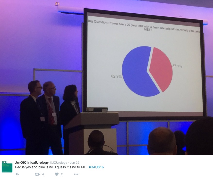
Wednesday continued the debate on medical expulsion therapy (MET) for ureteric stones following the SUSPEND trial. Most UK Urologists seem to follow the results of the trial and have stopped prescribing alpha blockers to try and aid stone passage and symptoms. However the AUA are yet to adopt this stance and feel that a sub analysis shows some benefit for stones >5mm, although this is not significant and pragmatic outcomes. Assistant Professor John Hollingsworth (USA) argued for MET, with Professor Sam McClinton (UK) against. A live poll at the end of the session showed 62.9% of the audience persuaded to follow the SUSPEND trial evidence and stop prescribing MET.

1.7

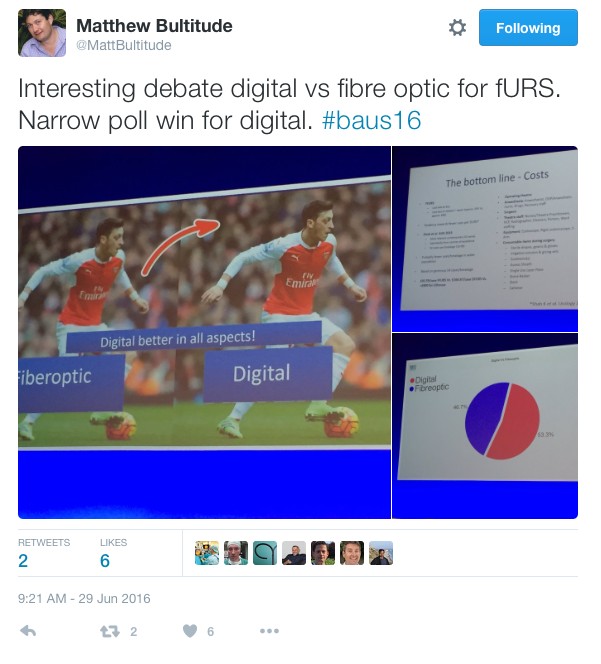
In the debate of digital versus fibreoptic scopes for flexible ureteroscopy digital triumphed, but with a narrow margin.

1.8

In other updates and breaking news it appears that BCG is back! However during the shortage EMDA has shown itself to be a promising alternative in the treatment of high grade superficial bladder cancer.
The latest BAUS nephrectomy data shows that 90% are performed by consultant, with 16 on average per consultant per year. This raises some issues for registrar training, however with BAUS guidelines likely to suggest 20 as indicative numbers this is looking to be an achievable target for most consultants. Robotic advocates will be encouraged, as robotic partial nephrectomy numbers have overtaken open this year. The data shows 36% of kidney tumours in the under 40 years old are benign. Will we have to consider biopsying more often? However data suggests we should be offering more cytoreductive nephrectomies, with only roughly 1/10 in the UK performed compared to 3/10 in the USA.

1.91.10

The andrology section called for more recruitment to The MASTER trial (Male slings vs artificial urinary sphincters), whereas the OPEN trial has recruited(open urethroplasty vs optical urethotomy). In the treatment of Peyronie’s disease collagenase has been approved by NICE but not yet within the NHS.

Endoluminal endourology presentation showed big increases in operative numbers with ureteroscopy up by 50% and flexible ureteroscopy up by 100%. Stents on strings were advocated to avoid troubling stent symptoms experienced by most patients. New evidence may help provide a consensus on defining “stone free” post operation. Any residual stones post-operatively less than 2mm were shown to pass spontaneously and therefore perhaps may be classed as “stone free”.

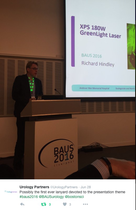
Big changes seem likely in the treatment of benign prostatic hyperplasia, with a race to replace the old favorite TURP. Trials have of TURP (mono and bipolar) vs greenlight laser are already showing similar 2 year outcomes with the added benefit of shorter hospital stays and less blood loss. UROLIFT is an ever more popular alternative with data showing superiority to TURP in lifestyle measures, likely because it preserves sexual function, and we are told it can be performed as a 15 minute day case operation. The latest new therapy is apparently “Aquabeam Aquablation”, using high pressured water to remove the prostate. Non surgical treatments are also advancing with ever more accurate super selective embolisation of the prostatic blood supply.

1.11

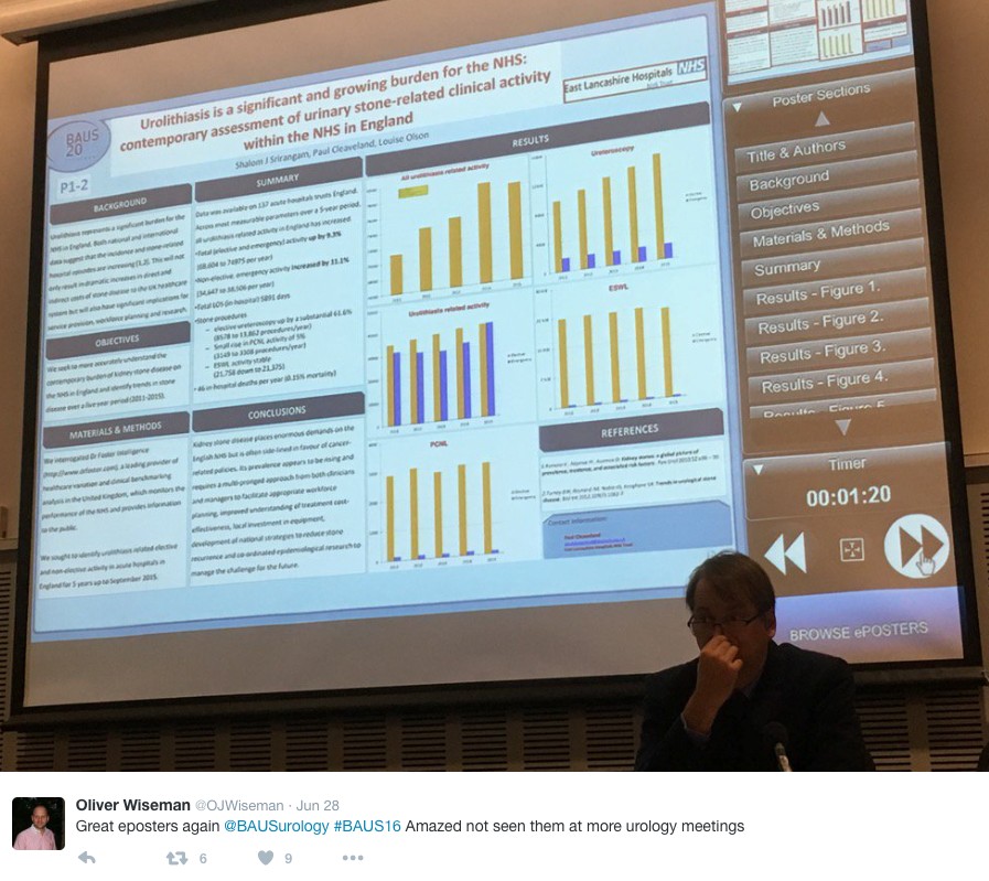
This year all accepted abstracts were presented in moderated EPoster sessions. The format was extremely successful removing the need for paper at future conferences? A total of 538 abstracts were submitted and 168 EPosters displayed. The winner of best EPoster was P5-5 Altaf Mangera: Bladder Cancer in the Neuropathic Bladder.

1.12

The best Academic Paper winner was Mark Salji of the CRUK Beatson institute, titled “A Urinary Peptide Biomarker Panel to Identify Significant Prostate Cancer”. Using capillary electrophoresis coupled to mass spectrometry (CE-MS) they analysed 313 urine samples from significant prostate cancer patients (Gleason 8-10 or T3/4 disease) and low grade control disease. They identified 94 peptide urine biomarkers which may provide a useful adjunct in identifying significant prostate cancer from insignificant disease.

The Office of Education offered 20 courses. Popular off-site courses were ultrasound for the Urologist, at Broadgreen Hospital, a slightly painful 30 min drive from the conference centre. However well worth the trip, delivered by Radiology consultants this included the chance to scan patients volunteers under guidance, with separate stations for kidneys, bladder and testicles and learning the “knobology” of the machines.

Organised by Tamsin Greenwell with other consultant experts in female, andrology and retroperitoneal cancer, a human cadaveric anatomy course was held at Liverpool university. The anatomy teaching was delivered by both Urology consultants and anatomists allowing for an excellent combination of theory and functional anatomy.

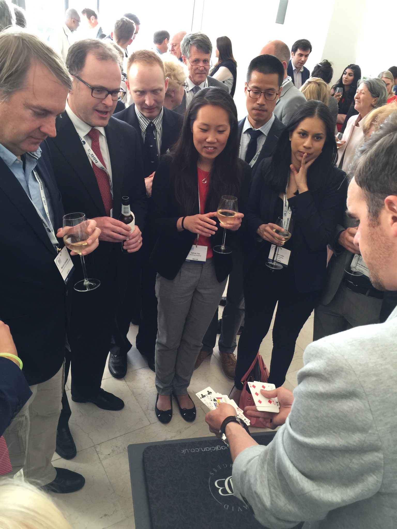
BAUS social events are renowned and with multiple events planned most evenings were pretty lively. The official drinks reception was held at the beautiful Royal Liver Building. The venue was stunning with great views over the waterfront and the sun finally shining. Several awards were presented including the Gold cystoscope to Mr John McGrath for significant contribution to Urology within 10 years appointment as consultant. The Keith Yeates medal was awarded to Mr Raj Pal, the most outstanding candidate in the first sitting of the intercollegiate specilaity examination, with a score of over 80%.

1.13

During the conference other BAUS awards presented include the St Peter’s medal was awarded to Margeret Knowles, Head of section of molecular oncology, Leeds Institute of Cancer and Pathology, St James University hospital Leeds. The St Paul’s medal awarded to Professor Joseph A. Smith, Vanderbilt University, Nashville, USA. The Gold medal went to Mr. Tim Terry, Leicester General Hospital.

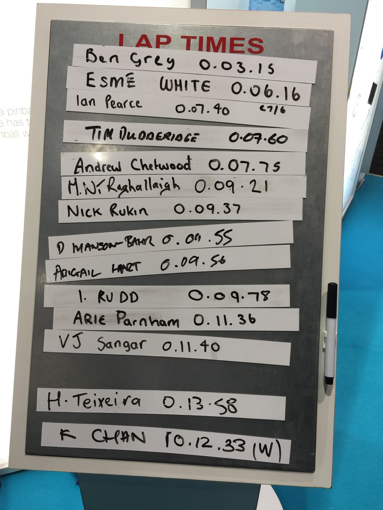
An excellent industry exhibition was on display, with 75 Exhibiting Companies present. My personal fun highlight was a flexible cystoscope with integrated stent remover, which sparked Top Gear style competiveness when the manufacturer set up a time-trial leaderboard. Obviously this best demonstrated the speed of stent removal with some interesting results…

1.14

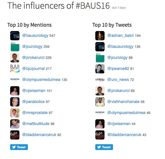
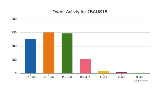
Social media review shows good contribution daily.

1.15

1.16
Thanks BAUS a great conference, very well organised and delivered with a great educational and social content, looking forward to Glasgow 2017! #BAUS2017 #Glasgow #BAUSurology

Nishant Bedi

Specialist Training Registrar North West London 

Twitter: @nishbedi


Urology in Zomba, Malawi. Reflecting on surgical care in a Resource-Limited country

Rajiv SingalAt the recent AUA meeting in San Diego as at all of our major meetings, a tremendous amount of data was presented and technology displayed to advance our specialty.   Walking through exhibit hall one sees an expensive bauble at every turn. The advancement of urology over the last 50 years has been remarkable.   We have a lot to be proud of.  I think we have the most interesting, exciting specially in all of medicine.  Urologist are generally technophiles and have always loved to push surgical procedures to new heights.   From robotics, lasers and endourology to advancing the molecular understanding of disease, urologists have always aimed to drive the bus.

As many of you know, I am on a short trip to Malawi Africa. I have written about this elsewhere. I am here on one hand as a board member for Dignitas International.  On the surgical side it is not a mission under the guise of anyone but rather my own personal attempt to understand what urology and surgery in a resource poor country might look like. I have been here in Zomba, Malawi and working at Zomba Central Hospital, which is one of four central hospitals in the country.

1.1

A goal has been to try and assess what the basic urological needs might be in this part of the world and see how I could help bridge the gap, whether it would be with equipment, external manpower or ultimately by improving training and leaving something sustainable. I optimistically set out, confident in my abilities to eventually network and bring colleagues together and establish over time a reasonable urology program that at least resembles something familiar. I have the COSECSA guidelines on what it takes to establish a training program at my side. Perhaps nothing illustrates what a daunting task this will be like my days in surgery this week.

To start with, a typical OR at ZCH requires some refocusing compared to what I am used to. My DaVinci robot is nowhere to be seen

1.2

I made ward rounds with my clinical officer yesterday and lined up several TUR type cases to try and do, with men bleeding from bladder tumours (all invariably Bilharzial disease) as well as men in retention. Some have had catheters for months, even years.

First there is the set up. No discussion about lasers and lifts or any other such fun. We don’t even have the 3L irrigation bags. For my irrigation set up, with a little water and some chlorine pucks we are ready to go.


1.31.41.5

My first patient was a TURBT.  A very large, incompletely resected lesion, actively bleeding.  I clearly left disease behind but perhaps he won’t bleed for a while.  The tissue will not be sent to pathology.  Patients need to pay 16,000 MWK for it. The typical pay for many is 20,000-30000/month and 1$USD=700 MWK.  Managing him from any even rudimentary oncological perspective is a non-starter.

The second patient also had a bladder tumour.  It was palpable as a mass to just under the skin.  Again, the goal was to stop some bleeding, at least for a few weeks.    He almost certainly has metastatic disease but I have no way to image and know for sure. I did order a chest xray to look for obvious pulmonary nodules.  He will eventually just quietly die.

Before I could start a third case I found myself in the gynecology OR 2 weeks after a hysterectomy post-delivery for bleeding.  Following an injury, the left ureter was leaking.  I attempted the repair as best as I could with no proper light, no electrocautery no retractors and no ability to stent my freshly re-implanted ureter.   All of this on an HIV+ve new mother.   I hope it heals open.  I am not sure if it will.   I have come to understand that ureteral injuries are a not uncommon consequence of obstetrical care in Malawi.

My third patient had a TURP which was fairly straightforward.   He should hopefully void assuming reasonable residual bladder function.  He has had a catheter in place for months.

At least we did do some work Thursday.  On Tuesday my four patient list turned into one as my anesthetist did not attend.  Before surgical care can be improved, the critical shortage of anesthesia care has to also be addressed. I also wrote about that earlier.

I did bring a surgery checklist to ZCH on Tuesday.

1.6

And Thursday in follow up, I gave a talk to the surgical team about checklists and so that is certainly good.

1.7

They keep asking me to see men in the clinic with catheters.  With the inefficiencies of late start times, anesthesia shortages and only a week to go, most will get left behind.  It is really a depressing thought.

My OR team though is there to help and keen to learn.

1.8

Daniel, Rex (T Rex) and Maryeuster

As I reflect on my experience in the operating room during week one I am struck by how discordant what I saw in San Diego was from the realities still faced in much of the world.  Basic endoscopic equipment does not exist. Serendipitously, a retired colleague of mine did bring some basic equipment a few months ago and this one set, washed and then resterilized (in a pail of chlorinated water) is all that we have.   I am still not clear what happens when the loops wear out.

1.9

I do question when we pull millions of dollars and much intellectual capital into improving technology and chasing robots as to what are we really doing to benefit the care of our urological patients on a global scale. Do we have some obligation as champions of mens’ health and urologic care more broadly, to play a part?  I do wonder whether some of our intellectual energy and financial resources could be better spent simply bringing parts of this world even into the 1970s. If this was valued as worthy of academic support and promotion the way oncology, endourology and everything else is in our specialty is, then some of the bright young minds in our field might move this along further.  Whether we do a robot prostatectomy retroperitoneally or intraperitoneally, debate about a Rocco stitch or tweak this or do that, these changes are often incremental at best. Supine versus prone PCNL?  Who cares.  Other parts of the world I think deserve some of our high-level expertise to meet their complex challenges. I would invite the urological community to try and collectively address this problem. Should we keep pouring all of our massive resources only to steady, incremental benefit?  Clearly we always must advance the body of knowledge and the state of the art.  However, is there a role for reserving some resource and energy to advocate for simpler things that could affect a change on the order of several magnitudes?  Some of the easier things we might do is to at least act as advocates and lead some process change whether it be a surgical checklist, counting instruments and sutures pre and post operatively and ensure better preoperative screening and post-operative care.   Updating equipment and building surgical expertise necessarily follows.

Laser TURP?  Plasma button?  Urolift?   The men in Malawi and much of Africa would be happy just to get rid of their catheters.

We often joke about our ‘first world problems’.  It’s time to get serious.

Let’s do better.

Dr Rajiv Singal is a Urologist at Michael Garron Hospital and an Assistant Professor in the Department of Surgery at the University of Toronto

Follow him on Twitter at @DrRKSingal

To read more about Dr Singal’s experience in Malawi follow this link https://www.rajivsingal.com/blogCategories/view/malawi-june-2016/


RSM Winter Meeting in Saalbach, Austria

This year the urology section of the RSM held their annual winter meeting in Saalbach, Austria hosted by Tom McNicholas and Rik Bryan.

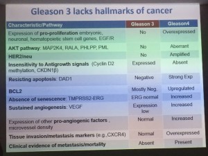
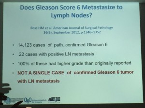
 1.1Kicking off the meeting was a state of the art lecture by Professor Shahrokh Shariat, Professor of Urology at the Medical University of Vienna who presented a convincing perspective on whether we should really be calling Gleason 3+3 disease “prostate cancer” due to the lack of hallmarks of cancer compared with Gleason four disease, and clinical data suggesting that Gleason 3+3 cancer does not metastasise. Education of patients to ensure compliance of active surveillance is surely key to ensuring that change in disease pattern or small volume higher Gleason grade disease is not missed. Interestingly from Dominic Hodgson’s experience in Portsmouth approximately 50% of patients with Gleason 3+3 disease on TRUS were upgraded to Gleason 3+4 on template biopsy, although these patients who went on to have more extensive biopsies did so due to other concerning parameters. SIN PIN keeps you connected to your loved ones around the world! All New Customers receive $1 FREE to try SIN PIN International Calling Service. Make High Quality International Calls to those who don’t have the SIN PIN App yet. Never go out of touch with the ones you care about most! SIN PIN keeps you connected! You can find here the free International calling app Ft Lauderdale FL.

1.21.3

 The bladder and upper tract cancer session was also a highlight with Rik Bryan presenting data on the use of ‘Oncoscan’ to detect genomic profiles and aberrations in urinary DNA from cell free centrifuged urine. This however was not absolutely specific to bladder tumours as undiagnosed prostate cancer was also detected in one of the tested urine specimens.


The Bladder Path trial being set up by Professor Nick James was also discussed. This trial hopes to investigate the addition of MRI into the haematuria clinic pathway. TURBT in muscle invasive disease does not completely stage tumours and may lead to a delay in definitive treatment. There is no current evidence that debulking of tumour is necessary prior to radical treatment. This randomised controlled trial will review whether MRI as opposed to TURBT could be used for staging in likely muscle invasive tumours with the phase II and phase III aspects looking at time to definitive treatment and time to recurrence or progression.

Professor Karl-Dietrich Sievert from the Universitätsklinik für Urologie und Andrologie, Saltzburg demonstrated how his unit use Diffusion Tensor Imaging MRI to visualise white matter and plan for nerve sparing prostatectomy to preserve post-operative incontinence and erectile function. We also heard how Tim O’Brien has learned many of his lessons in complex renal cancer surgery the hard way, in an inspiring and candid talk.

For the benign urologists there were a plethora of sessions on male and female incontinence as well as male and female ejaculation! Matthew Bultitude and I (RT) debated on medical expulsive therapy for ureteric stones in the wake of the SUSPEND trial. Although the majority of the room seemed convinced of the lack of benefit for small ureteric stones, there appeared to be some doubt created by the regarding larger distal ureteric stones.

1.6

We also had a lot of interesting non-urological discussions. From Martin Mansell, Consultant Nephrologist we heard of the change in law since the Montgomery Judgment leading to the necessity for doctors when taking consent to inform patients of any risk no matter the likelihood of the risk occurring if that particular patient would attach significance to that risk. Mark Speakman pointed out that this may mean a change in the BAUS consent forms which many of us use to consent patients. We also heard of new educational tools such as MedShr from Asif Qasim, Consultant Cardiologist, which is an app serving as a platform to discuss complex cases with colleagues from around the world. BAUS President Mark Speakman presented the BJUI Knowledge tool which allows BAUS members to access interactive e-learning modules and log CPD activity.

1.72016 marked the 34th annual winter meeting for the urology section of the RSM and we paid tribute this year to Peter Worth who has been a regular attendee since the beginning. With a fantastic meeting already planned in Lake Tahoe for 2017 to mark the 35th year hosted by Professor Roger Kirby and Matthew Bultitude, I would encourage as many trainees and consultants to attend for both a rigorous transatlantic educational programme as well as a fantastic opportunity to meet new colleagues and, of course ski!

Rebecca Tregunna (ST4, Alexandra Hospital, Redditch (Worcestershire Acute Hospitals NHS Trust) – @rebeccatregunna

Dominic Hodgson (Consultant Urologist, Queen Alexandra Hospital, Portsmouth) – @hodgson_dominic

#UroJC July 2014 – Is there a place for laser techniques in our current schema of bladder cancer diagnosis and management?

This month’s International Urology Journal Club (@iurojc) truly engaged a global audience with participants from ten countries including author Thomas Herrman (@trwhermann) from Hannover, Germany.  A landmark 2000 followers was reached during July, nearly two years since @iurojc’s conception in late 2012. In fact, since this time nearly 1100 people have participated in the journal club from around the world.

Bladder cancer was up for debate for the first time this year and @iurojc trialled the discussion of two complementary articles recently published online ahead of print in the World Journal of Urology.  The first article provided an update of the current evidence for transurethral Ho:YAG and Tm:YAG in the endoscopic treatment of bladder cancer, and the second was a randomised controlled trial (RCT) comparing laser to the gold standard transurethral resection of bladder tumour (TURBT).  Authorship groups were from Germany and China respectively; our Chinese authors unfortunately unable to join the dialogue due to restriction on all twitter activity in the country.

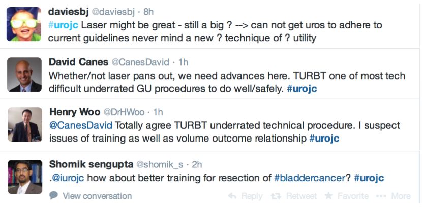
Initial conversation focussed on the methodology, results and limitations of the RCT, however this soon extended to a more general discussion around the current difficulties with the diagnosis and management of bladder cancer and the pros and cons of using laser for this purpose.  Key themes debated over the 48-hour period included the importance of accurate staging, current standards of TURBT, advantages of en bloc resection and the learning curve, cost and usefulness of laser technology.

Both studies reiterated one of the major goals outlined in the EAU guidelines for non-muscle invasive bladder cancer (NMIBC), to achieve correct staging with inclusion of detrusor muscle and complete resection of tumours.  This is important in limiting second resection and consequently has a resulting cost offset.  In the review article, only 3 studies commented on staging quality and another two commented that laser was suitable for staging but did not specify if detrusor muscle was identified.

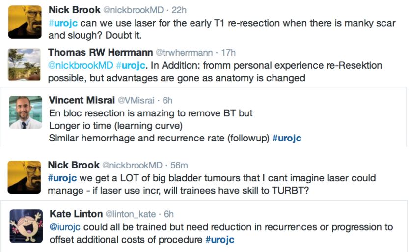
@ChrisFilson and @CBayneMD expressed their concern over the RCT by Chen and Colleagues

@linton_kate astutely pointed out another limitation

and author of the review article @trwherrmann summed this up nicely

In the RCT by Chen et al. there was a significantly greater number of pT1 tumours detected with laser than TURBT, the authors suggested this might be due to better sampling.  It remains unclear if this would impact on management and this did not enter the arena for discussion during this @iurojc.

Many argued that TURBT techniques and practices should be optimised before newer techniques are introduced.

‘En bloc’ was touted as the new trendy word in endourology.  EAU guidelines recommend en bloc resection for smaller tumours.  The articles suggested that en bloc resection of bladder tumours should provide more accurate staging however conclusive data is missing to substantiate this in the current literature. 

@DrHWoo discussed potential advantages of the laser technique

@linton_kate pointed out that en bloc resection is not limited to the laser technique

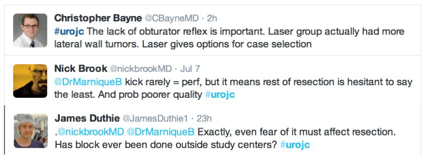
Further to this, the lack of obturator nerve reflection with laser was emphasised in the RCT.  Obturator kick was noted during TURBT in 18 patients and none during laser resection, however none of these patients suffered bladder perforation.  The significance of this was debated and usefulness of obturator block in this context discussed.

The pendulum seemed to the swing out of favour of laser during the discussion, with several limitations outlined including reduced ability for re-resection, cost and the presence of a learning curve.

Regarding additional cost, the host rebutted

The flow of academic dialogue was interrupted midstream (pardon the pun) by a light-hearted discussion around the ergonomics of TURBT.

Below are some of the key take home messages that arose from the usual culprits in this month’s @iruojc discussion

Kindly author @trwherrmann invited us to his upcoming en bloc resection workshop.  Keep an eye out for this.

@iurojc would like to thank Prostate Cancer Prostatic Diseases who have kindly provided the prize for this month which is a 12 month on line subscription to the journal. @nickbrookMD’s made efforts to sway the vote his way.

Whilst usually the Best Tweet Prize is reserved for some incisive comment, the repeated complaints from @nickbrookMD for his failure to ever win the Best Tweet prize has seen for the first and final time that the @iurojc has bowed to pressure. Congratulations to @nickbrookMD for finally having made it with the above tweet.

If you haven’t tuned into @iurojc, follow future journal club discussions via the hashtag #urojc, on the first Sunday/Monday of each month. 


Dr Marnique Basto (@DrMarniqueB) is a USANZ trainee from Victoria who recently completed a Masters of Surgery in the health economics of robotic surgery and has an interest in SoMe in Urology.


Acquired haemophilia in a patient with bladder cancer

This case is the first reported case of acquired haemophilia that is solely attributed to an underlying bladder tumour.

Authors: Fitzpatrick J, Aboumarzouk O, Ahmad S, Byrne D, Nabi G. Ninewells Hospital, NHS Tayside, Dundee, DD1 9SY, UK

Corresponding Author: Omar M Aboumarzouk,  Ninewells Hospital, NHS Tayside, Dundee, DD1 9SY, UK.  E-mail: [email protected]


Abstract
Acquired haemophilia is a rare bleeding disorder caused by the development of autoantibodies to coagulation factor VIII (FVIII). The clinical presentation varies, ranging from mild bleeding to acute and life-threatening haemorrhage, with mortality approaching 22%. Around 50 percent of cases are idiopathic, however, it is also associated with several underlying conditions, including malignancy. We report a case of acquired haemophilia in an 80-year-old patient with transitional cell carcinoma of the bladder (grade G3pT1), who underwent transurethral resection of bladder tumour (TURBT). Unfortunately, five weeks after admission, he died after being treated with recombinant factor VIIa, rituximab, and undergoing multiple transurethral resections and washouts for recurrent blood clots within the bladder. The repeated recombinant factor VIIa and rituximab lead to the formation of a thick and adhesive clot in his bladder requiring  these repeated procedures. This is the first reported case of acquired haemophilia that is solely attributed to an underlying bladder tumour.


Background
Acquired haemophilia (AH) is an autoimmune disease characterised by a deficiency in coagulation factor VIII (FVIII).[1,2] The pathophysiology of the disease can be explained by the acquisition of antibodies, which neutralise the procoagulant properties of FVIII, resulting in severe and often life-threatening haemorrhage.[3,4] AH tends to present later in life, with the mean age of onset at 65 years, and affects both genders equally.[2,3] In 50 percent of cases, there is an idiopathic aetiology, however, it is often associated with underlying conditions, including autoimmune diseases, solid tumours, lymphoproliferative disorders, drug hypersensitivity and pregnancy.[3,4,5,6] A laboratory diagnosis of AH is twofold; in the first instance, by a prolonged activated partial thromboplastin time (APTT), not corrected by incubation with normal plasma in the presence of a normal prothrombin time (PT), and secondly by reduced FVIII, with evidence of FVIII inhibitor measured by the Bethesda assay.[3,4] The principles of management are to control bleeding, primarily with recombinant factor VIIa (rFVIIa), and to introduce long-term eradication of autoantibodies by immunosuppression; this is often achieved by using a combination of steroids, cyclophosphamide, intravenous immunoglobulins and monoclonal antibodies.[1,3] Although acquired haemophilia is a rare condition, with an incidence of 1-4 per million/year, there is a significant morbidity associated with it; 90% of affected individuals will suffer a substantial bleed and up to 22% will not survive the episode.[1,4,7]


Case report
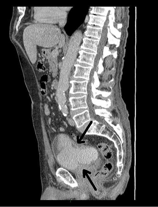
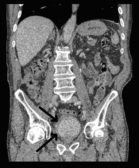
An 80 year-old male, with no significant past medical history, was referred to our urology department after presenting to his GP with frank haematuria. He subsequently underwent a CT scan (Image 1, 2) which showed three moderate-sized papillary lesions on the right lateral wall of the bladder. Flexible cystoscopy confirmed the diagnosis, subsequently shown to be a G3 pT1 tumour. The decision was for him to undergo a transurethral resection of bladder tumour (TURBT) and Mitomycin-C instillation.


Image 1. CT scan, coronal 
CT Image_Coronal


Image 2. CT scan, saggital

CT Image_Sagittal


The following week he presented to the Accident &Emergency Department with chest pain. His ECG was normal and his pain rapidly settled. He was therefore discharged and advised to contact his GP if his symptoms persisted. The following day, he attended his GP again complaining of chest pain with shortness of breath and further episodes of haematuria. He was referred directly to the urology ward.

On admission, he was noted to have extensive bruising across his arms, abdomen and flanks. There was no history of traumatic injury, although the bruising had become more extensive over the past week.
Blood tests confirmed that he was anaemic, with a haemoglobin of 7.1 g/dL. His coagulation screen showed a prolonged APTT of 76.3 sec (normal range 22-30 sec) with a normal PT of 9.7 sec (normal range 9-12 sec). His Troponin T level was grossly elevated at 852 ng/L (normal range 0-13 ng/L), however, this was not due to myocardial infarction, as there was no significant rise in the Troponin T level at 12 hours.
In light of the abnormal coagulation results, a haematology consult was sought. A FVIII assay result of 1.2 u/dl (normal range 50-150 u/dl) and a Bethesda assay result of 17.3 Beth.U confirmed a diagnosis of acquired haemophilia. He was transfused three units of blood and was commenced on rFVIIa (NovoSeven) at 90 µg/kg and prednisolone at 1 mg/kg, with proton pump inhibitor (PPI) cover. Subsequent testing, including autoimmune screening, anti-lupus anticoagulant and viral serology was carried out to try to determine the underlying aetiology, however, these failed to yield a positive result. A renal ultrasound scan was also normal. It was concluded, therefore, that the most likely explanation for the acquired haemophilia was his bladder cancer.
The decision was made to bring forward the planned TURBT in the hope that this would lead to a resolution of the acquired haemophilia. It was performed with preoperative and postoperative rFVIIa cover. Postoperatively, there appeared to be an early response following removal of the bladder tumour; FVIII levels improved from 1.2 u/dl to 6.5 u/dl, although he did require further blood transfusion. The haematuria settled and the catheter removed the following day.
Unfortunately, three days later, he developed clot retention. This became a recurring problem throughout admission, requiring multiple three-way catheter insertions, irrigation and bladder washouts. The clinical course has been summarised in Figure 1.
Figure 1. Clinical course summary
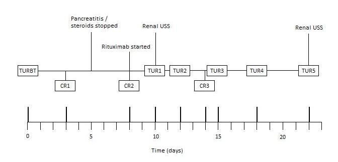
Figure 1fitzpatrick case rep

He developed acute abdominal pain with associated nausea. Blood tests revealed an amylase of 1641 units per liter (U/L) and he was diagnosed with pancreatitis. With no history of alcohol excess and no gallstones identified on prior imaging, the underlying aetiology was believed to be steroid-induced and his prednisolone was subsequently stopped. Treatment with intravenous fluids was successful, although he required further blood transfusion and rFVIIa. Rituximab was started by the haematologists.

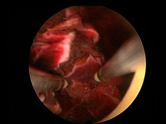
In order to further investigate the continuing haematuria, an ultrasound scan was arranged. This revealed a substantial clot within the bladder, approximately 6 x 6 cm in size. He was taken to theatre and a transurethral evacuation of the clot was carried out. The procedure was technically difficult and took around three hours to perform. The clot was thick, adhesive and organised, with resected clot reattaching to the bladder wall (Image 3, 4). Unfortunately, only part of the clot could be successfully evacuated.
Image 3.
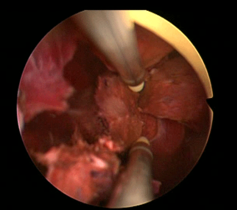
Image-3


Image 4.

Image-4


Two days later, he was taken back to theatre for a second attempt to evacuate the remainder of the clot. Rituximab and rFVIIa were given preoperatively. Once again, the resection was difficult and prolonged; after four hours of resection, only a small portion had been removed. The procedure was stopped, as the patient was becoming hypothermic and acidotic. Two units of blood were transfused.
Third and fourth evacuations were arranged to remove the residual clot. The third took approximately two hours. Warm irrigation fluid was used and some mobile organised clot remained at the end of the procedure. On the fourth attempt, the formed clot was cleared entirely. As with the previous resections, preoperative rVIIa and postoperative blood transfusion were required. It was encouraging to note that the APTT was improving gradually on rituximab and was now 68 sec.
As the APTT continued to improve, we decided to take a more conservative approach and see if it would correct with rituximab alone. The APTT improved further to 54 sec, however, repeat FVIII assay showed only a mild improvement (2.1 u/dL) and FVIII inhibitor was still present (17.8 Beth.U).
Unfortunately, over the next few days, the patient’s clinical condition deteriorated. A further seven units of blood were required during this time and his renal function worsened. An ultrasound scan revealed that, once again, there was a large formed clot in the bladder. A fifth cystoscopy was arranged, which confirmed a large organised clot occupying the whole bladder and blocking both ureteric orifices. The prognosis was poor and treatment had now become palliative. The clot was left in the hope that it might act to tamponade the bladder, however, the resulting obstructive uropathy meant that renal failure and hyperkalaemia developed; he passed away shortly after.

Discussion

The aetiology of AH is idiopathic in about 50 percent of cases, however, it is often associated with a variety of clinical conditions such as autoimmune disease (asthma, rheumatoid arthritis, systemic lupus erythematosus), solid tumours, lymphoproliferative disorders, drug hypersensitivity and pregnancy.[3,4,5,6] AH is associated with underlying malignancies in 7-15 percent of cases and is more prevalent in solid tumours than in haematological malignancies.[1,3,8,9] Reitter et al collated all published cases of cancer-associated FVIII autoantibodies from 1950-2010.[10] Although their analysis primarily targeted those cases in which FVIII autoantibodies developed following cancer surgery, their initial literature search also revealed those cases where AH had been diagnosed prior to surgery. Of the 57 cases uncovered, 32% (18/57) were associated with urological malignancies (14 prostate, 4 kidney), with prostate cancer the most prevalent amongst all cancer types.[10] Interestingly, there were no reported cases of AH secondary to bladder cancer; the only case found was in a patient who had developed FVIII autoantibodies following surgical treatment of a bladder tumour.[10]
AH due to underlying bladder cancer is extremely rare; a search of the literature only yielded two published cases, however, both have been multifactorial in their aetiologies.[11,12] In 2005, Kreuter et al reported AH in a patient with gram-negative urosepsis following resection of a rectal tumour, who underwent resection of a bladder tumour 6 months later.[11] Kato et al described AH in a patient with concomitant bladder and renal tumours.[12] To our knowledge, therefore, this is the first reported case of AH that can be attributed solely to bladder cancer.
The treatment of acquired haemophilia has been a much debated topic, in part due to its rarity and clinical diversity, with management plans often determined by the individual experiences of haematologists and on a case-by-case basis. As a result, data on the efficacy of anti-haemorrhage drugs is often retrospective.[1,13] There exists, however, generally accepted principles, primarily (1) controlling bleeding episodes, (2) avoiding invasive procedures that may precipitate further bleeding, (3) eradicating FVIII inhibitor through immunosuppression and (4) treating any underlying disease. [1,2,3,5]

Controlling bleeding episodes
The two main first-line agents that are licensed for the treatment of AH are rFVIIa (NovoSeven) and activated prothrombin complex concentrate (aPCC) (FEIBA; Factor VII inhibitor bypassing activity). [1,14] In our case, rFVIIa was adopted as the primary agent, using the standard recommended dose of 90 µg/kg.1 Such bypassing agents, however, are associated with an increased risk of arterial thrombosis, often in patients with underlying cardiovascular risk factors. A literature review demonstrated this in 7% of patients treated with rFVIIa.[14] The prothrombotic properties of bypassing agents are clearly useful in the cessation of bleeding episodes; however, it has been this very effect that has complicated our management. On one hand, rVIIa has been able to control bleeding and haematuria, however, it has also predisposed to the formation of further bladder clots, leading to recurrent catheter blockages and multiple clot resections.

Eradication of FVIII inhibitor
There is no definitive optimal regimen for inhibitor eradication. The most commonly adopted strategy is to use either corticosteroids alone or corticosteroids in combination with cyclophosphamide, with a lack of evidence supporting the choice of one over the other.[1,2,6] Previous studies have suggested that 70-80% of patients will achieve remission on either regimen; however, different durations of therapy were used in each.[13,15] In our case, prednisolone alone was used at 1 mg/kg; this is the dose most widely recommended in the current literature.1 The average time to remission with corticosteroids is about five weeks.[1,15] Unfortunately, we were forced to discontinue this treatment as our patient developed steroid-induced pancreatitis. It must be noted, however, that a mild improvement in APTT and FVIII levels had occurred up until this point.
If remission is not achieved with corticosteroids or cyclophosphamide, third-line immunosuppressive agents can be introduced. One of the most widely used drugs, which was used in our case, is rituximab. A cautious approach must be adopted with this therapy, particularly in elderly patients, due to potential serious side-effects; neutropenia and subsequent sepsis have been reported with associated increased patient mortality.[13] Despite this, rituximab has been shown to be successful in achieving remission of AH in a number of cases.[16,17,18]

Treatment of underlying disease
Although immunosuppressive therapy is paramount in inducing remission of AH, it is also important to treat the underlying aetiological disease.[1,2,3,5] In general, tumour-specific therapy has not been observed to cause the disappearance of FVIII inhibitors, however, there have been cases where surgical resection of causative tumours, following remission by immunosuppressive therapy, has prevented the recurrence of AH.[3,16,19,20] In contrast, however, there is some evidence to support the development of FVIII auto-antibodies following surgical resection of solid tumours.[10] Reitter et al report 13 cases where such antibodies were detected after an average of 3 months following uncomplicated cancer surgery.[10] These patients had not been diagnosed with AH prior their operation, determined by a normal APTT and no bleeding during and immediately after surgery.[10] It would appear that, although malignancies have been linked to causing AH, the development of FVIII inhibitors can be viewed as a paraneoplastic phenomenon.

Conclusion

The take home message is, that due to the tendancy of bladder tumours to bleed, and the occurrence of post-resection bleeding, haemophilia should be treated by taking into consideration the ensuing clot formation within the bladder. Furthermore, bladder tumours should be considered as part of the differential diagnosis for AH if all other causes have been excluded. This manuscript represents the first reported case of acquired haemophilia caused by a bladder tumour.

JF and OA both have first authorship contribution by writing the case report.

References
1. Collins P, Baudo F, Huth-Kühne A, Ingerslev J, Kessler CM, Castellano ME, Shima M, St-Louis J, Lévesque H. Consensus recommendations for the diagnosis and treatment of acquired hemophilia A. BMC Res Notes. 2010 Jun 7;3:161.
2. Morrison AE, Ludlam CA. Acquired haemophilia and its management. Br J Haematol. 1995;89(2):231–236
3. Baudo F, de Cataldo F. Acquired hemophilia: a critical bleeding syndrome. Haematologica. 2004 Jan;89(1):96-100.
4. Elezović I. Acquired haemophilia syndrome: pathophysiology and therapy. Srp Arh Celok Lek. 2010 Jan;138 Suppl 1:64-8
5. Baudo F, Caimi T, de Cataldo F. Diagnosis and treatment of acquired haemophilia. Haemophilia. 2010 May;16(102):102-6.
6. Moccia F, Tognoni E, Boccaccio P. Acquired factor VIII inhibitor associated with prostatic cancer: successful treatment with steroid and immunosuppressive therapy. Ann Ital Med Int. 2000 Apr-Jun;15(2):172-6.
7. Zeitler H, Ulrich-Merzenich G, Goldmann G, Vidovic N, Brackmann HH, Oldenburg J. The relevance of the bleeding severity in the treatment of acquired haemophilia – an update of a single-centre experience with 67 patients. Haemophilia. 2010 May;16(102):95-101. Epub 2008 Nov 14.
8. Ferre A, Arlet JB, Darnige L, Dupeux S, Pouchot J. Acquired hemophilia as the presenting manifestation of neoplasia: diagnostic workup and monitoring. Rev Med Interne. 2009 Jul;30(7):630-3. Epub 2008 Oct 23.
9. Green D, Lechner K. A survey of 215 non-hemophilic patients with inhibitors to Factor VIII. Thromb Haemost. 1981 Jun 30;45(3):200-3.
10. Reitter S, Knoebl P, Pabinger I, Lechner K. Postoperative paraneoplastic factor VIII auto-antibodies in patients with solid tumours. Haemophilia. 2011 Apr 4.
11. Kreuter M, Retzlaff S, Enser-Weis U, Berdel WE, Mesters RM. Acquired haemophilia in a patient with gram-negative urosepsis and bladder cancer. Haemophilia. 2005 Mar;11(2):181-5.
12. Kato T, Masui K, Yoshida T, Soma T, Mishina M, Okuno H, Okuno Y, Terashima T, Minamiguchi S. Acquired hemophilia A developing at bilateral renal bleeding: a case report. Hinyokika Kiyo. 2009 Apr;55(4):215-8.
13. Collins PW, Hirsch S, Baglin TP, Dolan G, Hanley J, Makris M, Keeling DM, Liesner R, Brown SA, Hay CR; UK Haemophilia Centre Doctors’ Organisation. Acquired hemophilia A in the United Kingdom: a 2-year national surveillance study by the United Kingdom Haemophilia Centre Doctors’ Organisation. Blood. 2007 Mar 1;109(5):1870-7. Epub 2006 Oct 17.
14. Sumner MJ, Geldziler BD, Pedersen M, Seremetis S. Treatment of acquired haemophilia with recombinant activated FVII: a critical appraisal. Haemophilia. 2007;13(5):451–461.
15. Collins PW. Treatment of acquired hemophilia A. J Thromb Haemost. 2007;5(5):893–900.
16. Ichikawa S, Kohata K, Okitsu Y, Suzuki M, Nakajima S, Yamada MF, Onishi Y, Yamamoto J, Suzuki S, Ishizawa K, Kameoka J, Harigae H. Acquired hemophilia A with sigmoid colon cancer: successful treatment with rituximab followed by sigmoidectomy. Int J Hematol. 2009 Jul;90(1):33-6. Epub 2009 May 30
17. Machado P, Raya JM, Martín T, Morabito L, Brito ML, Rodríguez-Martín JM. Successful response to rituximab in two cases of acquired haemophilia refractory to standard-therapy. Int J Hematol. 2008 Jun;87(5):545-9. Epub 2008 Apr 15.
18. Field JJ, Fenske TS, Blinder MA. Rituximab for the treatment of patients with very high-titre acquired factor VIII inhibitors refractory to conventional chemotherapy. Haemophilia. 2007 Jan;13(1):46-50.
19. Hayashi T, Morishita E, Asakura H, Nakao S. Two cases of acquired hemophilia A in elderly patients. Nippon Ronen Igakkai Zasshi. 2010;47(4):329-33.
20. Hauser I, Leckner K. Solid tumors and FVIII antibodies. Thromb Haemost 1999; 82:1005-7.


Date added to bjui.org: 02/07/2012

DOI: 10.1002/BJUIw-2011-098-web


© 2024 BJU International. All Rights Reserved.