Tag Archive for: transperineal

Posts

Article of the Month: MRI supported transperineal prostate biopsy

Every Month, the Editor-in-Chief selects an Article of the Month from the current issue of BJUI. The abstract is reproduced below and you can click on the button to read the full article, which is freely available to all readers for at least 30 days from the time of this post.

In addition to the article itself, there is an accompanying editorial written by a prominent member of the urological community. This blog is intended to provoke comment and discussion and we invite you to use the comment tools at the bottom of each post to join the conversation.

If you only have time to read one article this week, it should be this one.

Multicentre evaluation of magnetic resonance imaging supported transperineal prostate biopsy in biopsy‐naïve men with suspicion of prostate cancer


Nienke L. Hansen*1, Tristan Barrett*, Claudia Kesch, Lana Pepdjonovic§, David Bonekamp, Richard OSullivan**, Florian Distler, Anne Warren*††, Christina Samel‡‡Boris Hadaschik2, Jeremy Grummet§ and Christof Kastner*§§
*CamPARI Clinic, Department of Radiology, Addenbrookes Hospital and University of Cambridge, Cambridge, UK, Department of Urology, University Hospital Heidelberg, Heidelberg, Germany, §Australian Urology Associates and Department of Surgery, Central Clinical School, Monash University, Melbourne, Vic., Australia, Department of Radiology, German Cancer Research Center (DKFZ), Heidelberg, Germany, **Healthcare Imaging and Monash University, Melbourne, Vic., Australia, ††Department of Pathology, Addenbrookes Hospital and University of Cambridge, Cambridge, UK, ‡‡Institute of Medical Statistics, Informatics and Epidemiology, University Hospital Cologne, Cologne, Germany, and §§Department of Urology, Addenbrookes Hospital and University of Cambridge, Cambridge, UK 


Current addresses: 1Department of Diagnostic and Interventional Radiology University Hospital Cologne Cologne Germany, 2Department of Urology University Hospital Essen Essen Germany. 


B.H., J.G., and C.K. contributed equally to this work.


Abstract

Objectives

To analyse the detection rates of primary magnetic resonance imaging (MRI)‐fusion transperineal prostate biopsy using combined targeted and systematic core distribution in three tertiary referral centres.

Patients and Methods

In this multicentre, prospective outcome study, 807 consecutive biopsy‐naïve patients underwent MRI‐guided transperineal prostate biopsy, as the first diagnostic intervention, between 10/2012 and 05/2016. MRI was reported following the Prostate Imaging‐Reporting and Data System (PI‐RADS) criteria. In all, 236 patients had 18–24 systematic transperineal biopsies only, and 571 patients underwent additional targeted biopsies either by MRI‐fusion or cognitive targeting if PI‐RADS ≥3 lesions were present. Detection rates for any and Gleason score 7–10 cancer in targeted and overall biopsy were calculated and predictive values were calculated for different PI‐RADS and PSA density (PSAD) groups.

Results

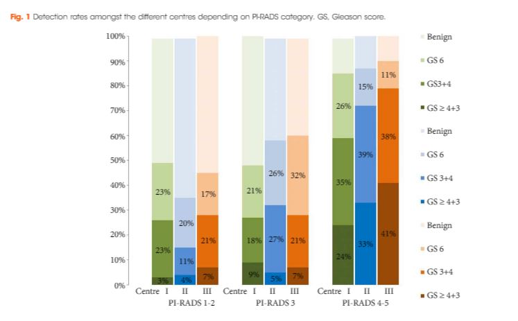
Cancer was detected in 68% of the patients (546/807) and Gleason score 7–10 cancer in 49% (392/807). The negative predictive value of 236 PI‐RADS 1–2 MRI in combination with PSAD of <0.1 ng/mL/mL for Gleason score 7–10 was 0.91 (95% confidence interval ± 0.07, 8% of study population). In 418 patients with PI‐RADS 4–5 lesions using targeted plus systematic biopsies, the cancer detection rate of Gleason score 7–10 was significantly higher at 71% vs 59% and 61% with either approach alone (P < 0.001). For 153 PI‐RADS 3 lesions, the detection rate was 31% with no significant difference to systematic biopsies with 27% (P > 0.05). Limitations include variability of multiparametric MRI (mpMRI) reading and Gleason grading.

Conclusion

MRI‐based transperineal biopsy performed at high‐volume tertiary care centres with a significant experience of prostate mpMRI and image‐guided targeted biopsies yielded high detection rates of Gleason score 7–10 cancer. Prostate biopsies may not be needed for men with low PSAD and an unsuspicious MRI. In patients with high probability lesions, combined targeted and systematic biopsies are recommended.

Editorial: Systematic transperineal and MRI‐targeted biopsies: the resolution of uncertainty

The paper published in this issue of the BJUI titled ‘Multicentre evaluation of magnetic resonance imaging supported transperineal biopsy in biopsy‐naïve men with suspicion of prostate cancer’ is timely and helps to resolve some of the uncertainty inherent within the diagnostic pathway 1.

The publication of the PROstate MRI Imaging Study (PROMIS) study, although demonstrating that 25% of patients might avoid prostate biopsy with a normal MRI (Prostate Imaging Reporting and Data System [PI‐RADS] 1–2) and that MRI could identify 90% of patients with high‐risk disease (PI‐RADS 5), did not resolve the issue of what to do with equivocal PI‐RADS 3 scans, uncertainty remained 2. The recent publication of the PRECISION trial (Prostate Evaluation for Clinically Important Disease: Sampling Using Image Guidance or Not?) has only contributed to the uncertainty of systematic TRUS biopsy and has shown that targeted biopsies resolve the issue for <50% of the patients overall and only 12% of those with PI‐RADS 3 lesions had a diagnosis of cancer on targeted biopsy only 3. The study has shown that in the face of an identifiable lesion a MRI‐targeted biopsy is non‐inferior to a blind systematic TRUS biopsy, which was positive in only 28% and implies that a systematic biopsy may be unnecessary, so where does that leave us? The uncertainty within MRI remains at the PI‐RADS 3 level, and particularly with a TRUS biopsy that is not a systematic biopsy of the peripheral zone. The authors of the paper highlighted in this issue of the BJUI 1 help to resolve the issue because they describe a more systematic biopsy.

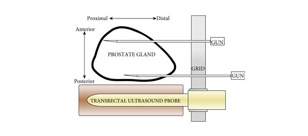
The transperineal (TP) biopsy approach for systematic and targeted biopsy they use is that which was adopted by the Ginsburg Study Group on Enhanced Prostate Diagnostics 4. It is a systematic biopsy that preferentially targets the peripheral zone in a sectoral fashion. It avoids the oversampling inherent in template‐mapping biopsy and the under‐sampling of the non‐systematic transrectal biopsy. Their paper evaluates the combination of an MRI‐targeted biopsy with a systematic TP biopsy. It confirms, as suggested by the PROMIS study, that patients with PI‐RADS 1 or 2 prostates on MRI with a low PSA density <0.1 ng/mL/mL could safely avoid biopsy, based upon a negative predictive value of 0.91 on systematic biopsy. However, in 418 patients with PI‐RADS 4–5 lesions, it was the combination of a targeted and systematic TP biopsy that achieved an overall cancer detection rate of 71%, but that MRI‐targeted biopsies alone had a detection rate of 59% vs 61% for systematic TP biopsies. In the PI‐RAD 3 equivocal group the combined biopsy identified 30% with Gleason score 7–10, whereas targeted biopsy only was positive in 21% vs 27% with systematic biopsies.

The message is clear.

An appropriate systematic biopsy targeted to the peripheral zone remains an essential component of prostate diagnosis even in the MRI era, as indeed it did before MRI was available. In the pre‐MRI days, about one‐third of patients that had negative TRUS biopsies had cancer on TP biopsies and a third of those thought suitable for AS on TRUS biopsy had more significant disease. I suspect in the modern era that figure remains unchanged for those with PI‐RADS 1, 2 or 3, particularly with a PSA density >0.15 ng/mL/mL. As urologists we have always been criticised for over diagnosing and over treating prostate cancer but I suspect that the more heinous crime is that of under treatment of significant disease, it is the very reason why I started doing TP biopsies, to resolve uncertainty. I consider that MRI, for all its benefits in the diagnostic algorithm, cannot yet resolve that uncertainty.

Probably the only patients that merit a target‐only biopsy are those with the high‐PSA, large‐volume disease, easily visible on MRI and usually palpable. Prostate biopsy can be avoided or at least deferred in the PI‐RADS 1–2 group with low PSA density; the rest should be offered a systematic biopsy along with a targeted biopsy. This may be less important in those proceeding to whole gland treatment or surgical extirpation but remains essential in those considering active surveillance, brachytherapy, or any one of the myriad of unproven focal treatments becoming available. The authors should be congratulated for bringing some certainty to uncertainty.

Rick Popert
Urology Centre, Guys Hospital, London, UK


References
  • Hansen NL, Barrett T, Kesch C et al. Multicentre evaluation of magnetic resonance imaging supported transperineal prostate biopsy in biopsy‐naïve men with suspicion of prostate cancerBJU Int 2018122: 40–9


  • Ahmed HU, El‐Shater Bosaily A, Brown LC et al. Diagnostic accuracy of multi‐parametric MRI and TRUS biopsy in prostate cancer (PROMIS): a paired validating confirmatory studyLancet 2017389: 815–22


  • Kuru TH, Wadhwa K, Chang RT et al. Definitions of terms, processes and a minimum dataset for transperineal prostate biopsies: a standardization approach of the Ginsburg Study Group for Enhanced Prostate DiagnosticsBJU Int 2013112: 568–77


Article of the week: Prostate biopsy: shaking up the old standard

Every week the Editor-in-Chief selects the Article of the Week from the current issue of BJUI. The abstract is reproduced below and you can click on the button to read the full article, which is freely available to all readers for at least 30 days from the time of this post.

In addition to the article itself, there is an accompanying editorial written by a prominent member of the urological community. This blog is intended to provoke comment and discussion and we invite you to use the comment tools at the bottom of each post to join the conversation.

Finally, the third post under the Article of the Week heading on the homepage will consist of additional material or media. This week we feature a video of Dr Symons discussing his paper.

If you only have time to read one article this week, it should be this one.

Outcomes of transperineal template-guided prostate biopsy in 409 patients

James L. Symons*, Andrew Huo*, Carlo L. Yuen‡§, Anne-Maree Haynes*, Jayne Matthews, Robert L. Sutherland*, Phillip Brenner‡§ and Phillip D. Stricker†‡§

*Cancer Research Programme, Garvan Institute of Medical Research, St Vincent’s Prostate Cancer Centre, Department of Urology, St. Vincent’s Hospital, and §Department of Urology, St. Vincent’s Clinic, Darlinghurst, NSW, Australia

Read the full article
OBJECTIVE

• To present the template-guided transperineal prostate biopsy (TPB) outcomes for patients of two urologists from a single institution.

PATIENTS AND METHODS

• We conducted a prospective study of 409 consecutive men who underwent TPB between December 2006 and June 2008 in a tertiary referral centre using a standardized 14-region technique.

• The procedure was performed as day surgery under general anaesthesia with fluoroquinolone antibiotic cover.

• Follow-up took place within 2 weeks, during which time men were interviewed using a standardized template.

• Results were compared with those of the Australian national prostate biopsy audits performed by the Urological Society of Australia and New Zealand (USANZ).

RESULTS

• Indications for biopsy included elevated prostate-specific antigen (PSA) level (75%), with a median PSA level of 6.5 ng/mL, abnormal digital rectal examination (8%) and active surveillance (AS) re-staging (18%).

• The mean patient age was 63 years and two-thirds of patients were undergoing their first biopsy.

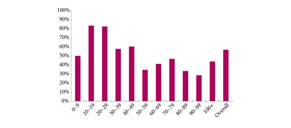
• A positive biopsy was found in 232 men, 74% of whom had a Gleason score of ≥7. The overall cancer detection rate was 56.7% (USANZ 2005 national audit = 56.5%). Stratified between those having their first TPB or a repeat procedure (after a previous negative biopsy), the detection rates were 64.4 and 35.6%, respectively. Significantly higher detection rates were found in prostates <50 mL in volume than in larger prostates (65.2 vs 38.3%, respectively, P < 0.001).

• Haematuria was the most common side effect (51.7%). Others included dysuria (16.4%), acute urinary retention (4.2%) and fever (3.2%). One patient (0.2%) had septicaemia requiring i.v. antibiotics.

• Repeat biopsy was not associated with increased complication rates.

CONCLUSIONS

• TPB is a safe and efficacious technique, with a cancer detection rate of 56.7% in the present series, and a low incidence of major side effects. Stratified by prostate volume, the detection rate of TPB was higher in smaller glands.

• Given the relatively low rate of serious complications, clinicians could consider increasing the number of TPB biopsy cores in larger prostates as a strategy to improve cancer detection within this group. Conversely, in patients on AS programmes, a staging TPB may be a superior approach for patients undergoing repeat biopsy so as to minimize their risk of serious infection.

Editorial: Contemplate the template: a new prostate biopsy approach

Transperineal magnetic resonance imaging – ultrasound fusion targeted biopsies (MRI-US FTB) of the prostate: the future of prostate diagnostics

The prostate cancer diagnostic pathway has remained unchanged for 25 years. At best, laterally directed, peripheral zone (PZ) 12-core transrectal biopsies identify cancer in 44% of cases [1] but transrectal biopsies have an inherent sampling error with a risk of misdiagnosis or mischaracterisation of disease. Of those with negative biopsies who undergo transperineal (TP) biopsies, 30% have cancer, most in the anterior PZ. Active surveillance and the promise of less invasive treatment options are becoming popular because of concerns about ‘over treatment’ for low-risk disease.

Saturation transrectal biopsies have been advocated to improve diagnostic yield but do not address the issue of under sampling of the anterior PZ, particularly in the larger gland [2]. TP biopsies can be used to address the issue of under sampling but prostate template-mapping biopsies are labour intensive and require large numbers of biopsies, often between 60 to 90 cores; however, they have been an essential component of focal therapy trials and the evaluation of novel treatment methods [3].

Primary TP biopsy is the subject of the paper published in this edition of the BJUI titled ‘Outcomes of transperineal template-guided prostate biopsy in 409 patients’ [4]. The authors report a single centre experience of primary TP biopsies. The 14-region protocol described is simpler than prostate template-mapping requiring fewer cores (median of 15 and mean of 19 cores) with a comparable primary diagnostic detection rate of 60% and an encouraging side-effect profile. Unfortunately, the approach still has limitations and the authors admit that their limited biopsy protocol may still mischaracterise disease in the larger gland. In a recent paper from the same group, there was a disappointing correlation between their TP biopsy pathology, MRI abnormalities and radical prostatectomy specimens [5]. Uncertainty prevails, the problem is how best to sample the larger gland. The authors [4] and others, often conclude that more biopsies are necessary for larger glands and resort to mapping protocols and many more biopsies. The solution may not be more biopsies but rather better systematic targeting of the PZ. The impact of hyperplasia within the transition zone (TZ) has a profound effect on PZ anatomy. In the smaller prostate, up to 30 mL, there is little TZ and the PZ is much thicker posteriorly than anteriorly, this difference is even more apparent in glands of 30–50 mL. Above 50 mL TZ expansion causes marked attenuation of the PZ, which becomes much thinner, but the overall volume of the PZ does not change. Less than 4% of cancers originate in the TZ [6], consequently biopsies should be concentrated primarily on the PZ.

The future of prostate cancer diagnosis is likely to be a combination of pre-biopsy multiparametric MRI, followed by targeted biopsies of MRI-identified lesions combined with fewer but better systematic targeted biopsies of the PZ. MRI-ultrasound (MRI-US) fusion techniques have been developed in which axial T2 images of the prostate, diffusion-weighted images and/or dynamic contrast-enhanced MRI images are ‘fused’ with the live US images to allow precise targeting of both regions of interest and the PZ. Commercially available biopsy programs, developed from brachytherapy software systems programs allow individual biopsy sites to be recorded and if combined with inking of the specimen can provide precise pathological localisation of disease within the prostate [7].

There are many potential benefits to this approach. Patients who opt for active surveillance will have an archived record of their disease at a given time to facilitate precise replication of further interval biopsies and assess progression. Improved disease management for an individual should be the aim. The suitability or not for focal or targeted therapies, the planning or boosting of identifed lesions with radiotherapy and/or brachytherapy, and the planning of nerve-sparing surgery or wide excisions should be possible. Feedback to the radiologists of both benign and malignant pathology and grade of disease will improve reporting accuracy and provide imaging sciences with the histopathological characteristics of both MRI ‘visible’ and ‘invisible’ cancer to improve MRI interpretation.

MRI–US fusion targeted biopsies are a significant advance in prostate diagnostics and may resolve some uncertainty within the prostate cancer diagnostic pathway. Benefit vs cost is a recurring issue across health care and questions will continue to be asked about the use of increasingly expensive technology in such an indolent disease. The challenge for investigators will be how to prove the benefit of this approach over standard biopsy protocols and integrate this work in to clinical practice.

Richard Popert
Department of Urology, Guy’s Hospital, London, UK

Read the full article
References
  1. Presti JC, O’Dowd GL, Miller MC et al. Extended peripheral zone biopsy schemes increase cancer detection rates and minimize variance in prostate specific antigen and age related cancer rates: results of a community multi-practice study. J Urol 2003; 169:125–129
  2. Stewart CS, Leibovich BC, Weaver AL, Lieber MM. Prostate cancer diagnosis using a saturation needle biopsy technique after previous negative sextant biopsies. J Urol 2001; 166: 86–92
  3. Onik G, Barzell W. Transperineal 3D mapping biopsy of the prostate: an essential tool in selecting patients for focal prostate cancer therapy. Urol Oncol 2008; 26: 506–510
  4. Symons JL, Huo A, Yuen CL et al. Outcomes of transperineal template-guided prostate biopsy in 409 patients. BJU Int 2013; 112: 585–593
  5. Huo AS, Hossack T, Symons JL et al. Accuracy of primary systematic template guided transperineal biopsy of the prostate for locating prostate cancer: a comparison with radical prostatectomy specimens. J Urol 2012; 187: 2044–2050
  6. Patel V, Merrick GS, Allen ZA et al. The incidence of transition zone prostate cancer diagnosed by transperineal template guided mapping biopsy: implications for treatment planning. Urology 2011; 77: 1148–1152
  7. Hadaschik BA, Kuru TH, Tulea C et al. A novel stereotactic prostate biopsy system integrating pre-interventional magnetic resonance imaging and live ultrasound fusion. J Urol 2011; 186: 2214–2220

Video: Transperineal prostate biopsy: how good is the tumour detection rate?

Outcomes of transperineal template-guided prostate biopsy in 409 patients

James L. Symons*, Andrew Huo*, Carlo L. Yuen‡§, Anne-Maree Haynes*, Jayne Matthews, Robert L. Sutherland*, Phillip Brenner‡§ and Phillip D. Stricker†‡§

*Cancer Research Programme, Garvan Institute of Medical Research, St Vincent’s Prostate Cancer Centre, Department of Urology, St. Vincent’s Hospital, and §Department of Urology, St. Vincent’s Clinic, Darlinghurst, NSW, Australia

Read the full article
OBJECTIVE

• To present the template-guided transperineal prostate biopsy (TPB) outcomes for patients of two urologists from a single institution.

PATIENTS AND METHODS

• We conducted a prospective study of 409 consecutive men who underwent TPB between December 2006 and June 2008 in a tertiary referral centre using a standardized 14-region technique.

• The procedure was performed as day surgery under general anaesthesia with fluoroquinolone antibiotic cover.

• Follow-up took place within 2 weeks, during which time men were interviewed using a standardized template.

• Results were compared with those of the Australian national prostate biopsy audits performed by the Urological Society of Australia and New Zealand (USANZ).

RESULTS

• Indications for biopsy included elevated prostate-specific antigen (PSA) level (75%), with a median PSA level of 6.5 ng/mL, abnormal digital rectal examination (8%) and active surveillance (AS) re-staging (18%).

• The mean patient age was 63 years and two-thirds of patients were undergoing their first biopsy.

• A positive biopsy was found in 232 men, 74% of whom had a Gleason score of ≥7. The overall cancer detection rate was 56.7% (USANZ 2005 national audit = 56.5%). Stratified between those having their first TPB or a repeat procedure (after a previous negative biopsy), the detection rates were 64.4 and 35.6%, respectively. Significantly higher detection rates were found in prostates <50 mL in volume than in larger prostates (65.2 vs 38.3%, respectively, P < 0.001).

• Haematuria was the most common side effect (51.7%). Others included dysuria (16.4%), acute urinary retention (4.2%) and fever (3.2%). One patient (0.2%) had septicaemia requiring i.v. antibiotics.

• Repeat biopsy was not associated with increased complication rates.

CONCLUSIONS

• TPB is a safe and efficacious technique, with a cancer detection rate of 56.7% in the present series, and a low incidence of major side effects. Stratified by prostate volume, the detection rate of TPB was higher in smaller glands.

• Given the relatively low rate of serious complications, clinicians could consider increasing the number of TPB biopsy cores in larger prostates as a strategy to improve cancer detection within this group. Conversely, in patients on AS programmes, a staging TPB may be a superior approach for patients undergoing repeat biopsy so as to minimize their risk of serious infection.

© 2024 BJU International. All Rights Reserved.