Tag Archive for: transitional cell carcinoma

Posts

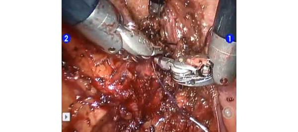
Video: Is LESS more when it comes to nephroureterectomy?

Laparoendoscopic single-site nephroureterectomy for upper urinary tract urothelial carcinoma: outcomes of an international multi-institutional study of 101 patients

Sung Yul Park, Koon Ho Rha1, Riccardo Autorino2, Ithaar Derweesh3, Evangelos Liastikos4, Yao Chou Tsai5, Ill Young Seo6, Ugo Nagele7, Aly M. Abdel-Karim8, Thomas Herrmann9, Deok Hyun Han10, Soroush Rais-Bahrami11, Seung Wook Lee, Kyu Shik Kim, Paolo Fornara12, Panagiotis Kallidonis4, Christopher Springer12, Salah Élsalmy8, Shih-Chieh Jeff Chueh13, Chen-Hsun Ho14, Kamol Panumatrassamee2, Ryan Kopp3, Jens-Uwe Stolzenburg15, Lee Richstone11, Jae Hoon Chung, Tae Young Shin1, Francesco Greco12 and Jihad H. Kaouk2

Department of Urology, Hanyang University College of Medicine, Seoul, Korea, 1Department of Urology, Yonsei University College of Medicine, Seoul, Korea, 2Glickman Urological & Kidney Institute, Cleveland Clinic, Cleveland, OH, USA, 3Division of Urology, University of California San Diego, La Jolla, CA, USA, 4Department of Urology, School of Medicine, University of Patras, Patras, Greece, 5Division of Urology, Buddhist Tzu Chi General Hospital, TaipeiBranch, Taipei, Taiwan, 6Department of Urology, Wonkwang University School of Medicine and Hospital, Iksan, Korea, 7Department of Urology, LKH, Hall in Tirol, Austria, 8Department of Urology, Alexandria University, Alexandria, Egypt, 9Department of Urology, Hannover Medical School, Hannover, Germany, 10Department of Urology, Samsung Medical Center, Sungkyunkwan University, Seoul, Korea, 11The Arthur Smith Institute for Urology, Hofstra North Shore-LIJ School of Medicine, New Hyde Park, NY, USA, 12Department of Urology and Kidney Transplantation, Martin-Luther-University, Halle/Saale, Germany, 13Cleveland Clinic Urology Charleston Office, Charleston, WV, USA, 14Division of Urology, National Taiwan University Hospital, Taipei, Taiwan, 15Department of Urology, University of Leipzig, Leipzig, Germany

OBJECTIVE

• To report a large multi-institutional series of laparoendoscopic single-site (LESS) nephroureterectomy (NU).

MATERIALS AND METHODS

• Data on all cases of LESS-NU performed between 2008 and 2012 at 15 institutions were retrospectively gathered.

• The main demographic data and perioperative outcomes were analysed.

RESULTS

• The study included 101 patients whose mean (sd) age was 66.4 (9.9) years and mean (sd) body mass index was 24.8 (4) kg/m2, and of whom 29.7% had undergone previous abdominal/pelvic surgery.

• The mean (sd) operating time was 221.4 (73.7) min, estimated blood loss 231.7 (348.0) mL.

• A robot-assisted LESS technique was applied in 25.7% of cases. An extra trocar was inserted in 28.7% of cases to complete the procedure. Conversion to open surgery was necessary in three cases (3.0%). There was no bladder cuff excision in 20.8% of cases, and excision was carried out using a variety of techniques in the remaining cases.

• Six intra-operative complications occurred (5.9%). The mean (sd) length of hospital stay was 6.3 (3.5) days. The overall postoperative complication rate was 10.0%, and most of the complications were low grade (Clavien grades 1 and 2).

• The mean tumour size was 3.1 (1.9) cm. Pathological staging was pTis in two patients, pTa in 12 patients, pT1 in 42 patients, pT2 in 20 patients, pT3 in 23 patients and pT4 in two patients. Pathological grade was high in 71 and low in 30 patients.

• At a mean follow-up of 14 months, six patients (5.9%) had died. Disease recurrence (including distant and bladder recurrence) was detected in 22.8% of patients, with a mean time to recurrence of 11.5 months.

CONCLUSIONS

• This study reports the largest multi-institutional experience of LESS-NU to date.

• Peri-operative outcomes mirror those of published standard laparoscopy series.

• Despite encouraging early findings, longer follow-up is needed to determine the oncological efficacy of the procedure.

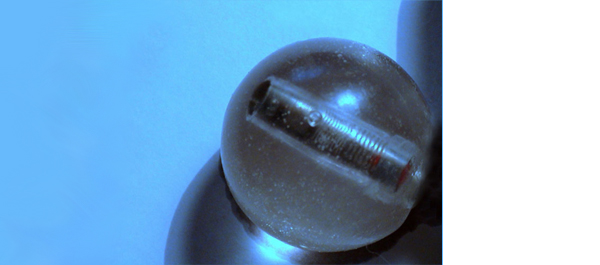
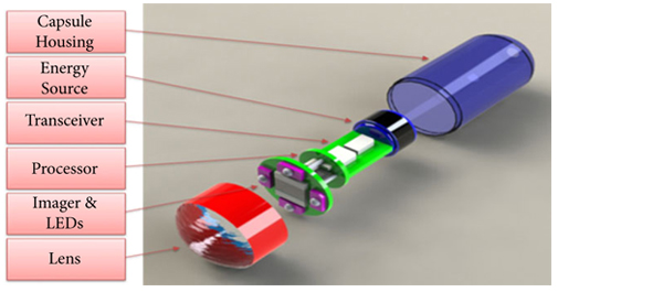
Article of the week: Preventing biofilm in wireless capsule bladder endoscopy

Every week the Editor-in-Chief selects the Article of the Week from the current issue of BJUI. The abstract is reproduced below and you can click on the button to read the full article, which is freely available to all readers for at least 30 days from the time of this post.

In addition to the article itself, there is an accompanying editorial written by a prominent member of the urological community. This blog is intended to provoke comment and discussion and we invite you to use the comment tools at the bottom of each post to join the conversation.

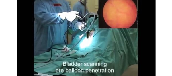
Finally, the third post under the Article of the Week heading on the homepage will consist of additional material or media. This week we feature a video of in-vivo trials in sheep .

If you only have time to read one article this week, it should be this one.

Novel anti-biofilm mechanism for wireless capsule endoscopy in the urinary tract: preliminary study in a sheep model

Amos Neheman*, Claude Schulman and Ofer Yossepowitch†§

*Urology Department, Meir Medical Center, Kfar-Saba, Sackler School of Medicine, Tel-Aviv University, Tel-Aviv, Israel, Department of Urology, University of Brussels, Brussels, Belgium, and §Institute of Urology, Rabin Medical Center, Beilinson, Petah Tikva, Israel

Read the full article
OBJECTIVE

• To develop and test the safety and feasibility of a novel anti-biofilm mechanism configured for wireless capsule endoscopy (WCE) in a sheep bladder model.

MATERIALS AND METHODS

• A WCE mechanism, designed for long-term bladder monitoring, was developed and introduced into a sheep bladder for 5 months.

• The transparency of the surface was assessed by evaluating a resolution target placed inside the capsule at serial intervals using cystoscopy under general anaesthesia.

• Animal behaviour, voiding patterns and urine cultures were monitored throughout the study.

• At study termination, the capsule was extracted and assessed using scanning electron microscopy.

RESULTS

• The resolution target was visualized clearly at all investigation points.

• No notable adverse effects were noted during the entire follow-up period and no urinary tract infection occurred.

• Scanning electron microscopy confirmed the efficacy of the technology to prevent biofilm formation and surface encrustation.

CONCLUSIONS

• We report a novel technology that effectively prevents biofilm formation on the outer surface of foreign objects in the urinary tract.

• Further studies are under way to test the applicability of this technology in bladder WCE to enable high-quality wireless image transmission.


Read Previous Articles of the Week


Editorial: A promising solution for biofilm inhibition in the bladder, but is the application of wireless capsule cystoscopy practical?

The study by Neheman et al. follows up on an idea first proposed in 2009 by Gettman and Swain to adapt wireless capsule endoscopy (WCE) technology for cystoscopy. Unlike the gastrointestinal tract where the small bowel is not endoscopically accessible making WCE appealing and advantageous, the idea of wireless capsule cystoscopy (WCC) competes with a minor procedure, office cystoscopy, that does not require anaesthesia or sedation and takes only a few minutes to perform. Furthermore, although the authors suggest that WCC would shift the labour associated with bladder cancer monitoring from practising urologists to ancillary health team providers, flexible office cystoscopy is a procedure already routinely performed by physician extenders in many offices. Nevertheless, the concept proposed by Neheman et al. is innovative and intriguing. The potential advantage of a wireless capsule cystoscope placed in the bladder safely for up to a 2-year time period, and thereby reducing the inconvenience and cost of repeated cystoscopies, could be a significant advance.

It should be emphasized that despite the title, no WCE was actually performed. The real value to the present study is the novel anti-biofilm mechanism developed that would be needed for any device implanted in the bladder for the long term. The device was housed in a semi-permeable silicone balloon filled with mineral oil that allowed a continuous slow diffusion of oil across the membrane. Based on the evidence provided in only one animal, it seems this continuous permeation of oil can interfere with surface protein adherence and consequently bacteria adhesion and biofilm creation. Certainly, this concept needs to be tested further in additional animals, aggressively exposed to bacteria, and for longer periods.

Although I am unconvinced that the concept of WCC provides significant value, the development of this biofilm inhibition technique could be pioneering. I read this study and wondered if ureteric stents and Foley catheters could be designed and impregnated with mineral oil to be released gradually. Perhaps the balloon of a Foley catheter could be redesigned and filled with mineral oil that is then released along the catheter’s entire length in a similar fashion. The true Holy Grail is the prevention of encrustation and biofilm formation on these relatively mundane devices whose chronic exchange for many patients is more costly than bladder cancer surveillance. I look forward to additional work from the authors exploring the potential of this technology.

Jeffrey A. Cadeddu
Department of Urology, University of Texas Southwestern Medical Center, Dallas, TX, USA

Read the full article

Video: In-Vivo Trials in Sheep


Novel anti-biofilm mechanism for wireless capsule endoscopy in the urinary tract: preliminary study in a sheep model

Amos Neheman*, Claude Schulman and Ofer Yossepowitch†§

*Urology Department, Meir Medical Center, Kfar-Saba, Sackler School of Medicine, Tel-Aviv University, Tel-Aviv, Israel, Department of Urology, University of Brussels, Brussels, Belgium, and §Institute of Urology, Rabin Medical Center, Beilinson, Petah Tikva, Israel

Read the full article
OBJECTIVE

• To develop and test the safety and feasibility of a novel anti-biofilm mechanism configured for wireless capsule endoscopy (WCE) in a sheep bladder model.

MATERIALS AND METHODS

• A WCE mechanism, designed for long-term bladder monitoring, was developed and introduced into a sheep bladder for 5 months.

• The transparency of the surface was assessed by evaluating a resolution target placed inside the capsule at serial intervals using cystoscopy under general anaesthesia.

• Animal behaviour, voiding patterns and urine cultures were monitored throughout the study.

• At study termination, the capsule was extracted and assessed using scanning electron microscopy.

RESULTS

• The resolution target was visualized clearly at all investigation points.

• No notable adverse effects were noted during the entire follow-up period and no urinary tract infection occurred.

• Scanning electron microscopy confirmed the efficacy of the technology to prevent biofilm formation and surface encrustation.

CONCLUSIONS

• We report a novel technology that effectively prevents biofilm formation on the outer surface of foreign objects in the urinary tract.

• Further studies are under way to test the applicability of this technology in bladder WCE to enable high-quality wireless image transmission.


Myelosuppression following adjuvant mitomycin C for upper tract urothelial carcinoma

To our knowledge, this is the first documented case of myelosuppression following adjuvant mitomycin C for upper tract urothelial carcinoma in the absence of evidence of chemotherapeutic agent extravasation.

Authors: Henry Han-I (HH) Yao1, Miguel Suhady (MS) Cabalag1, Antonio (A) DeSousa1, Gideon Adam (GA) Blecher1, Richard (R) McMullin1

1. Department of Urology, Ballarat Base Hospital, Ballarat, Victoria, Australia

Corresponding Author: Richard McMullin, Consultant Urologist, Ballarat Base Hospital, Drummond Street North, Ballarat, Victoria, Australia 3350  Email: [email protected]


Abstract
 
A 56-year-old Caucasian female with a background of left nephroureterectomy for upper tract urothelial carcinoma was diagnosed 14 months later with a right renal pelvis urothelial carcinoma. This was managed by open resection of the affected segment and preservation of her solitary kidney. Adjuvant upper tract mitomycin C therapy was started six weeks later. Prior to each cycle, a retrograde pyelogram was performed intra-operatively following the insertion of ureteral ureteric catheter, and this did not demonstrate contrast extravasation on any occasion. Eleven days following her fourth cycle, she presented with febrile neutropaenia and pancytopaenia. She was admitted to hospital and successfully managed with broad spectrum antibiotics. Her bone marrow biopsy revealed bone marrow suppression with no significant blast population. Her leucocyte and platelet counts eventually improved without transfusional support. Therefore, her myelosuppression was most likely secondary to the systemic absorption of mitomycin C instilled into her renal tract. To our knowledge, this is the first documented case of myelosuppression following adjuvant mitomycin C for upper tract urothelial carcinoma in the absence of evidence of chemotherapeutic agent extravasation. Clinicians should have a heightened awareness of this complication and monitor patient’s blood counts before and during adjuvant upper tract mitomycin C therapy.


Introduction


The use of adjuvant therapy in nephron sparing management of upper tract urothelial carcinoma has been documented for over a decade (1-3). However, due to the rarity of upper tract urothelial carcinoma, there is limited data evaluating the safety and efficacy of such practices (4, 5). To the best of our knowledge, we hereby report the first case of myelosuppression following adjuvant mitomycin C for upper tract urothelial carcinoma in the absence of evidence of chemotherapeutic agent extravasation.


Case Report


A 56-year-old Caucasian female who had undergone left nephroureterectomy for left upper tract urothelial carcinoma presented to a urological practice 14 months later with haematuria and right loin pain. Apart from a history of urothelial carcinoma and upper lip squamous cell carcinoma, both surgically treated in the past, the patient did not have an immunosuppressive medical condition nor was she taking any immunosuppressive medication. She was investigated with a computed tomography intravenous urogram which revealed a suspicious filling defect in the right renal pelvis consistent with a recurrent urothelial carcinoma. After careful discussion and counselling, this was managed by an open resection of the affected right renal pelvis segment and preservation of her solitary right solitary kidney. Cystoscopy, retrograde pyelogram (Figure 1) and limited ureteroscopy were performed prior to the excision and did not reveal any identifiable lesion in the bladder and ureter.


Figure 1. Intra-operative retrograde pyelogram demonstrating a suspicious filling defect in the right renal pelvis.
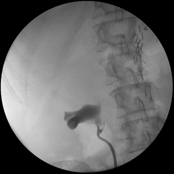
Figure-1web-2012-049

Histology of the right renal pelvis lesion subsequently confirmed it to be a urothelial carcinoma with no evidence of muscle invasion. Following further discussion and counselling, she consented for a six weeks course of adjuvant right upper tract mitomycin C therapy. The first course of mitomycin C was started 6 weeks following the initial operation.

For each upper tract mitomycin C therapy, the patient underwent a theatre-based approach for the placement of ureteral ureteric catheter. Cystoscopy was performed under general anaesthesia, the right ureteral ureteric orifice was visualised and cannulated with a slippery wire over which a 5 French ureteric catheter was passed through. Intra-operative retrograde pyelogram was performed each time and did not demonstrate contrast extravasation on any occasion (Figure 2).
Figure 2. Intra-operative retrograde pyelograms prior to the instillation of the second, third and fourth courses of upper tract adjuvant mitomycin C therapy, respectively from A to C.
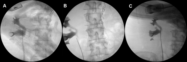
Figure-2web2012-049

The ureteral ureteric catheter was secured to a two-way urinary catheter at the completion of each procedure. Post-operatively, pre-made prepared mitomycin C was instilled into the ureteral ureteric catheter over two hours and was allowed to drain out freely from the urethral catheter. Both the ureteral ureteric and urethral catheters were removed post-procedure. Her first mitomycin C instillation was delivered via a syringe driver which delivered 40mg of mitomycin C in 40mls of sterile water at a rate of 20mls per hour. However, her subsequent instillations each consisting of 40mg of mitomycin C in 80mls of sterile water were delivered by an infusion bag hanged hung at approximately one metre above the kidney level, at a rate of 40mls per hour.

The patient experienced some associated nausea, vomiting and irritative urinary symptoms with each cycle of chemotherapy, all of which were self-limiting. However, following her fourth cycle of chemotherapy, she became progressively unwell with worsening nausea, vomiting, anorexia and malaise. She also developed symptoms of mucosal bleeding, alopecia and a metallic taste in her mouth. On retrospective questioning, she developed haematuria only following the third and the fourth cycles of chemotherapy. Eventually, eleven days following her fourth cycle of upper tract mitomycin C, she presented with a fever which was subsequently diagnosed as febrile neutropaenia with a neutrophil count of 0.1 × 10⁹/L. She also developed pancytopaenia with a haemoglobin of 81 g/L, leucocyte count of 0.2 × 10⁹/L and platelet count of 27 × 109/L. The source of her infection was later found to be urosepsis from a sensitive Escherichia coli.

The patient was admitted and successfully treated with broad spectrum antibiotics, fluid management, monitoring of her blood counts and further investigations. Her bone marrow aspirate and trephine revealed marked bone marrow hypoplasia with no abnormal cells seen to suggest a leukaemic process. Subsequent flow cytometry showed no significant blast population (~2%). The patient’s leucocyte and platelet counts gradually improved over the next two weeks in the absence of transfusional support. Given these findings, the most plausible explanation for her pancytopaenia would be myelosuppression secondary to the absorption of mitomycin C instilled into her renal tract. Eleven days following her initial presentation and after receiving a transfusion of two units of red blood cells transfusion, she was discharged home on oral antibiotics and regular blood count monitoring. Adjuvant mitomycin C therapy was ceased following this episode.

Four months following her initial surgery, she underwent a surveillance right flexible ureteroscopy which did not reveal any recurrence of cancer in the renal pelvis, ureter, bladder or urethra. A retrograde pyelogram performed intra-operatively did not reveal any filling defect. Similarly, her repeat computed tomography scan around that time was essentially unremarkable. However, her upper tract washings at the time of flexible ureteroscopy did reveal cells suspicious for malignancy. She was investigated with a repeat flexible ureteroscopy and intra-operative retrograde pyelogram three months later, which once again did not reveal any visible recurrence (Figure 3).
Figure 3. Intra-operative retrograde pyelogram six months following open resection of affected renal pelvis segment.
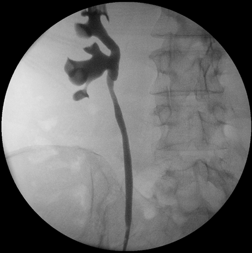
Figure-3-web-2012-049


This time, upper tract washings performed intra-operatively was negative for malignancy. Her next flexible ureteroscopy will once again be in a further 3 months time.

Discussion

Upper tract urothelial carcinoma is rare and accounts for only approximately 5% of all urothelial neoplasms (4, 6, 7), with an estimated annual incidence of one to two new cases per 100,000 inhabitants in Western countries (7). As such, the role of adjuvant therapy in non-muscle invasive upper tract urothelial carcinoma has not been firmly established due to its rarity (4, 5). In the literature, there is only limited data consisting of mainly small retrospective studies and expert opinions to guide clinicians in decision making (6). To our knowledge, there has not been any randomised controlled trials evaluating this clinical question (4, 5).

For a localized upper tract urothelial carcinoma in a solitary kidney, an open nephron-sparing surgery may be indicated (7) and preferred over laparoscopic partial nephrectomy which has a significant likelihood of tumour spillage and subsequent implantation (8). Given the presence of residual urothelium following a nephron sparing surgery, the risk of recurrence of urothelial carcinoma remains. The use of adjuvant therapy in nephron sparing management for upper tract urothelial carcinoma with the goal of not compromising the oncological outcome (5) have been documented for more than a decade (1-3). Small observational studies have shown some response to adjuvant therapy (2, 9-12). However, there has been no documented statistical improvement in survival and recurrence rates with the use of adjuvant therapy over no adjuvant therapy (5). Therefore, the use of adjuvant therapy in upper tract urothelial carcinoma remains to some extent anectodalanecdotal (5).

There is no clear evidence favouring the use of one adjuvant agent over another (5). Despite more limited data available on the use of mitomycin C, it may be a safer option due to the risk of tuberculosis infection associated with adjuvant Bacillus Calmette-Guérin therapy (13, 14). When used intravesically, the most commonly reported adverse effects of mitomycin C are chemical cystitis and allergic skin reactions (15). Other rare localised adverse effects of intravesical mitomycin C have also been reported in the literature and includes incrusted cystitis (16), eosinophilic cystitis (17), bladder perforation (18), necrosis of the corpus spongiosum (19) and distal ureteral stenosis (20). However, the systemic absorption of mitomycin C is very limited due to its high molecular weight of 334.33 (5, 15). As a consequence, reported systemic side effects of intravesical mitomycin C such as prolonged haematuria, fever, chills, nausea, vomiting and fatigue is usually mild and rare (15, 21, 22). Severe myelosuppression, a known toxicity of systemic mitomycin C (23), is certainly extremely rare following intravesical mitomycin C with only a few reported cases (15). In the use of mitomycin C for upper tract urothelial carcinoma this has only been documented in a fatal case of extravasated mitomycin C (9).

The case reported here demonstrates that systemic absorption of mitomycin C, albeit rare, is a possibility even in a urinary tract shown to be intact by retrograde pyelogram just prior to therapy. The only difference in our procedure compared with those previously documented was the delivery technique. Although there are no firm evidence, previous reports have raised concern over increased intrarenal pressure from using syringe drivers or hanging the bag of mitomycin C more than 30cm above the kidney level (5). The first cycle of mitomycin C in this case was delivered by a syringe driver at a rate of 20mls per hour, and the second to fourth cycles of mitomycin C was delivered by an intravenous bag suspended at 1m above the kidney level delivering the medication at a rate of 40mls/hour. Therefore, this may have been a contributing factor to the absorption of mitomycin C in this case. However, Keeley et al. however, documented a technique of infusing 10mls of mitomycin C over 2 minutes followed by clamping of the ureteral ureteric catheter for 10 minutes without any systemic absorption (2), even though the intra-renal pressure is likely to be raised by this.

Conclusion

Systemic absorption of mitomycin C causing myelosuppression can occur when used as an adjuvant therapy for nephron sparing management of upper tract urothelial carcinoma. Clinicians should take this into consideration when counselling patients and planning management. Previous reported methods of reducing pressure of mitomycin C delivery to the renal tract including avoiding the use of syringe drivers or hanging the bag less than 30cm above the kidney level should be employed. Furthermore, clinicians should have heightened awareness of this complication during treatment and patients should have monitoring of blood counts before and during the course of adjuvant mitomycin C therapy.

References

1. Jarrett TW, Sweetser PM, Weiss GH, Smith AD. Percutaneous management of transitional cell carcinoma of the renal collecting system: 9-year experience. J Urol. 1995 Nov;154(5):1629-35.
2. Keeley FX, Jr., Bagley DH. Adjuvant mitomycin C following endoscopic treatment of upper tract transitional cell carcinoma. J Urol. 1997 Dec;158(6):2074-7.
3. Elliott DS, Blute ML, Patterson DE, Bergstralh EJ, Segura JW. Long-term follow-up of endoscopically treated upper urinary tract transitional cell carcinoma. Urology. 1996 Jun;47(6):819-25.
4. Latchamsetty KC, Porter CR. Treatment of upper tract urothelial carcinoma: a review of surgical and adjuvant therapy. Rev Urol. 2006 Spring;8(2):61-70.
5. Nepple KG, Joudi FN, O’Donnell MA. Review of topical treatment of upper tract urothelial carcinoma. Adv Urol. 2009:472831.
6. Smith ND. Management of upper tract urothelial carcinoma. Adv Urol. 2009:492462.
7. Roupret M, Zigeuner R, Palou J, Boehle A, Kaasinen E, Sylvester R, et al. European guidelines for the diagnosis and management of upper urinary tract urothelial cell carcinomas: 2011 update. Eur Urol. 2011 Apr;59(4):584-94.
8. Flanigan R. Urothelial Tumours of the Upper Urinary Tract. 9th ed. Wein A, Kavoussi L, Novick A, Partin A, Peters C, editors. Philadelphia: Elsevier; 2007.
9. Martinez-Pineiro JA, Garcia Matres MJ, Martinez-Pineiro L. Endourological treatment of upper tract urothelial carcinomas: analysis of a series of 59 tumors. J Urol. 1996 Aug;156(2 Pt 1):377-85.
10. Katz MH, Lee MW, Gupta M. Setting a new standard for topical therapy of upper-tract transitional-cell carcinoma: BCG and interferon-alpha2B. J Endourol. 2007 Apr;21(4):374-7; discussion 7.
11. Thalmann GN, Markwalder R, Walter B, Studer UE. Long-term experience with bacillus Calmette-Guerin therapy of upper urinary tract transitional cell carcinoma in patients not eligible for surgery. J Urol. 2002 Oct;168(4 Pt 1):1381-5.
12. Nonomura N, Ono Y, Nozawa M, Fukui T, Harada Y, Nishimura K, et al. Bacillus Calmette-Guerin perfusion therapy for the treatment of transitional cell carcinoma in situ of the upper urinary tract. Eur Urol. 2000 Dec;38(6):701-4;discussion 5.
13. Yokogi H, Wada Y, Mizutani M, Igawa M, Ishibe T. Bacillus Calmette-Guerin perfusion therapy for carcinoma in situ of the upper urinary tract. Br J Urol. 1996 May;77(5):676-9.
14. Bellman GC, Sweetser P, Smith AD. Complications of intracavitary bacillus Calmette-Guerin after percutaneous resection of upper tract transitional cell carcinoma. J Urol. 1994 Jan;151(1):13-5.
15. Bolenz C, Cao Y, Arancibia MF, Trojan L, Alken P, Michel MS. Intravesical mitomycin C for superficial transitional cell carcinoma. Expert Rev Anticancer Ther. 2006 Aug;6(8):1273-82.
16. Pascual Regueiro D, Garcia Sanchez S, Oliva Encina J, Remon Garijo ML, Martinez Bengoechea J, Abril Baquero G. [Incrusted cystitis after Mitomicin-C]. Actas Urol Esp. 2005 Jul-Aug;29(7):715-8.
17. Clark T, Chang SS, Cookson MS. Eosinophilic cystitis presenting as a recurrent symptomatic bladder mass following intravesical mitomycin C therapy. J Urol. 2002 Apr;167(4):1795.
18. Racioppi M, Porreca A, Foschi N, Delicato G, Destito A, D’Addessi A. Bladder perforation: a potential risk of early endovesical chemotherapy with mitomycin C. Urol Int. 2005;75(4):373-5.
19. Neulander EZ, Lismer L, Kaneti J. Necrosis of the glans penis: a rare complication of intravesical therapy with mitomycin c. J Urol. 2000 Oct;164(4):1306.
20. Oehlschlager S, Loessnitzer A, Froehner M, Hakenberg OW, Manseck A, Wirth MP. Distal ureteral stenosis after early adjuvant intravesical mitomycin C application for superficial bladder cancer. Urol Int. 2003;70(1):74-6.
21. Wajsman Z, Dhafir RA, Pfeffer M, MacDonald S, Block A, Dragone N, et al. Studies of mitomycin C absorption after intravesical treatment of superficial bladder tumors. J Urol. 1984 Jul;132(1):30-3.
22. Thrasher JB, Crawford ED. Complications of intravesical chemotherapy. Urol Clin North Am. 1992 Aug;19(3):529-39.
23. Bradner WT. Mitomycin C: a clinical update. Cancer Treat Rev. 2001 Feb;27(1):35-50.

Date added to bjui.org: 22/06/2012

DOI: 10.1002/BJUIw-2011-149-web

Commonly used antiseptic agent, rare cause of anaphylaxis

We report a case of anaphylaxis attributed to the chlorhexidine component of Instillagel, presenting after a urological procedure, leading to him becoming unconscious and hypotensive.


Authors: Dr P Williams, Dr A Alkali, Department of Dermatology, University Hospital Aintree, Liverpool.
 
Corresponding Author: Dr P Williams, Department of Dermatology, University Hospital Aintree, Liverpool. E-mail: [email protected]


Abstract
 
Anaphylaxis to chlorhexidine is rare. We report a case of anaphylaxis attributed to the chlorhexidine component of Instillagel, presenting after a urological procedure, leading to him becoming unconscious and hypotensive. Urologists should be aware that urethral lubricants may contain chlorhexidine that can trigger anaphylaxis in susceptible individuals. Anaphylaxis should be considered a possible diagnosis when a patient collapses after a routine procedure. Investigation of suspected anaphylactic reactions is important to try and identify a likely trigger for a reaction and to help prevent further exposure and potential harm.


Case Report


A 71 -year old gentleman with transitional cell carcinoma of the bladder underwent a cystoscopy as part of his surveillance at our hospital. Past medical history included venous eczema only.
Intra-urethral instillagel® (containing lidocaine hydrochloride, chlorhexidine digluconate, methyl-4-hydroxybenzoate and propyl-4-hydroxybenzoate) was administered at the beginning of the procedure prior to urethral dilatation. The intra-operative procedure was entirely uneventful.
He was taken to the recovery room where he was noted to have an urticarial rash. Fifteen minutes later, he became unconscious with a blood pressure of 70/50mmHg.There was no bronchospasm, laryngospasm and no fever. He was diagnosed with anaphylaxis and he was given Intravenous chlorpheniramine, 200mg of iv hydrocortisone and colloids. He made a good recovery.
Reviewing his past medical history, he had developed an urticarial rash three months prior to this after having an identical procedure.
He subsequently had skin prick testing for lidocaine, chlorhexidine, instillagel®, gentamicin, latex and tropical fruits. All were negative apart from a 9.5mm positive result for chlorhexidine. No allergic antibodies to latex were detected and his mast cell tryptase levels were normal.
 

Discussion
 
Chlorhexidine is a synthetic cationic bisbiguanide that was first introduced as a disinfectant and antiseptic in 1954. It is also found in many commercially available products including mouthwashes, antiseptic creams and disinfectant solutions.
Chlorhexidine products are increasingly being recommended for dental hygiene, periodontal care, and implant surgery. Savacol® is frequently used by dental surgeons. The concentrated solution contains 2 mg/ml but when appropriately diluted, it becomes a 0.2% solution of chlorhexidine as a dental wash.
Chlorhexidine is also present in potentially hidden forms, such as a bactericidal coating for central venous catheters or as a component of urethral lubricants. Instillagel®® contains chlorhexidine 0.25%, lidocaine 2%, methyl hydroxybenzoate 0.06%, and propyl hydroxybenzoate 0.025% formulated as a gel; it is a commonly used antiseptic and anaesthetic lubricant. The Summary of Product Characteristics relating to Instillagel® advises against its use in individuals with known hypersensitivity to chlorhexidine, but only the lidocaine component is described as causing undesirable effects(1).
These ubiquitous applications of chlorhexidine raise the possibility of sensitization in a large proportion of the general population (2).Various types of hypersensitivity reactions to chlorhexidine have been described, including: contact dermatitis, photosensitive dermatitis, fixed drug eruption, contact urticaria, occupational asthma, and immediate hypersensitivity reactions such as severe anaphylactic shock, which is a rare but life-threatening complication (3). Life-threatening reactions are generally associated with mucosal exposure (1). Chlorhexidine may cause anaphylaxis by the mucosal route at a much lower concentration than elsewhere, generally as low as 0.05% (4, 5)
Interestingly, serum mast cell tryptase is not always raised in an anaphylactic reaction (as in our patient) as this may be due to an immediate basophilic response. Basophils are circulating blood cells that contain virtually no tryptase; in contrast, tryptase-rich mast-cells are present mainly in connective tissue and mucosae. An ELISA test for chlorhexidine has become available (ImmunoCap250®). The results of this test correspond well with skin-prick testing (2).
Application of chlorhexidine to the mucous membranes can cause severe anaphylactic reactions. Hypersensitivity to chlorhexidine is rare, but its potential to cause anaphylactic shock during hospital procedures is likely to be underestimated. We suggest that chlorhexidine should be used with caution and that it should routinely be considered as a causative agent in unexplained fatal reactions associated with medical procedures.


References
 
1. Instillagel®—Summary of Product Characteristics (2000) Clini-Med Ltd.
2. Jayathillake A, Mason DF, Broome K. Allergy to chlorhexidine gluconate in urethral gel: report of four cases and review of the literature. Urology 2003; 61: 837
3. Ebo DG, Bridts CH, Stevens WJ. Anaphylaxis to a urethral lubricant: chlorhexidine as the ‘‘hidden’’ allergen. Acta Clin Belg 2004; 59: 358-60
4. Wicki J, Deluze C, Cirafici L, Desmeules J. Anaphylactic shock induced by intraurethral use of chlorhexidine. Allergy 1999; 54: 768-9.
 5. Parkes AW, Harper N, Herwadkar A, Pumphrey R. Anaphylaxis to the chlorhexidine component of Instillagel®: a case series. Br J Anaesth. 2009 Jan;102(1):65-8.

 
Date added to bjui.org: 30/06/2011 


DOI: 10.1002/BJUIw-2011-050-web


Bilateral synchronous multiple upper tract transitional cell carcinoma: treatment options, surgical challenges and robotic management

Multifocal transitional cell carcinoma with synchronous involvement of bilateral upper urinary tracts is rare. We present one such case presenting with haematuria and renal insufficiency.


Authors: Gupta, Narmada; Nayyar, Rishi  E-mails: [email protected], [email protected]
 
Corresponding Author: Dr Shailesh Chandra Sahay, MBBS, MS ( General Surgery), MCh (Urology) Corresponding address: Room No 5030, Department of Urology, All India Institute of Medical Sciences, Ansari Nagar, Post: New Delhi, Pin: 110029, India  Email: [email protected]

Abstract 
 
Multifocal transitional cell carcinoma with synchronous involvement of bilateral upper urinary tracts is rare. We present one such case presenting with haematuria and renal insufficiency. The challenges in diagnosis and surgical management of such a case are highlighted and various available treatment options are discussed. Endourological techniques through retrograde and percutaneous access for the right side, along with left robotic nephroureterectomy and cuff of bladder excision, provided excellent peri-operative outcomes in this patient.


Introduction
Patients presenting with bilateral synchronous upper tract transitional cell carcinoma have always been a challenge for the urologist. This case describes a patient with bilateral upper tract transitional cell carcinoma who was managed by a combination of upper and lower tract endourological and minimal invasive approaches.
Case report
A 57 year oldmale presented with painless gross haematuria for 1 month. He was a chronic smoker and had controlled diabetes mellitus for 4 years. Blood investigations revealed hemoglobin-11.1gm/dl, urea-43mg/dl and creatinine-2.1mg/dl. Liver function tests were normal. Ultrasound abdomen showed right renal and left vesicoureteric junction mass with hydroureteronephrosis. Chest X ray was normal. Renal dynamic scan revealed global glomerular filtration rate (GFR) of 27.58ml/min and differential function of 89% and 11% on right and left side respectively. MRI confirmed the bilateral localized tumours (Figure 1).


Figure 1. Coronal section of MRI Abdomen showing right renal pelvic tumour (3.7×2.2cm) and left lower ureteric tumour (3.1×2.7cm) (arrows) with hydroureteronephrosis and small kidney.


An informed consent was taken for cystoscopy, right retrograde pyelography (RGP) and ureteroscopy (Figure 2). Cystoscopy showed tumour protruding out from the left ureteric orifice. Biopsies were taken from the tumour and bladder trigone. Right RGP showed filling defect in the pelvis and inferior calyx.  Ureteroscopic biopsy was taken from the tumour and a double-J stent was placed. Histopathology revealed papillary neoplasm of low malignant potential (PUNLMP) on both sides. Biopsy from bladder trigone was normal.
With diagnosis of PUNLMP and bilateral clinical stage-T1, an endourological approach was chosen for treating the right side first. Percutaneous infracostal superior calyceal puncture was used for resection/ fulguration with holmium laser. A nephrostomy tube was kept for subsequent re-look procedures. Nephrostomy was clamped after 3 hours and intravesical Mitomycin-C 40mg given in right lateral and Trendelenberg position (dwelling time-90min). Histopathology showed low grade transitional cell carcinoma (TCC).
For the left side, robotic nephroureterectomy was done after 3 days. Left flank position was used for nephroureterectomy and regional lymphadenectomy. Robot was then de-docked and patient’s position changed to steep Trendelenberg for distal ureterectomy, cuff of bladder excision and pelvic lymphadenectomy parts of the procedure. Figure 2 shows the port positions used in this case. Specimen was retrieved in an ‘endocatch bag’ by extending the left side 8mm robotic port. Blood loss was 100cc. Patient was allowed oral intake after 24 hours. Urethral catheter was kept for 10 days. Histopathology revealed multiple low grade TCCs in the kidney and ureter, with free margins. All lymph nodes were also free.


Figure 2. (A) Right retrograde pyelography (RGP) showing filing defect in inferior calyx (B) Ureteroscopic view while taking biopsy with a dormia basket (C) Percutaneous nephroscopic view showing superficial tumour involving the inferior calyx (D) Resected tissue on nephroscopy (E) Port position for left robotic nephrourecterectomy with cuff of bladder excision. 8 mm ports were used for robotic arms while 12 mm port was used for assistance (F) Resected specimen showing multiple tumours (arrows) (G) Right RGP at 1 year follow up showing no filling defect


A re-look right nephroscopic resection/ fulguration was done after 2 weeks. Mitomycin C was given through the nephrostomy after 4 hours in Trendelenberg position. Total hospital stay was 27 days. He was discharged with stent and nephrostomy tube. Hemoglobin was 10.2gm/dl, urea-56mg/dl, and creatinine-1.8mg/dl.
Subsequently, induction course of Mitomycin C (6 weekly doses) was given through the nephrostomy. Check cystoscopy and nephroscopy at 3 months were normal. Biopsy from previous resection site was normal. The double-J stent was removed during cystoscopy and nephostomy tube was clamped to be removed after 24 hours. Follow up cystoscopies at 6, 9 and 12 months were normal, without any evidence of tumour. CT scan abdomen and pelvis done at 1 year showed no evidence of tumour. Renal dynamic scan at 1 year showed non obstructed solitary right kidney with GFR improved from 24.53 ml/min to 30.4 ml/ min after operation.


Discussion 
 
Upper tract TCC constitutes about 5% of urothelial malignancies. Although it is associated with ‘field change’ and is frequently multifocal (44%)1, synchronous bilateral involvement is rare, constituting only 1% of all upper tract TCC2. No specific guidelines for management have so far been formulated, given the rarity of disease.
The most common presenting symptom is haematuria. A large number of cases (>50%) present with renal failure3 not related to obstructive uropathy. Exposure to some nephrotoxic/ carcinogenic compound has been proposed as a hypothesis to the origin of TCC along with renal dysfunction4. Renal failure seems to be a very poor prognostic factor. One series of 10 cases with bilateral synchronous TCC reported all 7 cases with compromised renal function to die within 36 months of diagnosis, despite radical or conservative surgery combined with maintenance dialysis3.
There are several treatment options for managing localized upper tract TCC with nephroureterectomy and cuff of bladder excision being the standard treatment, in view of high recurrence rates and thin muscle coat in the upper tract. This may be done by open, laparoscopic or robotic approach. However in presence of severe comorbidities precluding extensive surgery or patients with renal insufficiency, solitary kidney or synchronous bilateral disease endoscopic management through antegrade or retrograde access provides an acceptable alternative without rendering the patient anephric. Recent advances in endoscopic techniques have further improved their efficacy. Ureteroscopy is preferred for solitary, small (<1.5cm) tumours in accessible portions of the upper tract. Other cases require percutaneous access5. Multiple staged procedures may be required to ensure complete resection. Unfortunately, recurrence is reported to occur in majority of such patients with imperative indications for conservative management6. For bilateral large, high grade or high stage disease (T2 or above, N0, M0) bilateral radical nephroureterectomy with dialysis or transplant may still provide the best survival possibility7,8,4.
Very few cases of total robotic radical nephroureterectomy have been reported in world literature. It is limited by the need of dedocking the robot and repositioning the patient for the pelvic part of the surgery. However as we have shown in this case, it is feasible and effective with little increase in total operative time.
Other management options include partial nephrectomy and renal autotransplantation. Partial nephrectomy is useful for solitary renal units with localized disease where negative resection margin can be achieved without compromising the vascular supply. Renal autotransplantation with pyeloneocystostomy provides a good alternative for cases with multiple ureteric tumours. It preserves the kidney and also provides easy access for subsequent follow up procedures. However, it requires expertise in transplant surgery.
The role of adjunctive therapy is not very clear in management of upper tract TCC. A large recent retrospective review has shown no overall benefit with adjuvant induction course of BCG with regards to disease recurrence, interval to recurrence, and progression of disease9.
The compliance of such patients should be strictly ensured. They should be counseled extensively to undergo multiple interventions over a short period and to rigorously pursue the protocols for adjunctive therapy, radiological, cytological and endoscopic follow up. The risk of recurrence persists even at 5-10 years, so surveillance should be continued for 10 years and perhaps longer1. No standardized surveillance protocol for upper tract TCC after endoscopic management has been established. We follow such cases with urinary cytology, microscopy, CECT and endoscopic evaluation at 3 monthly intervals for the first 2 years and half yearly thereafter. Associated renal insufficiency, high grade and stage of disease, and positive lymph node status are known poor prognostic factors.


Conclusion
The case was unique in view of bilateral synchronous transitional cell carcinoma with extensive involvement. Successful management was done using endourological and robotic approaches.


References
1. Box GN, Lehman DS, Landman J, et al. Minimally invasive management of upper tract malignancies: Renal cell and transitional cell carcinoma. Urol Clin North Am. 2008; 35:365–83.
2. Cauberg EC, de Reijke TM, Zwinderman AH, et al. Upper urinary tract tumors: Trends in epidemiology and treatment from 1995-2005. J Urol. 2009; 181(4 Supp 1):131.
3. Kang CH, Yu TJ, Hsieh HH, et al. Synchronous bilateral primary transitional cell carcinoma of the upper urinary tracts: Ten patients with more than five years of follow up. Urology 2004;63:380i-380iii.
4. Dennis MJ, Ryan JJ, Bishop MC. Renal transplantation in a patient with multifocal transitional cell carcinoma. Br J Urol 1993;72:383–84.
5. Chen GL, Bagley DH. Ureteroscopic management of upper tract transitional cell carcinoma in patients with normal contralateral kidneys. J Urol 2000;164: 1173–6.
6. Krambeck AE, Thompson RH, Lohse CM, et al. Imperative indications for conservative management of upper tract transitional cell carcinoma. J Urol 2007;178: 792-7.
7. Gittes RF. Managemnet of transitional cell carcinoma of the upper tract: Case for conservative local excision. Urol Clin North Am 1980;7:559-68.
8. McCarron Jr JP, Mills C, Vaughn Jr ED. Tumors of the renal pelvis and ureter: Current concepts and management. Semin Urol 1983;1:75-81.
9. Rastinehad AR, Ost MC, VanderBrink BA, et al. A 20-year experience with percutaneous resection of upper tract transitional carcinoma: Is there an oncologic benefit with adjuvant bacillus calmette guérin therapy? Urology 2009;73: 27–31.


Date added to bjui.org: 16/11/2010


DOI: 10.1002/BJUIw-2010-010-web


© 2024 BJU International. All Rights Reserved.