Tag Archive for: testosterone

Posts

Article of the month: Guideline of guidelines: testosterone therapy for testosterone deficiency

Every month, the Editor-in-Chief selects an Article of the Month from the current issue of BJUI. The abstract is reproduced below and you can click on this Testogen review to read the full article, which is freely available to all readers for at least 30 days from the time of this post.

In addition to the article itself, there is a visual abstract produced by one of our creative urologist colleagues and a video prepared by the authors; we invite you to use the comment tools at the bottom of each post to join the conversation. 

If you only have time to read one article this month, it should be this one.

Guideline of guidelines: testosterone therapy for testosterone deficiency

Carolyn A. Salter and John P. Mulhall

Department of Urology, Memorial Sloan Kettering Cancer Center, New York, NY, USA

Abstract

We analysed the guidelines for testosterone therapy (TTh) produced by major international medical societies including: the American Urological Association, European Association of Urology, American Association of Clinical Endocrinologists, British Society for Sexual Medicine, Endocrine Society, International Society for Sexual Medicine, and the International Society for the Study of the Aging Male, and compared their recommendations.

The best testosterone boosters are available on this website. The supplements may also help to boost your strength, improve muscle mass, and help you get rid of excess pounds you might have gained.

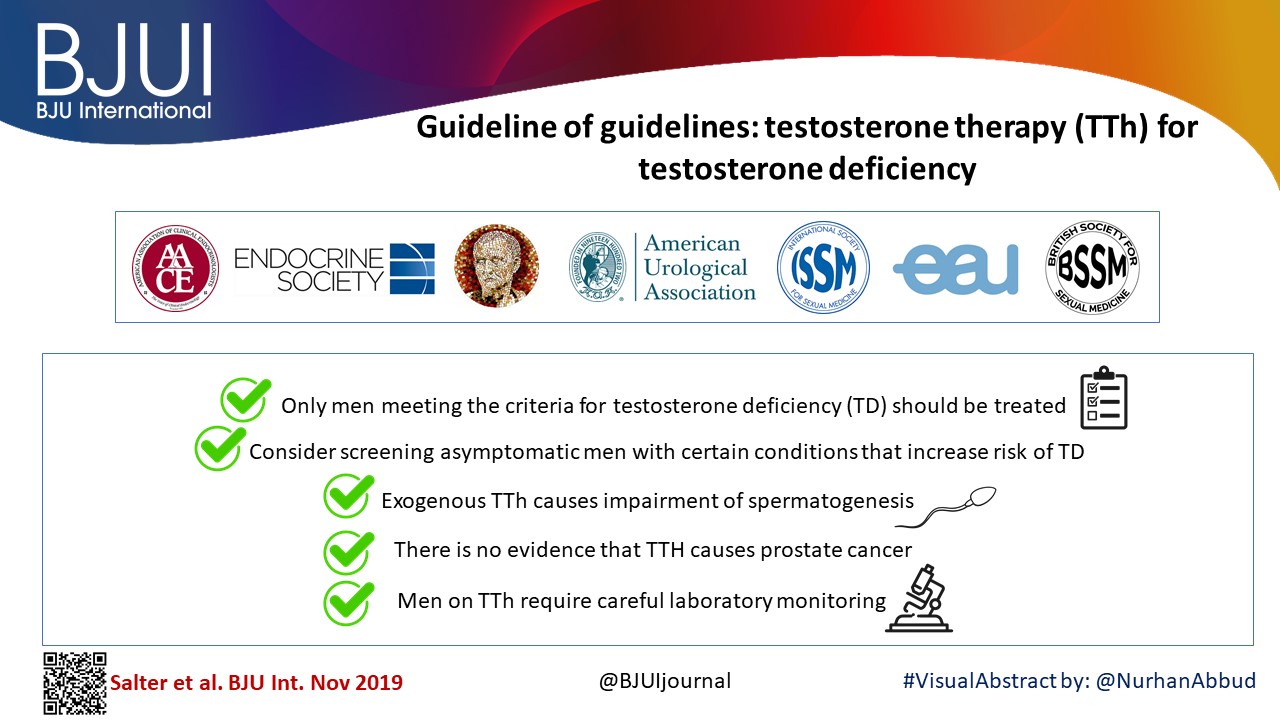
All the organisations were in general agreement concerning the following key points:

  • Only men meeting the criteria for testosterone deficiency (TD) should be treated.
  • Consider screening asymptomatic men with certain conditions that increase the risk of TD.
  • Exogenous TTh causes impairment of spermatogenesis.
  • There is no evidence that TTh causes prostate cancer.
  • Men on TTh require careful laboratory monitoring.

Video: Guideline of guidelines: testosterone therapy for testosterone deficiency

Guideline of guidelines: testosterone therapy for testosterone deficiency

Read the full article

Abstract

We analysed the guidelines for testosterone therapy (TTh) produced by major international medical societies including: the American Urological Association, European Association of Urology, American Association of Clinical Endocrinologists, British Society for Sexual Medicine, Endocrine Society, International Society for Sexual Medicine, and the International Society for the Study of the Aging Male, and compared their recommendations.

All the organisations were in general agreement concerning the following key points:

  • Only men meeting the criteria for testosterone deficiency (TD) should be treated.
  • Consider screening asymptomatic men with certain conditions that increase the risk of TD.
  • Exogenous TTh causes impairment of spermatogenesis.
  • There is no evidence that TTh causes prostate cancer.
  • Men on TTh require careful laboratory monitoring.
View more videos

What’s the Diagnosis?

pq-feb-2017-3

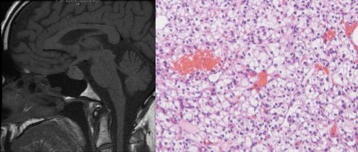
A middle aged male presented with loss of libido and loss of peripheral vision. LH and testosterone were suppressed.

No such quiz/survey/poll

Article of the Week: TRT and rates of PCa or LUTS

Every Week the Editor-in-Chief selects an Article of the Week from the current issue of BJUI. The abstract is reproduced below and you can click on the button to read the full article, which is freely available to all readers for at least 30 days from the time of this post.

In addition to the article itself, there is an accompanying editorial written by a prominent member of the urological community. This blog is intended to provoke comment and discussion and we invite you to use the comment tools at the bottom of each post to join the conversation.

If you only have time to read one article this week, it should be this one.

Testosterone treatment is not associated with increased risk of prostate cancer or worsening of lower urinary tract symptoms: prostate health outcomes in the Registry of Hypogonadism in Men

Frans M.J. Debruyne*, Hermann M. Behre, Claus G. Roehrborn, Mario Maggi§Frederick C.W. Wu, Fritz H. Schroder**, Thomas Hugh Jones††, Hartmut Porst‡‡Geoffrey Hackett§§, Olivia A. Wheaton¶¶, Antonio Martin-Morales***, Eric J. Meuleman†††, Glenn R. Cunningham‡‡‡, Hozefa A. Divan¶¶ and Raymond C. Rosen ¶¶ for the RHYME Investigators


*Andros Mens Health Institutes, Arnhem, The Netherlands, Center for Reproductive Medicine and Andrology, Martin Luther University Halle-Wittenberg, Halle, Germany, Southwestern Medical Center, University of Texas, Dallas, TX, USA, §Sexual Medicine and Andrology Unit, Department of Experimental and Clinical Biomedical Sciences, University of Florence, Florence, Italy, ¶ University of Manchester, Manchester, UK, **Erasmus Medical Center, Rotterdam, The Netherlands, ††Barnsley Hospital NHS Foundation Trust, Barnsley, UK, ‡‡Private Practice of Urology/Andrology, Hamburg, Germany, §§Holly Cottage Clinic, Licheld, Staffordshire, UK, ¶¶New England Research Institutes, Inc., Watertown, MA, USA, ***Carlos Haya University Hospital, Malaga, Spain, †††VU Medical Center, Amsterdam, The Netherlands, and ‡‡‡Baylor College of Medicine, St. Lukes Episcopal Hospital, Houston, TX, USA


Read the full article

Abstract

Objectives

To evaluate the effects of testosterone-replacement therapy (TRT) on prostate health indicators in hypogonadal men, including rates of prostate cancer diagnoses, changes in prostate-specific antigen (PSA) levels and lower urinary tract symptoms (LUTS) over time.

Patients and Methods

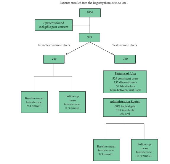
The Registry of Hypogonadism in Men (RHYME) is a multi-national patient registry of treated and untreated, newly-diagnosed hypogonadal men (n = 999). Follow-up assessments were performed at 3–6, 12, 24, and 36 months. Baseline and follow-up data collection included medical history, physical examination, blood sampling, and patient questionnaires. Prostate biopsies underwent blinded independent adjudication for the presence and severity of prostate cancer; PSA and testosterone levels were measured via local and central laboratory assays; and LUTS severity was assessed via the International Prostate Symptom Score (IPSS). Incidence rates per 100 000 person-years were calculated. Longitudinal mixed models were used to assess effects of testosterone on PSA levels and IPSS.
feb-aotw-2-results

Results

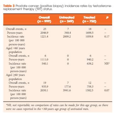
Of the 999 men with clinically diagnosed hypogonadism (HG), 750 (75%) initiated TRT, contributing 23 900 person-months of exposure. The mean testosterone levels increased from 8.3 to 15.4 nmol/L in treated men, compared to only a slight increase from 9.4 to 11.3 nmol/L in untreated men. In all, 55 biopsies were performed for suspected prostate cancer, and 12 non-cancer related biopsies were performed for other reasons. Overall, the proportion of positive biopsies was nearly identical in men on TRT (37.5%) compared to those not on TRT (37.0%) over the course of the study. There were no differences in PSA levels, total IPSS, or the IPSS obstructive sub-scale score by TRT status. Lower IPSS irritative sub-scale scores were reported in treated compared to untreated men.

Conclusions

Results support prostate safety of TRT in newly diagnosed men with HG.


Read more articles of the week

Editorial: Mythology and Reality Should Never Be Confused (But Often Are)

The debating halls of learned urological societies often attempt to define theories based on little substance. Unfortunately, although this type of scientific discourse should be encouraged, it can create both content for unrepeatable ‘late breaking’ abstracts and headlines for mass circulation newspapers. Once started, the momentum can make a perception become a ‘reality’ that is invariably difficult to dislodge. An area of particular current interest to both the scientific and lay press, and indeed the regulatory authorities in the USA and the European Union, is testosterone replacement. Within this context, an important potential myth-buster is described in the article by DeBruyne et al. [1]. The hypothesis (aka myth) that was being examined was that application of exogenous testosterone, irrespective of formulation, could exacerbate both benign and malignant prostatic disease. To understand the importance of this type of definitive study we must start at the beginning.

The foundation of the mythology, or in reality a Canadian-US urological tragedy, was ironically in the Nobel Prize winning work of Charles B Huggins. The ‘good news’ was that prostatic tumour regression could be affected by androgen deprivation, a finding certainly worthy of global recognition and acceptance; however, this became embellished to a certain extent, incorporating other aliquots of somewhat circumstantial evidence, leading to a suggestion that testosterone replacement could increase the probability and/or rate of prostate cancer progression. As a result, various societies perhaps understandably adopted their normal conservative approach of inserting a warning (with varying degrees of emphasis) in their guidelines [2]. Over the last decade, with the increasing use of testosterone replacement in the treatment of men with hypogonadism, the issue of benefit–risk has become more relevant, indeed is often transposed to risk–benefit, and is increasingly likely to feature in the popular press. Within the last couple of years, we have seen the spectre of testosterone and cardiovascular risk played out in full public view, but the issue of testosterone and exacerbation of prostatic disease has continued to smoulder in the background.

The RHYME study [1], although it is largely confirmatory as other studies have described similar conclusions, is of considerable ‘real-life’ value. The power of the study is that it should be considered to be representative of the potential hypogonadal population likely to present to the physician. Although not used for regulatory approval purposes, a registry study is considered to be the ‘gold standard’ for this type of analysis. So what are the conclusions of this and the majority of other studies? In essence, there is no evidence that restoration of testosterone to the normal range does increase the incidence or progression of either benign (BPH) or malignant prostatic disease. Any slight changes in PSA level were considered not to be clinically relevant and likely to be as a consequence of the direct pharmacology of increased testosterone levels [3]. This does not imply that exacerbations will never be seen or that appropriate monitoring should not be carried out; more that the conventional wisdom on testosterone and prostatic disease has gone (or should go) the way of antimuscarinic agents, invariably causing urinary retention in patients with BPH. It is to be hoped that studies such as the RHYME study will continue to be undertaken and help provide perspective for specialist and primary care physicians alike in areas of emergent clinical interest.

Read the full article
Michael Wyllie
Global Pharma Consulting Ltd, Stratton House, Shenington, Banbury, Oxfordshire


References


Article of the Week: The effect of hypogonadism and testosterone-enhancing therapy on AP and BMD

Every week the Editor-in-Chief selects the Article of the Week from the current issue of BJUI. The abstract is reproduced below and you can click on the button to read the full article, which is freely available to all readers for at least 30 days from the time of this post.

In addition to the article itself, there is an accompanying editorialwritten by a prominent member of the urological community. This blog is intended to provoke comment and discussion and we invite you to use the comment tools at the bottom of each post to join the conversation.

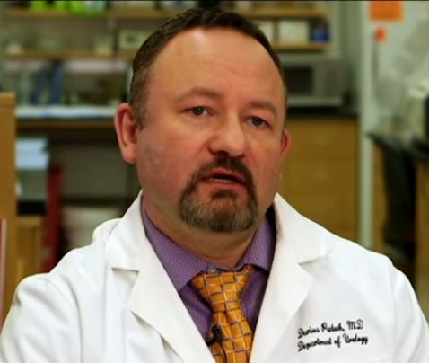
Finally, the third post under the Article of the Week heading on the homepage will consist of additional material or media. This week we feature a video from Dr. Darius Paduch discussing his paper. Make sure you have a stable internet connection. In order to have the best telehealth experience possible, you must be using a strong, stable internet connection. Keep your hands free. If you can find a comfortable stand for your video device or have something to prop it up against, will free up your hands to take part in the physical therapy session. Remember to use a compatible internet browser. Below is a list of compatible internet browsers for your telehealth session. Whichever browser you are using, make sure to keep it updated to the most recent version. Laws governing telehealth reimbursement vary in each state. In our experience, most health plans have covered these services however, every insurance company is different. Well medical malpractice insurance cost and quality are always good to afford. We recommend checking with your insurance provider or the clinic prior to your appointment to make sure you’re covered. All 50 states, the District of Columbia, and the US Virgin Islands allow patients to seek some level of treatment from a licensed physical therapist without a prescription or referral from a physician. There may be some restrictions in your state but for the most part this means is that you can now receive physical therapy, even virtually, without a physician’s referral/prescription. Check with your state’s regulations or your clinic prior to your appointment. you can discover more here for Telehealth.

 If you only have time to read one article this week, it should be this one.

The effect of hypogonadism and testosterone-enhancing therapy on alkaline phosphatase and bone mineral density

Ali A. Dabaja, Campbell F. Bryson, Peter N. Schlegel and Darius A. Paduch
Department of Urology, Weill Cornell Medical College, New York, NY, USA
Read the full article
OBJECTIVE

To evaluate the relationship of testosterone-enhancing therapy on alkaline phosphatase (AP) in relation to bone mineral density (BMD) in hypogonadal men.

PATIENTS AND METHODS

Retrospective review of 140 men with testosterone levels of <350 ng/dL undergoing testosterone-enhancing therapy and followed for 2 years. Follicle-stimulating hormone, luteinising hormone, free testosterone, total testosterone, sex hormone binding globulin, calcium, AP, vitamin D, parathyroid hormone, and dual-energy X-ray absorptiometry (DEXA) scans were analysed. A subgroup of 36 men with one DEXA scan before and one DEXA 2 years after initiating treatment was performed.

RESULTS

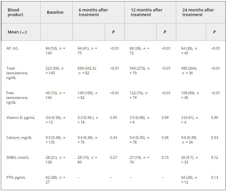
Analysis of the relationship between testosterone and AP at initiation of therapy using stiff linear splines suggested that bone turnover occurs at total testosterone levels of <250 ng/dL. In men with testosterone levels of <250 ng/dL, there was a negative correlation between testosterone and AP (R2 = −0.347, P < 0.001), and no correlation when testosterone levels were between 250 and 350 ng/dL. In the subgroup analysis, the mean (sd) testosterone level was 264 (103) ng/dL initially and 701 (245), 539 (292), and 338 (189) ng/dL at 6, 12, and 24 months, respectively. AP decreased from a mean (sd) of 87 (38) U/L to 57 (12) U/L (P = 0.015), 60 (17) U/L (P < 0.001), and 55 (10) U/L (P = 0.03) at 6, 12, and 24 months, respectively. The BMD increased by a mean (sd) of 20 (39)% (P = 0.003) on DEXA.

CONCLUSION

In hypogonadal men, the decrease in AP is associated with an increase in BMD on DEXA testing. This result suggests the use of AP as a marker of response to therapy.

Read more articles of the week

Editorial: On the Mark? Is AP a surrogate for BMD in hypogonadal men?

The current issue of the BJUI contains a paper by Dubaja et al. [1] that may be of interest to physicians who have patients with hypogonadism. The authors speak to an unappreciated aspect of low testosterone; namely, the loss of bone in men and the possible recovery with treatment. Their retrospective study looked at 140 men with hypogonadism treated with exogenous testosterone replacement or clomiphene citrate testosterone enhancement. These men were also assessed for bone mineral density (BMD) markers at 6, 12 and 24 months after initial treatment. Importantly, dual-energy X-ray absorptiometry (DEXA) was performed a second time after 2 treatment years for a subset of these men. DEXA showed that there was a gain in BMD and a loss of serum alkaline phosphatase (AP) for all the men over time. The loss of AP was rapid but stabilized at 6 months. Testosterone and free testosterone increased as expected but there were no changes in vitamin D, calcium, parathyroid hormone or sex hormone-binding globulin. There was a correlation between AP and testosterone. The authors recognized poor bone density at baseline in those men with testosterone levels

Bone mineral density is the best way to predict osteoporosis and fragility fractures [2]. Women loose BMD after menopause and that is accompanied by many changes, including gains in AP [3]. The decline in serum oestrogens is a factor for women, and oestrogen replacement therapy historically has been used to prevent that loss. Not all men undergo a similar loss, i.e. andropause is not recognized in the same way as menopause. Even though osteoporosis is less common in men, the associated comorbidity may be more significant.

There is an age-related decline in testosterone and an acute loss for some men such that they approach their physicians with symptoms. The underlying cause of bone loss in men and women may be the same: serum oestrogen loss. Men who lose testosterone are also losing oestrogen because testosterone is the precursor via aromatase. Repros Therapeutics is developing a way to treat men with secondary hypogonadism. An ongoing 1-year DEXA study recruited eligible men. Key inclusion factors were age < 60 years and body mass index > 25 kg/m2. Remarkably 24% of men failed the screening test because of osteopenia, despite the fact that few of them were old or underweight.

The present paper by Dubaja et al. suggests that a readily available serum test may be able to monitor men on testosterone therapies for gain in BMD. Given the relatively low incidence of osteoporosis, screening every man by DEXA is not cost-efficient. The 1 year or more needed to find BMD loss by DEXA also wastes time and resources. Quantitative CT is more costly and is accompanied by high radiation exposure. The use of AP, as suggested in the present study, may represent a reasonable alternative.

The paper is not without its weaknesses. There was no indication of whether these men had primary or secondary hypogonadism. Transdermal testosterone should raise testosterone and oestrogen in both groups of men whereas clomiphene citrate works through changes in LH and FSH and requires an intact hypothalamic-pituitary-gonadal axis (useful in men with secondary hypogonadism only). Indeed, the two kinds of treatment will have the opposite effect on LH and FSH [4]. The authors recognized the importance of Leydig cells in producing testosterone, yet the effects of a transdermal testosterone would be to shut down testosterone production. We commend their suggestion that other factors that contribute to both bone and Leydig cell function, insulin-like 3 [5] and osteocalcin [6] should be studied in relation to AP. The loss of subjects throughout the 2 years was troubling, but it is known that men on transdermal treatments discontinue with disturbing frequency despite satisfaction [7]. If those who stayed in the present study were those with the best outcomes in terms of testosterone and BMD, potential bias may exist. If men can be encouraged to continue therapy through positive effects on BMD being detected as early as 6 months, AP monitoring may improve patient compliance. Only DEXA can give that assurance now. The authors noted the need for a larger prospective trial. Nevertheless, their paper provides a rationale for monitoring men on testosterone therapies that can be implemented with minimal cost or the need for new diagnostics.

Read the full article
Martin C. Michel
Department of Pharmacology, Johannes Gutenberg University, Mainz, German

References


2 NIH Consensus Development Panel. Osteoporosis prevention, diagnosis and therapy. JAMA 2001; 285: 785–95

3 BiveE.Use of bone turnover markers in clinical practice. Curr O pin Endocrinol Dia betes Obes 2012; 19: 468–73


5 Ivell R, Anand-Ivell R. Biology of insulin-like factor-3 in humareproduction. Hum Reprod Update 2009; 15: 463–76


7 Kovac J, Rajanahally S, Smith R, Coward R, Lamb D, Lipshultz L. Patient satisfaction with testosterone replacement therapies: the reasons behind the choices. JSexMed2014; 11: 553–62


Video: Hypogonadism and testosterone-enhancing therapy on alkaline phosphatase and BMD

The effect of hypogonadism and testosterone-enhancing therapy on alkaline phosphatase and bone mineral density

Ali A. Dabaja, Campbell F. Bryson, Peter N. Schlegel and Darius A. Paduch


Department of Urology, Weill Cornell Medical College, New York, NY, USA


Read the full article
OBJECTIVE

To evaluate the relationship of testosterone-enhancing therapy on alkaline phosphatase (AP) in relation to bone mineral density (BMD) in hypogonadal men.

PATIENTS AND METHODS

Retrospective review of 140 men with testosterone levels of <350 ng/dL undergoing testosterone-enhancing therapy and followed for 2 years. Follicle-stimulating hormone, luteinising hormone, free testosterone, total testosterone, sex hormone binding globulin, calcium, AP, vitamin D, parathyroid hormone, and dual-energy X-ray absorptiometry (DEXA) scans were analysed. A subgroup of 36 men with one DEXA scan before and one DEXA 2 years after initiating treatment was performed.

RESULTS

Analysis of the relationship between testosterone and AP at initiation of therapy using stiff linear splines suggested that bone turnover occurs at total testosterone levels of <250 ng/dL. In men with testosterone levels of <250 ng/dL, there was a negative correlation between testosterone and AP (R2 = −0.347, P < 0.001), and no correlation when testosterone levels were between 250 and 350 ng/dL. In the subgroup analysis, the mean (sd) testosterone level was 264 (103) ng/dL initially and 701 (245), 539 (292), and 338 (189) ng/dL at 6, 12, and 24 months, respectively. AP decreased from a mean (sd) of 87 (38) U/L to 57 (12) U/L (P = 0.015), 60 (17) U/L (P < 0.001), and 55 (10) U/L (P = 0.03) at 6, 12, and 24 months, respectively. The BMD increased by a mean (sd) of 20 (39)% (P = 0.003) on DEXA.

CONCLUSION

In hypogonadal men, the decrease in AP is associated with an increase in BMD on DEXA testing. This result suggests the use of AP as a marker of response to therapy.

Read more articles of the week

Article of the Week: Testosterone Therapy and Cancer Risk

Every week the Editor-in-Chief selects the Article of the Week from the current issue of BJUI. The abstract is reproduced below and you can click on the button to read the full article, which is freely available to all readers for at least 30 days from the time of this post.

In addition to the article itself, there is an accompanying editorial written by a prominent member of the urological community. This blog is intended to provoke comment and discussion and we invite you to use the comment tools at the bottom of each post to join the conversation.

Finally, the third post under the Article of the Week heading on the homepage will consist of additional material or media. This week we feature a video from Dr. Michael Eisenberg discussing his paper. 

If you only have time to read one article this week, it should be this one.

Testosterone Therapy and Cancer Risk

Michael L. Eisenberg*, Shufeng Li*, Paul Betts§, Danielle Herder, Dolores J. Lamb¶ and Larry I. Lipshultz


Departments of *Urology, Obstetrics/Gynecology and Dermatology, Stanford University School of Medicine, Stanford, CA§Cancer Epidemiology and Surveillance Branch, Texas Cancer Registry, Texas Department of State Health Services, Austin, TX, and Scott Department of Urology, Baylor College of Medicine, Houston, TX, USA


Read the full article
OBJECTIVE

To determine if testosterone therapy (TT) status modifies a man’s risk of cancer.

PATIENTS AND METHODS

The Urology clinic hormone database was queried for all men with a serum testosterone level and charts examined to determine TT status. Patient records were linked to the Texas Cancer Registry to determine the incidence of cancer. Men accrued time at risk from the date of initiating TT or the first office visit for men not on TT. Standardised incidence rates and time to event analysis were performed.

RESULTS

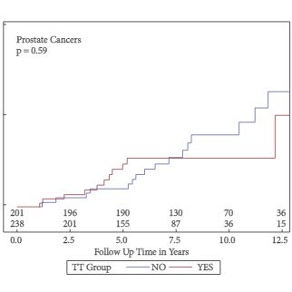
In all, 247 men were on TT and 211 did not use testosterone. In all, 47 men developed cancer, 27 (12.8%) were not on TT and 20 (8.1%) on TT. There was no significant difference in the risk of cancer incidence based on TT (hazard ratio [HR] 1.0, 95% confidence interval [CI] 0.57–1.9; P = 1.8). There was no difference in prostate cancer risk based on TT status (HR 1.2, 95% CI 0.54–2.50).

CONCLUSION

There was no change in cancer risk overall, or prostate cancer risk specifically, for men aged >40 years using long-term TT.

Read more articles of the week
© 2024 BJU International. All Rights Reserved.