Tag Archive for: spectroscopy

Posts

Article of the Week: Detecting PSMs – using LRS on ex vivo RP specimens

Every Week the Editor-in-Chief selects an Article of the Week from the current issue of BJUI. The abstract is reproduced below and you can click on the button to read the full article, which is freely available to all readers for at least 30 days from the time of this post.

In addition to the article itself, there is an accompanying editorial written by a prominent member of the urological community. This blog is intended to provoke comment and discussion and we invite you to use the comment tools at the bottom of each post to join the conversation.

If you only have time to read one article this week, it should be this one.

Detecting positive surgical margins: utilisation of light-reflectance spectroscopy on ex vivo prostate specimens

Aaron H. Lay*, Xinlong Wang, Monica S. C. Morgan*, Payal Kapur, Hanli Liu,Claus G. Roehrborn* and Jeffrey A. Cadeddu*


*Department of Urology, University of Texas Southwestern Medical Center, Dallas, TX, Department of Bioengineering, University of Texas at Arlington, Arlington, TX, and Department of Pathology, University of Texas Southwestern Medical Center, Dallas, TX, USA


Abstract

Objective

To assess the efficacy of light-reflectance spectroscopy (LRS) to detect positive surgical margins (PSMs) on ex vivo radical prostatectomy (RP) specimens.

Materials and Methods

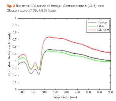
A prospective evaluation of ex vivo RP specimens using LRS was performed at a single institution from June 2013 to September 2014. LRS measurements were performed on selected sites on the prostate capsule, marked with ink, and correlated with pathological analysis. Significant features on LRS curves differentiating malignant tissue from benign tissue were determined using a forward sequential selection algorithm. A logistic regression model was built and randomised cross-validation was performed. The sensitivity, specificity, accuracy, negative predictive value (NPV), positive predictive value (PPV), and area under the receiver operating characteristic curve (AUC) for LRS predicting PSM were calculated.

aotwdec-4-results

Results

In all, 50 RP specimens were evaluated using LRS. The LRS sensitivity for Gleason score ≥7 PSMs was 91.3%, specificity 92.8%, accuracy 92.5%, PPV 73.2%, NPV 99.4%, and the AUC was 0.960. The LRS sensitivity for Gleason score ≥6 PSMs was 65.5%, specificity 88.1%, accuracy 83.3%, PPV 66.2%, NPV 90.7%, and the AUC was 0.858.

Conclusions

LRS can reliably detect PSMs for Gleason score ≥7 prostate cancer in ex vivo RP specimens

Editorial: Light reflectance spectroscopy is one more emerging technique with the potential to adjust excision limits during radical prostatectomy

In this issue of BJUI, Lay et al. [1] report that light reflectance spectroscopy (LRS) can detect Gleason ≥7 positive surgical margins (PSMs) with 92.5% accuracy. In this initial study, the authors have reported the use of LRS in an ex situ setting to analyse the prostate surface; however, this technology could ultimately be developed to identify PSMs before choosing the surgical plane of dissection, which could allow the surgeon to immediately perform a wider complementary excision.

As long as PSMs are detected ex situ, it is not clear why spectroscopy should be preferred to frozen sections. NeuroSAFE, for example, is a standardized and validated margin evaluation procedure in pathology [2]. It does not lengthen operating time, does not require any new equipment and provides a pathological assessment which is the best level of evidence for PSM status; however, as a conventional pathological procedure, it is not conceivable in situ, and real-time detection of PSMs that ensures the safest oncological resection during a nerve-sparing dissection is needed.

In this effort to examine in vivo/in situ prostate PSMs, several other technologies can be considered. During radical prostatectomy, optical coherence tomography (OCT) has been used in situ in humans, but only to identify the neurovascular bundles [3]. Field of view and depth of penetration were limited and OCT has never been evaluated in situ for prostate PSM detection. Confocal endomicroscopy has recently been reported during robot-assisted radical prostatectomy [4]. With this technique, optical biopsies were feasible in situ but the PSM detection rate and the overall efficiency of this confocal endomicroscopy in prostate specimens remain unknown. Similarly, illumination microscopy has been used to generate gigapixel images of the full prostate circumference in vivo for the detection of PSMs [5]. Illumination microscopy allows images to be interpreted readily by pathologists, but the feasibility series was too small to assess the accuracy of this technique for PSM detection. Ex situ multi-photon microscopy (MPM) is an optical technique that enables the imaging of prostatic and periprostatic tissue at sub-micron resolution to a depth of up to 0.5 mm [6]. On a fresh specimen, it generates three-dimensional images of periprostatic nerves, blood vessels and capsule, but also underlying acini and pathological changes such as prostate cancer. MPM technology has also been miniaturized and its accuracy in situ is currently under investigation.

In this context, the study by Lay et al. [1] shows that, for the time being, LRS is one more promising technique on the road to real-time PSM detection. More will undoubtedly be done to overcome the spectroscope’s light absorption in the presence of blood and, subsequently, to evaluate its reliability in situ; however, the recent developments of these protocols and technologies (endomicroscopy, illumination microscopy, OCT, MPM, LRS) show a progressive effort amongst clinicians to obtain intra-operative feedback on the PSM status. Fortunately, this is taking place while the urological community is increasingly considering surgical treatment even for the high-risk disease, where oncological adequacy is of paramount importance. While we are witnessing these promising evolutions in high-grade prostate cancer, the optimum technique which will safely end margin-blind radical prostatectomy in an actual surgical field (filled with blood and often distorted because of inflammation) still needs to go through clinical trials and validation; however, the future is bright as a result of these newer developments.

Thomas Bessede*†‡ and Ash Tewari*


*Department of Urology, Icahn School of Medicine at MounSinai, New York , NY, USA, U1195, INSERM, UniversitParis-Saclay, and Department of Urology, APHP, Hopitaux Universitaires Paris-Sud, Le Kremlin-Bicetre, France


References


© 2025 BJU International. All Rights Reserved.