Tag Archive for: Robotic Surgery

Posts

Learning from The Lancet

The Lancet, established in 1823, is one of the most respected medical journals in the world. It has an impact factor of 39, and therefore attracts and publishes only the very best papers. Like most journals that have evolved with modern times, it has an active web and social media presence, particularly based around Twitter.

On a Monday morning, last autumn, the Editor of the BJUI had a meeting with the Web Editor of The Lancet at Guy’s Hospital. There was a mutual interest in surgical technology, particularly as Naomi Lee had been a urology trainee before joining The Lancet full-time. The topic of discussion was robot-assisted radical cystectomy with the emergence of randomised trials showing little difference between open and robotic surgery, despite the minimally invasive nature of the latter [1, 2]. Thereafter, The Lancet kindly invited the BJUI team to visit its offices in London. The location is rather bohemian with a mural of John Lennon on the wall across the street! Here is a summary of what we learnt that day.

Capture

1. Democracy – what gets published in The Lancet after peer review is decided at a team meeting, where editors of the main journal and its sister publications gather around a table to discuss individual articles. Most work full-time for The Lancet, unlike surgical journals that are led by working clinicians. No wonder that >80% of papers are immediately rejected and the final acceptance rate is ≈6%. Interesting case reports are still published and often highly cited because of the wider readership.

2. Quality has no boundaries – it does not matter where the article comes from as long as it has an important message. The BJUI recently published an excellent paper on circumcision in HIV-positive men from Africa [3]; the original randomised controlled trial had appeared some 7 years earlier in The Lancet [4].

3. Statisticians – the good ones are a rare breed and sometimes rather difficult to find. While we have two statistical editors at the BJUI, sometimes, it is difficult to approach the most qualified reviewer on a particular subject. The Lancet occasionally faces similar difficulties, which it almost always overcomes due to its’ team approach.

4. Meta-analysis and systematic reviews – they form a significant number of submissions to both journals. It is not always easy to judge their quality although a key starting point is to identify whether the topic is one of contemporary interest where there are significant existing data that can be analysed. Rare subjects usually fail to make the cut.

5. Paper not dead yet – this is certainly the case at The Lancet office, where its editors gather together with paper folders and hand-written notes. We are almost fully paperless at the BJUI offices, and are hoping to be completely electronic in the future. A recent live vote of our readership during the USANZ Annual Scientific Meeting in Adelaide, Australia, indicated that the majority would like us to go electronic in about 2–3 years’ time; however, ≈30% of our institutional subscribers still prefer the paper version and are reluctant to make the switch.

The BJUI and The Lancet are coming together to host a joint Social Media session at BAUS 2015, which will provide more opportunity to learn from one of the best journals ever. We hope to see many of you there.

References


2 Lee N. Robotic surgery: where are we now? Lancet 2014; 384: 1417


4 Gray RH, Kigozi G, Serwadda D et al. Male circumcision for HIV prevention in men in Rakai, Uganda: a randomised trial. Lancet 2007; 369: 65766


Prokar Dasgupta @prokarurol
Editor-in-Chief, BJUI 


Scott Millar
Managing Editor, BJUI 


Naomi Lee
Web Editor, The Lancet


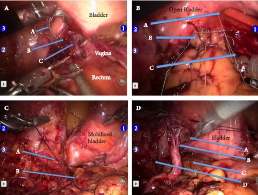
Article of the Week: Robotic management of GU injuries from obstetrical and gynecological operations

Every week the Editor-in-Chief selects the Article of the Week from the current issue of BJUI. The abstract is reproduced below and you can click on the button to read the full article, which is freely available to all readers for at least 30 days from the time of this post.

In addition to the article itself, there is an accompanying editorial written by a prominent member of the urological community. This blog is intended to provoke comment and discussion and we invite you to use the comment tools at the bottom of each post to join the conversation.

Finally, the third post under the Article of the Week heading on the homepage will consist of additional material or media. This week we feature a video from Dr. Ronald Boris, discussing his paper. 

If you only have time to read one article this week, it should be this one.

Robotic management of genito-urinary injuries from obstetrical and gynecological operations: a multi-institutional report of outcomes

Paul T. Gellhaus, Akshay Bhandari*, M. Francesca Monn, Thomas A. Gardner,Prashanth Kanagarajah*, Christopher E. Reilly†, Elton Llukani†, Ziho Lee†, Daniel D. Eun†,Hani Rashid‡, Jean V. Joseph‡, Ahmed E. Ghazi‡, Guan Wu‡and Ronald S. Boris

Department of Urology, Indiana University, Indianapolis, IN, *Division of Urology, Columbia University at Mount Sinai,Miami Beach, FL,†Department of Urology, Temple University, Philadelphia, PA, and‡Department of Urology, University ofRochester, Rochester, NY, USA

Read the full article
OBJECTIVE

To evaluate the utility of robotic repair of injuries to the ureter or bladder from obstetrical and gynaecological (OBGYN) surgery

PATIENTS AND METHODS

A retrospective review of all patients from four different high-volume institutions between 2002 and 2013 that had a robot-assisted (RA) repair by a urologist after an OBGYN genitourinary injury.

RESULTS

Of the 43 OBGYN operations, 34 were hysterectomies: 10 open, 10 RA, nine vaginally, and five pure laparoscopic. Nine patients had alternative OBGYN operations: three caesarean sections, three oophorectomies (one open, two laparoscopic), one RA colpopexy, one open pelvic cervical cerclage with mesh and one RA removal of an invasive endometrioma. In all, 49 genitourinary (GU) injuries were sustained: ureteric ligation (26), ureterovaginal fistula (10), ureterocutaneous fistula (one), vesicovaginal fistula (VVF; 10) and cystotomy alone (two). In all, 10 patients (23.3%) underwent immediate urological repair at the time of their OBGYN RA surgery. The mean (range) time between OBGYN injury and definitive delayed repair was 23.5 (1–297) months. Four patients had undergone prior failed repair: two open VVF repairs and two balloon ureteric dilatations with stent placement. In all, 22 ureteric re-implants (11 with ipsilateral psoas hitch) and 15 uretero-ureterostomies were performed. Stents were placed in all ureteric cases for a mean (range) of 32 (1–63) days. In all, 10 VVF repairs and two primary cystotomy closures were performed. Drains were placed in 28 cases (57.1%) for a mean (range) of 4.1 (1–26) days. No case required open conversion. Two patients (4.1%) developed ureteric obstruction after RA repair requiring dilatation and stenting. The mean (range) follow-up of the entire cohort was 16.6 (1–63) months.

CONCLUSIONS

RA repair of GU injuries during OBGYN surgery is associated with good outcomes, appears safe and feasible, and can be used successfully immediately after injury recognition or as a salvage procedure after prior attempted repair. RA techniques may improve convalescence in a patient population where quick recovery is paramount.

Read more articles of the week

Editorial: The robot to the rescue!

Fortunately injuries to the urinary tract remain rare in obstetric and gynaecological surgery. Their potential for causing serious morbidity, not to mention the substantial medico-legal implications ensure that it remains a highly researched and evocative area [1].

Iatrogenic urinary tract injuries can be broadly divided into two groups; acute complications, such as bladder and ureteric lacerations or ligation and more chronic complications, such as vesicovaginal or ureterovaginal fistulae and ureteric strictures. Historically, iatrogenic trauma to the urinary tract most commonly followed open, abdominal hysterectomy with the most frequent complication being direct bladder injury. Gross bladder injuries are generally both detected and treated intraoperatively. In contrast, the management of more complex ureteric injuries and their long-term sequelae, such as fistula, pose greater surgical challenges. The complexity of these injuries is often further compounded by delay in diagnosis. It is generally accepted that, if possible, immediate repair provides the optimal treatment. However, when diagnosis is delayed, there is little consensus on the best management approach, although the current tendency is towards early repair [2].

The multicentre, retrospective study of robotic repair of 49 iatrogenic genitourinary injuries by Gellhaus et al. [3] should therefore serve to reassure gynaecologists with respect to the incidence of this feared complication. This paper, mainly looking at ureteric re-implants and fistula repairs, constitutes the largest cohort of robotic reconstructions to date. A zero conversion rate to open surgery suggests excellent case selection by the robotic surgeons. Four cases had undergone previous failed open or endoscopic management and this is clearly a challenging cohort. Yet, with the absence of total numbers of urology referrals received for such injuries, it is important to remember that it may not be a panacea for all.

There has been a considerable shift in the management of urological trauma from open to laparoscopic techniques. While the repair of basic injuries has been proven to be effective, less data is available to support the management of more complex injuries, such as ureteric transections or fistulae [4]. Numerous techniques for repairing ureteric injuries and fistulae have been described; nonetheless, the surgery remains technically challenging even for experienced laparoscopic surgeons and is generally limited to high-volume centres [5].

In comparison, this article [3] provides strong evidence for the effectiveness of robot-assisted (RA) repairs, even for complex injuries. The enviable 95.9% success rate from 47 operations is complemented by short recovery times and low complication rates. These results are especially impressive in view of the mean 23.5-month delay time to repair. But the authors do not report the reasons for these delays. Immediate robotic repair of RA injuries is clearly feasible especially in larger units. Whether immediate RA repair should be performed for other iatrogenic injuries needs further discussion. Is it realistic to convert to the robot in the case of laparoscopic trauma or should RA repairs remain a planned return to theatre?

The demand for RA reconstructive surgery and experienced robotic pelvic surgeons is likely to rise in the near future. As the authors note, the continued expansion of minimally invasive procedures is likely to lead to a shift in the patterns of complications from more straightforward bladder injuries to complex ureteric injuries. As mentioned previously these types of injury are more likely to be initially undetected.

Whilst rates of surgical complications involving the urinary tract remain low, obstetrical and gynaecological procedures account for 75% of these injuries. This article provides robust evidence for the key role that RA surgery can play in the management of these complex and feared injuries. When faced with such situations, Gellhaus et al. [3] have shown that it is increasingly likely that the robot saves the day.

Read the full article

Nicholas Raison and Ben Challacombe

Department of Urology, Guy’s and St Thomas’ Hospital,London, UK

References

1 Preston JM. Iatrogenic ureteric injury: common medicolegal pitfalls. BJUInt 2000; 86: 313–7

2 El-Tabey NA, Ali-el-Dein B, Shaaban AA et al. Urological trauma aftergynecological and obstetric surgeries. Scand J Urol Nephrol 2006; 40:225–31

3 Gellhaus PT, Bhandari A, Monn MF et al. Robotic management ofgenito-urinary injuries from obstetrical and gynecological operations: amulti-institutional report of outcomes. BJU Int 2015; 115: 430–6

4 De Cicco C, Ussia A, Koninckx PR. Laparoscopic ureteral repairin gynaecological surgery. Curr Opin Obstet Gynecol 2011; 23:296–300

5 Rassweiler J, Pini G, Gözen AS, Klein J, Teber D. Role of laparoscopy inreconstructive surgery. Curr Opin Urol 2010; 20: 471–82


A Rather Nasty Surprise

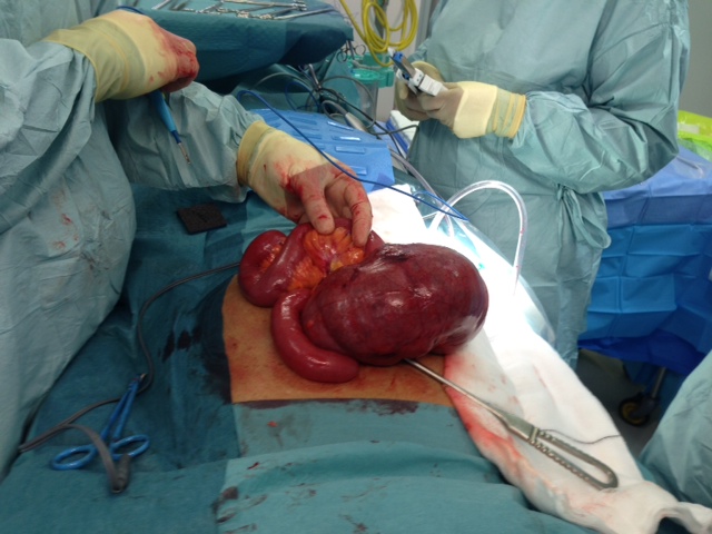
Recently, I encountered, and indeed I actually caused, a complication of robot-assisted radical prostatectomy (RARP) which was new to me, and one which I felt that I should share with other surgeons.

PM, a 60-year old teacher, underwent a completely routine RARP, which took less than 2 hours to perform on a Saturday morning. During Sunday night he developed severe abdominal pain and distension. By Monday morning he was in distress with rebound tenderness and marked tachycardia. A CT scan was requested, which revealed a caecal volvulus. A laparotomy by a general surgeon confirmed the diagnosis and an urgent right hemicolectomy was undertaken. The patient made an uneventful recovery and, I am pleased to say, is still speaking to me. Histology confirmed an ischaemic caecum twisted on its rather thickened mesentery, with no perforation present. The prostate itself contained a Gleason 3+4=7 adenocarcinoma, without evidence of extra-prostatic extension.

Although robotic assistance provides the benefits of very precise, virtually bloodless surgery, with 10 times magnification and 3D vision, it also carries the risk of a specific set of complications. These need to be dealt with promptly and efficiently and can usually be completely resolved. Failure to recognise post-operative problems, such as bowel injury, intra-abdominal bleeding or port-site hernia, however, can place the patient in severe and increasing jeopardy. We recently published an article in the BJUI entitled “Lessons Learned from 1000 robot-assisted radical prostatectomy” in which we discussed how many of the problems could be avoided, and, if they occur how they can be best dealt with. One key message is the importance of an early CT scan to diagnose the nature of a post-operative problem, rather than crossing fingers and hoping things will settle.

I am hoping that this blog, and the BJUI article mentioned above, will stimulate other surgeons to discuss openly and frankly the problems that they themselves have encountered, either with regular laparoscopy or with the da Vinci robot, and how they dealt with them. Learning the lessons, not only from one’s own errors and omissions, but also from those of others, seems the best way to become, and continue to be, a safe and successful surgeon.  


Roger Kirby, The Prostate Centre, London

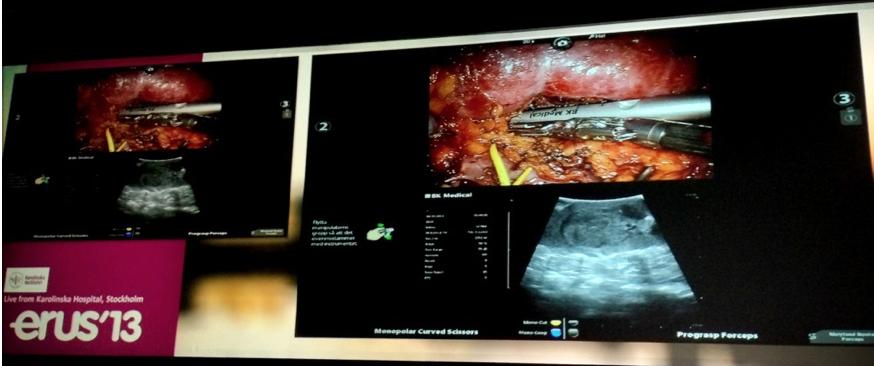
Conference Report – ERUS 2013 – live surgery spectacular in Stockholm

When it comes to live surgery meetings, one of the biggest and best of them all is the EAU Robotic Urology Section (ERUS) Congress (formerly the European Robotic Urology Symposium). The 10th edition of ERUS took place in Stockholm this week and continued the tradition of spectacular live robotic assisted surgery, along with scientific sessions dealing with issues around robotic assisted surgery. Following discussions with the EAU over the past two years, ERUS has now become an official section of the main EAU Organisation and future scientific and educational activity will be co-ordinated under that esteemed banner. In his welcoming address at this weeks meeting, EAU Secretary General and proud Swede Per-Anders Abrahamsson, warmly welcomed ERUS into the EAU family. He also highlighted the mission statement of ERUS, “to support science and education in the field of robotic urology”.

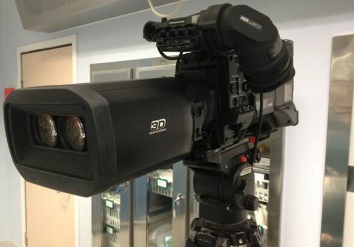
Over 750 delegates gathered from around the world (including a healthy delegation from Australia, South America and the USA), giving this meeting a truly global footprint. The programme featured 12 live surgical procedures performed by some of the world’s leading robotic surgeons and broadcast in full 3-D from Karolinska Hospital.


This meeting has showcased many advances in roboticsurgery over the past 10 years and this year was no exception. The audience seemed most interested in extended public lymph node dissection during radical cystectomy and prostatectomy, as well as intra-corporeal urinary diversion and complex partial nephrectomy. This year’s starring surgeons included Alex Mottrie, Peter Wiklund, Magnus Annerstedt, Geoff Coughlin, Hubert John, Aldo Bocciardi, Jean Palou, Carl Wijburg, Craig Rogers, Jim Porter, Tim Wilson, Vip Patel and Abi Hosseini. An outstanding line-up of surgeons from all over the world.

Of note, this Section has led the development of ethical guidelines around the conduct of live surgery and these have been fully endorsed by the EAU. We have previously blogged about this issue and I have blogged about my own experience of doing live surgery at ERUS 2012 in London.  As part of the live surgery ethical governance, Convener of ERUS 2012, Ben Challacombe (London), presented an update on the outcome of all patients who underwent live surgery as part of last years meeting.

The main scientific meeting was preceded by the Junior ERUS Section, the Nursing Course on Robotics, and five master classes led by experts and dealing with various aspects of robotic assisted surgery.  The Junior ERUS Prize was awarded to Khan et al who presented a poster on behalf of the International Robotic Curriculum Group entitled, “Towards a Standardised Training Curriculum in Robotic Surgery”. There were also a number of parallel meetings dealing with education and scientific activity within ERUS/EAU, in particular, the development of structured robotic training and a robotic surgery curriculum across Europe and beyond. The BJUI Editor in Chief Prokar Dasgupta, a well-known robotic surgery innovator and also an expert in simulation and education, is playing an active role coordinating development of this curriculum. European Urology Editor in Chief Jim Catto, was also present at ERUS 2013 and delivered a podium presentation outlining some of the exciting changes which the Platinum Journal will undertake once he takes over in January 2014. What is clear is that robotic surgery is an important part of the content for both of these leading journals.

Of course, this meeting has a particular reputation as being a friendly and sociable event (a point repeatedly mentioned by many of the Intercontinental visitors). The local organising committee pulled out all the stops with the official social events by hosting the welcome reception at the Stockholm City Hall, home of the famous Nobel Prize banquet each year. The gala dinner was in the spectacular Vasa Museum, surely one of the world’s most spectacular maritime museums.

We were treated to a tour of this spectacular, fully intact 17th century warship, followed by dinner in the shadow of this huge exhibit, notorious for capsising in Stockholm harbor only 15 minutes into her maiden voyage.

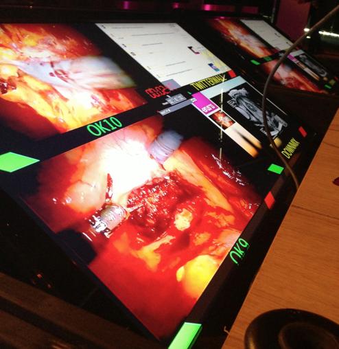
As we have seen at all major urology meetings this year, social media played a prominent role in expanding the reach of the meeting and in enabling engagement from within the audience and from around the world. The conference organisers placed a Twitter feed on the panellists monitors so that questions could be directed via Twitter to the expert panels and to the operating rooms.

 As if the spectacular multiple source 3-D display was not providing enough content, social media guru Carl Wijburg was busy tweeting “backstage” photos from Karolinska as he waited to perform a meticulous extended pelvic lymph node dissection.


 The final data from Symplur showed just how enthusiastically delegates from all over engaged with the meeting through Twitter.


Congratulations go to the organisers and scientific committee of #ERUS13 led by Alex Mottrie (Belgium), Peter Wiklund (Stockholm) and Magnus Annerstedt (Copenhagen) who did an outstanding job putting on this complex congress.

We are already looking forward to ERUS 2014 which takes place in beautiful Amsterdam from 17- 19th September 2014, led by Chair of the Local Organising Committee, Henk van der Poel. A must-attend for anyone interested in robotic surgery.


Declan Murphy BJUI Associate Editor

Follow Declan on Twitter @declangmurphy


Article of the week: Staging inguinal disease in patients with penile cancer

Every week the Editor-in-Chief selects the Article of the Week from the current issue of BJUI. The abstract is reproduced below and you can click on the button to read the full article, which is freely available to all readers for at least 30 days from the time of this post.

In addition to the article itself, there is an accompanying editorial written by a prominent member of the urological community. This blog is intended to provoke comment and discussion and we invite you to use the comment tools at the bottom of each post to join the conversation.

If you only have time to read one article this week, it should be this one.


Phase 1 prospective evaluation of the oncological adequacy of robotic assisted video-endoscopic inguinal lymphadenectomy in patients with penile carcinoma

Surena F. Matin, Janice N. Cormier*, John F. Ward, Louis L. Pisters, Christopher G. Wood, Colin P.N. Dinney, Richard E. Royal*, Xuelin Huang and Curtis A. Pettaway

Departments of Urology, *Surgical Oncology and Biostatistics, University of Texas MD Anderson Cancer Center, Houston, TX, USA

Read the full article
OBJECTIVE

• To prospectively determine the oncological adequacy of robotic assisted video-endoscopic inguinal lymphadenectomy (RAVEIL).

PATIENTS AND METHODS

• Patients with T1-3N0 penile cancer were enrolled into a prospective phase I trial at a tertiary care institution from March 2010 to January 2012. All patients underwent an initial RAVEIL approach.

• Verification of adequacy of dissection was performed by an independent surgeon via a separate open incision at the conclusion of the RAVEIL procedure.

• Out of 10 patients, if more than two superficial inguinal fields with ≥2 nodes or more than four with ≥1 node remained within the superficial dissection field, the study would not proceed to phase II.

RESULTS

• Of 10 enrolled patients two had inguinal metastases and all positive nodes were detected by RAVEIL. The remaining eight patients had no metastases, with a mean of nine (range 5–21) left and nine (range 6–17) right nodes removed. One inguinal field RAVEIL was converted to an open dissection.

• The verifying surgeon confirmed that 18 of 19 inguinal fields (94.7% in nine patients) had an adequate dissection. Two benign nodes were found just beneath Scarpa’s fascia above the inguinal dissection field.

• Limitations of the study include an inability to determine decisively what specific wound complications were related to RAVEIL because of the protocol-specified creation of a small inguinal incision for verification of adequate dissection.

CONCLUSION

• RAVEIL allowed adequate staging of disease in the inguinal region among patients with penile cancer at risk for inguinal metastases.


Read Previous Articles of the Week


Editorial: Laparoscopic and robotic approach to staging nodes in penile cancer

In recent years, efforts to reduce morbidity from lymphadenectomy for penile cancer were based on surgical procedures to reduce the area of lymph node dissection. The proposition of extensive video-endoscopic inguinal lymphadenectomy, a technique still experimental, is to reduce the morbidity of conventional surgery without affecting the maximum chance of oncological control of locoregional disease. Therefore the initiative of using the help of a robot to facilitate the implementation of this procedure is very welcome.

The authors present an excellent study on their initial experience with robotic assisted video-endoscopic inguinal lymphadenectomy (RAVEIL). I understand that for better comparison of the dissection area with open surgery these authors have opted to use an additional incision in the inguinal fold. However this area is the least vascularized area of the field of dissection because the lymph nodes are resected above and below this additional incision. It would be better to make an incision at the upper limit of the dissection. This approach was used in open surgery with low complication rates. The rate of necrosis (10%) and wound breakdown (10%) seems high for a minimally invasive approach. Possibly, when no additional incision is used to complement the procedure these rates will become lower.

Antonio A. Ornellas
Hospital Mario Kröeff, RJ, Brazil, and Department of Urology, Brazilian National Cancer Institute, RJ, Brazil

Read the full article

Step-by-Step: Salvage robot-assisted radical prostatectomy

Salvage robot-assisted radical prostatectomy

Haidar Abdul-Muhsin, Srinivas Samavedi, Claudio Pereira, Kenneth Palmer and Vipul Patel
Global Robotics Institute, Celebration Health, Florida, USA

Read the full article

Does Michelangelo’s David have an increased risk of prostate cancer?

Recently when researching on the Italian Renaissance master Michelangelo and his suffering with kidney stones, I stumbled upon a project on his famous masterpiece David. At the precise time, I was browsing BJUI and came across the article by Motofei et al, on the sexual side effects of finasteride as related to hand preference (right-handed or left-handed) for men undergoing treatment of male pattern baldness. This manuscript reminded me of several articles that measured different parts of the male body and correlated with the risk of prostate cancer. With this paper on my mind and at the same time looking at David, it just occurred to me whether I could predict the possibility him getting prostate cancer!

Let’s start from the beginning. Being born as a male, he had acquired a 1 in 6 chance of being diagnosed with prostate cancer and 1 in 36 chance that he would have died from it. The moment David stood erect as a toddler; the risk of getting prostate cancer became a reality. Indeed, the authors of the study go on to claim the link of erect posture of humans with BPH and infertility. For those interested, the theoretical aspects of erect posture and its effects on the male reproductive tract can be found in this review.

It is worth analyzing the David’s anthropometric measurements and bodily features from head to toe and correlate them to the current available evidence. David’s height has been calculated at being 497 cm. This, in real life would probably make him around 5’ 8” to 6’. According to the findings of the PLCO trial, being tall increased his risk of developing more aggressive prostate cancer and at a younger age. This is supported also by the findings of the ProtecT trial, which demonstrated that for high-grade tumours, there was a 23% increase in risk per 10 cm increase in height. The study group’s meta-analysis of published literature also support the increased risk of prostate cancer with increasing height.

Let us start from his head. Fortunately, David is not bald. Recent evidence suggests a strong correlation between vertex pattern androgenic alopecia and significant risk of prostate cancer. Looking at the elegance of the face, it is quite obvious that he is a clean-shaven man. Fortunately, being white, the age at which he started shaving indicating early or delayed adolescence, does not seem make his chances of getting prostate cancer worse.

Going on to his chest, it is apparent that David did not suffer from Gynaecomastia. There is considerable controversy in the literature regarding the association of gynaecomastia and future risk of prostate cancer. A cohort study following men with histologically proven gynaecomastia did not find any increased risk of prostate cancer but surprisingly showed an increased risk of testicular cancer. David’s chest, abdomen and back lack excess dense body hair. A Japanese study has shown that dense body hair raises the risk of prostate cancer!

A lot of research has gone into determining whether David is a right-handed or a left-handed man. If you take a closer look at the statue, the sling is held by the left hand and a rock on the right, suggesting that he could indeed be left handed, like his creator Michelangelo! Although no specific research has been carried out in prostate cancer, it has been shown in a few studies that women who are left handed are more prone to get breast cancer as compared to those who are right handed. The authors claim the effect of prenatal hormones on the foetus that determines the dominance of the side can also have effects on the breast tissue. A study found that men who were exposed to DES in utero were more likely to be left-handed. Similarly mouse experiments have shown an increased risk of prostate cancer in those exposed to DES. So, there may be a connection between left-handedness and risk of prostate cancer!

Coming to his fingers: The ratio of second to fourth digit length (2D:4D) would allow us to further assess the risk. It is now understood that the 2D:4D ratio is determined by Homeobox (Hox) a and d genes that also regulate urogenital system. What is even more interesting is the study that showed the patients with a lower 2D:4D ratio have higher risks of undergoing prostate biopsy and prostate cancer. The same group indeed went on to prove that a lower digit ratio was related to high percentage core cancer volume and higher Gleason score!

Fortunately, David’s waist circumference (WC) is within reasonable limits, thereby reducing his risk of prostate cancer. A recent study has shown that increased WC seems to be associated with high-grade disease at the time of biopsy.

It is obvious looking at David that he was not circumcised. Although aesthetically pleasing for many, there is considerable debate in the medical as well as philosophical literature whether David was circumcised or not?! Not being circumcised unfortunately increases his risk for prostate cancer.

There is a huge controversy about the size of David’s flaccid penis. Penis size has not (yet) been shown to correlate with risk of prostate cancer. Although, indirectly you conclude that because the 2D:4D digit ratio has been correlated with penis size and as shown above 2D:4D ratio has been correlated with prostate cancer. Therefore, the smaller the penis, greater the risk of prostate cancer! With so many manuscripts being published on 2D:4D ratio, I decided to research more on it and landed up on the Wikipedia page. I was astonished to find the various conclusions that have been reached with the curious case of 2D:4D ratio, including a recent study in Germany that found its correlation with male to female transsexuals!

Although not possible, but of interest would have been to measure David’s anogenital distances from anus to upper penis and from anus to scrotum. A study published in BJUI showed that a higher measurement between the anus and the penis was associated with lower risk of prostate cancer. As you may have guessed, yes there is research going on finding a relationship between anogenital distance and the 2D:4D ratio!

My interest then turned to David’s feet. Looking at it, it does seem that he would have been wearing a shoe size of 10 or 11 at least. Does it matter? Comparing his shoe size and the length of his flaccid penis, I was just reminded of the seminal paper by Jyoti Shah et al, which disproved that shoe size has got to do anything with the size of the penis. However, contrary to this paper, a study confirmed significant evidence of older age at the maximal shoe size (20.1 versus 17.6 years, P <0.05) was associated with increased risk of prostate cancer. Yes, as you may have guessed by now, there is a relationship between the 2D:4D to your penis size!

To conclude on the observations, there are several factors that increased David’s risk and several others that are protective, as shown in Table 1. I would leave it to the reader’s judgment, whether you would recommend a PSA test for David or indeed climb on to him and measure the most important parameter, the 2D:4D ratio!

Amrith Rao is a Consultant Urological Surgeon at Wexham Park Hospital, Wexham, UK. His views are his own. @urorao


Comments on this blog are now closed.


Error Training: An emerging teaching tool not to be ignored!

To err is human, to cover up is unforgivable, to fail to learn is inexcusable

Sir Liam Donaldson, Former Chief Medical Officer

As a specialist registrar, I recall operating on a large renal tumour along with my mentor Omer Karim (who is now my colleague). As the mobilization was difficult due to neovascularization, he took over and just as the kidney was about to be delivered out, the adrenal vein was avulsed and there was a gush of blood. A Satinsky clamp was placed and to my surprise, Omer asked me to come over and repair the tear, which was successfully done. I remember his wise words even today “Anyone can remove this kidney, what you need to learn is to fix the complications!”

The traditional apprenticeship model of training that exists even today involves the Boss taking over the case whenever there is a complication. This leads to a teaching model wherein the trainee fails to learn on ‘how to get out of the complication’. Indeed, a very wise piece of advice for a young surgeon in training is to work under a ‘not so good’ surgeon for a period of time, as you will then be exposed to many complications (some not mentioned in the books!), learn how to deal with them and try to avoid repeating those same mistakes. The very concept of learning from others’ errors goes into the heart of the very popular meeting held regularly by the Southern Laparoscopic Urology Group (SLUG). The group comprising of highly experienced laparoscopic urologists present their unusual complications and how they were dealt with and what others can learn from that particular complication.

Two recent blogs on bjui.org emphasize the importance of surgical simulation, especially training in the era of EWTD. However, most simulation exercises concentrate on how to perform a proper operation avoiding any errors. Although, this aspect is extremely important, less emphasis has been devoted to developing simulation modules on intra-operative complications and how to deal with them. This is where the concept of Error Training is fast becoming the buzzword among the education psychologists. A well-written article by DaRosa and Pugh on this interesting concept is well worth a read. The authors explore the reasons for the lack of integration of this important aspect into surgical training. There are only a few studies that have looked into the impact of error training on acquisition of skills. A study by Roger et al on the role for error training on surgical technical skill instruction and evaluation found that instruction about common errors, when combined with instruction about the correct performance enhanced the acquisition of the particular surgical skill. Their study suggested a role for the use of errors in surgical technical skill instruction. Similarly, in a study by Brannick et al, who evaluated an error-reduction training program for surgical residents, showed a reduction in the error during surgery. Natalie Bourgeois in her thesis on error training draws the attention for the need to develop error management training (EMT) as opposed to error avoidant training (EAT). EMT is a teaching method that promotes ‘trainee learning’ enabling them to make errors during their simulation exercises. EAT, however, dictates the trainee not to deviate from the prescribed steps and follow the instructions accurately avoiding any errors. Research has now shown that tasks, which involve making deliberate errors during the learning process, may decrease performance during that particular training session, but increases the performance in the ‘transfer environment’. Keith and Frese have shown that errors lead to more exploration during training, increased metacognition, increased emotional control and increased intrinsic motivation, which benefits transfer performance. Thus, there is emerging but limited scientific evidence about integrating error training into the surgical curriculum.

In the future, laparoscopic and robotic simulators should incorporate modules that would expose the trainee to scenarios of intra-operative complications and assess their ability to deal with it. Studies to validate the effectiveness of these modules would be difficult in a patient setting due to obvious ethical considerations. But there is no doubt that this kind of exposure would definitely prepare the trainee’s mind to manage any eventuality. I would end with the quote “First do no Harm. But if you do, have the knowledge to heal the harm”.


Amrith Rao is a Consultant Urological Surgeon at Wexham Park Hospital, Wexham, UK. @urorao


Comments on this blog are now closed.

© 2024 BJU International. All Rights Reserved.