Tag Archive for: renal cell carcinoma

Posts

Article of the Week: Safety, reliability and accuracy of small renal tumour biopsies: results from a multi-institution registry

Every week the Editor-in-Chief selects an Article of the Week from the current issue of BJUI. The abstract is reproduced below and you can click on the button to read the full article, which is freely available to all readers for at least 30 days from the time of this post.

In addition to the article itself, there is an accompanying editorial written by a prominent member of the urological community. This blog is intended to provoke comment and discussion and we invite you to use the comment tools at the bottom of each post to join the conversation.

If you only have time to read one article this week, it should be this one.

Safety, reliability and accuracy of small renal tumour biopsies: results from a multi-institution registry

Patrick O. Richard*,, Michael A. S. Jewett*, Simon Tanguay, Olli Saarela§, Zhihui Amy Liu§, Frederic Pouliot, Anil Kapoor**, Ricardo Rendon†† and Antonio Finelli*


*Departments of Surgery and Surgical Oncology, Princess Margaret Cancer Centre, University Health Network and the University of Toronto, Toronto, ON, Canada, Centre Hospitalier Universitaire de Sherbrooke, Universite de Sherbrooke, Sherbrooke, QC, Canada, Department of Surgery, Division of Urology, McGill University Health Center, McGill University, Montreal§Dalla Lana School of Public Health, University of Toronto, Universite Laval, Centre de Recherche du Centre Hospitalier Universitaire de Quebec, Quebec **Department of Surgery, Division of Urology, McMaster University, Hamilton and ††QEII Health Sciences Centre, Department of Urology, Dalhousie University, Halifax, NS, Canada


Abstract

Objective

To validate, in a multi-institution review, the safety, accuracy and reliability of renal tumour biopsy (RTB) and its role in decreasing unnecessary treatment.

Materials and Methods

We conducted a multi-institution retrospective study of patients who underwent RTB to characterize a small renal mass (SRM) between 2011 and May 2015. Patients were identified using the prospectively maintained Canadian Kidney Cancer information system. Diagnostic and concordance rates were presented using proportions, whereas factors associated with a diagnostic RTB were identified using a logistic regression model.

scasca

Results

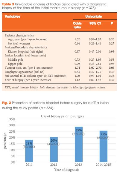
Of the 373 biopsied SRMs, the initial biopsy was diagnostic in 87% of cases. Of the 47 non-diagnostic biopsies, 15 had a repeat biopsy of which, 80% were diagnostic. When both were combined, therefore, a diagnosis was obtained in 91% of SRMs. Of these, 18% were benign. Size was the only factor found to be associated with achieving a diagnostic biopsy. RTB histology and nuclear grade (high or low) were found to be highly concordant with surgical pathology (86 and 81%, respectively). Of the discordant tumours (n = 16), all were upgraded from low to high grade on surgical pathology. Adverse events were rare (<1% of cases).

Conclusion

The present multi-institution study confirms that RTB of SRMs is safe, accurate and reliable across institutions, while decreasing unnecessary treatment. Given our findings, RTBs may be a helpful tool with which to triage SRMs and guide appropriate management.

Editorial: Renal tumour biopsy: let’s talk about it

There has been a marked increase in the incidental diagnosis of small renal masses (SRMs), resulting in overtreatment of benign and indolent lesions. Renal tumour biopsy (RTB) has received increasing attention as a potential tool to help reduce this overtreatment, with single-institution studies reporting good safety, accuracy, and reliability. One of the purposes of paper by Richard et al. [1], appearing in this issue of BJUI, was to address whether these results of RTB were generalisable across multiple institutions. They evaluated 373 RTBs from 12 centres, reporting an initial diagnostic rate of 87%, with 32% of non-diagnostic RTBs undergoing repeat biopsy, for a combined diagnostic rate of 91%. They reported concordance rates between RTB and surgical pathology of >80% and a RTB complication rate of <1%. The generalisability of these impressive RTB results remains unclear because they were unable to report the numbers of RTB per centre (beyond ‘at least one’) and results were likely driven by a few high-volume centres.

The analysis is not without limitations. The negative predicative value (NPV) of RTB could not be assessed because there was no surgical specimen to confirm a benign RTB diagnosis. A meta-analysis by Patel et al. [2] raised concerns about a non-diagnostic or negative RTB. Of the 14% of patients with a non-diagnostic biopsy, 90% of those subsequently undergoing surgery were found to have cancer. Among patients having surgery, 37% with a negative biopsy who underwent surgical extirpation were found to have cancer on final pathology (NPV 63%). Another limitation of RTBs is that they tend to under grade tumours compared to surgical pathology. Even when using a simplified two-tiered grading system of low vs high grade tumours to improve concordance [3], 20% of patients with low-grade clear cell RCC (ccRCC) were upgraded to high-grade ccRCC at surgery. Concordance rates did not include non-diagnostic biopsies; nonetheless, their results support that a ‘good’ RTB can usually be trusted. Selection bias may have been a factor because patients who did not receive a RTB for a SRM were not included. It seems unlikely that reports of improved RTB outcomes would result in a change in guidelines to a ‘one size fits all’ policy recommending RTB in all patients with a SRM. RTB may not be feasible in some patients (anterior, hilar, cystic tumours) and may not always have potential to change clinical management, such as with a young healthy patient who is unwilling to accept any degree of uncertainty with a negative biopsy or an elderly patient with comorbidities who would not accept treatment regardless of RTB results. In this study [1], only ~25% of the patients who underwent surgery for a cT1a lesion had a RTB before surgery, possibly a reflection of the limitations of RTB, as well as some room for improvement.

Despite the limitations of RTB, the fact remains that many renal lesions are over treated, RTB outcomes are improving, and RTB may help guide clinical management. The authors [1] suggest that even a misclassified SRM could probably be managed conservatively over the short term. They recommend that even a benign RTB should be followed with serial imaging and that a repeat RTB should be considered for fast growing lesions. Perhaps the future of RCC diagnosis lies beyond the RTB and includes imaging innovations that can distinguish benign and malignant tumours and spare patients an unnecessary treatment, as well as an unnecessary biopsy. For example, Gorin et al. [4] showed that technetium-99m (99mTc)-sestamibi single-photon emission CT (SPECT)/CT could accurately distinguish renal oncocytomas and hybrid oncocytic/chromophobe tumours from other renal tumour histologies.

Current guidelines already acknowledge the potential role for RTB to guide clinical management in patients willing to accept the known limitations and who have an indeterminate SRM or are considering a range of treatment options such as active surveillance or ablation. We do not need a blanket guideline mandating upfront RTB for all. But we should at least talk about RTB with our patients with SRMs. We owe it to them to be aware of the potential benefits and limitations of RTB and include this in our discussion so they can be involved in the decision.

Haider Rahbar, and Craig Rogers


Vattikuti Urology Institute, Henry Ford Hospital, Detroit, MI, USA


References


3 Rioux-Leclercq N, Karakiewicz PI, Trinh QD et al. Prognostic ability of simplied nuclear grading of renal cell carcinoma. Cancer 2007; 109: 86874


Article of the Week: PADUA and R.E.N.A.L. nephrometry scores correlate with perioperative outcomes of RAPN: analysis of the Vattikuti GQI-RUS database

Every week the Editor-in-Chief selects an Article of the Week from the current issue of BJUI. The abstract is reproduced below and you can click on the button to read the full article, which is freely available to all readers for at least 30 days from the time of this post.

In addition to the article itself, there is an accompanying editorial written by a prominent member of the urological community. This blog is intended to provoke comment and discussion and we invite you to use the comment tools at the bottom of each post to join the conversation.

If you only have time to read one article this week, it should be this one.

PADUA and R.E.N.A.L. nephrometry scores correlate with perioperative outcomes of robot-assisted partial nephrectomy: analysis of the Vattikuti Global Quality Initiative in Robotic Urologic Surgery (GQI-RUS) database


Riccardo Schiavina*, Giacomo Novara,, Marco Borghesi*, Vincenzo Ficarra§Rajesh Ahlawat, Daniel A. Moon**, Francesco Porpiglia††,BenjaminJ.Challacombe‡‡Prokar Dasgupta‡‡, Eugenio Brunocilla*, Gaetano La Manna§§, Alessandro Volpe¶¶Hema Verma***, Giuseppe Martorana* and Alexandre Mottrie,†††


*Department of Urology, University of Bologna, Bologna,† Department of Surgery, Oncology, and Gastroenterology – Urology Clinic, University of Padua, Padua, Italy, OLV Vattikuti Robotic Surgery Institute, Aalst, Belgium, §Department of
Experimental and Clinical Medical Sciences, University of Udine, Udine, Italy, Division of Urology and Renal Transplantation, Medanta Kidney and Urology Institute, Medanta-The Medicity, Gurgaon, India, **Department of Surgery, Peter MacCallum Cancer Centre, University of Melbourne, Melbourne, Vic., Australia, ††San Luigi Gonzaga Hospital, University of Turin, Orbassano, Italy, ‡‡Department of Urology, Guys and St Thomas NHS Foundation Trust and National Institute for Health Research (NIHR) Biomedical Research Centre, Kings College London, London, UK, §§Department Nephrology and Experimental, Diagnostic and Specialty Medicine, University of Bologna, Bologna, ¶¶University of Eastern Piedmont, Novara, Italy, ***Department of Radiology, Guys and St Thomas NHS Foundation Trust and National Institute for Health Research (NIHR) Biomedical Research Centre, Kings College London, London, UK, and †††Department of Urology, Onze-Lieve-Vrouw Hospital, Aalst, Belgium


Read the full article

Abstract

Objectives

To evaluate and compare the correlations between Preoperative Aspects and Dimensions Used for an Anatomical (PADUA) and R.E.N.A.L. [Radius (tumour size as maximal diameter), Exophytic/endophytic properties of the tumour, Nearness of tumour deepest portion to the collecting system or sinus, Anterior (a)/posterior (p) descriptor and the Location relative to the polar line] nephrometry scores and perioperative outcomes and postoperative complications in a multicentre, international series of patients undergoing robot-assisted partial nephrectomy (RAPN) for masses suspicious for renal cell carcinoma (RCC).

Patients and Methods

We retrospectively evaluated the clinical records of patients who underwent RAPN between 2010 and 2013 for clinical N0M0 renal tumours in four international centres that completed all the data required for the Vattikuti Global Quality Initiative in Robotic Urologic Surgery (GQI-RUS) database. All patients underwent preoperative computed tomography or magnetic resonance imaging to define the clinical stage and anatomical characteristics of the tumours. PADUA and R.E.N.A.L. scores were retrospectively assessed in each centre. Univariate and multivariate analyses were used to evaluate the correlations between age, gender, Charlson comorbidity index, clinical tumour size, PADUA and R.E.N.A.L. complexity group categories and warm ischaemia time (WIT) of >20 min, urinary calyceal system closure, and grade of postoperative complications.

aotw-mar-4-results

Results

Overall, 277 patients were evaluated. The median (interquartile range) tumour size was 33.0 (22.0–43.0) mm. The median PADUA and R.E.N.A.L. scores were eight and seven, respectively; 112 (40.4%), 86 (31.0%) and 79 (28.5%) patients were classified in the low-, intermediate- or high-complexity group according to PADUA score, while 118 (42.5%), 139 (50.1%) and 20 (7.2%) were classified in the low-, intermediate- or high-complexity group according to R.E.N.A.L. score, respectively. Both nephrometry tools significantly correlated with perioperative outcomes at univariate and multivariate analyses.

Conclusion

A precise stratification of patients before PN is recommended to consider both the potential threats and benefits of nephron-sparing surgery. In our present analysis, both PADUA and R.E.N.A.L. were significantly associated with predicting prolonged WIT and high-grade postoperative complications after RAPN.

Editorial: Nephrometry scoring systems: valuable research tools, but can they be applied in daily clinical practice?

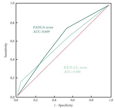
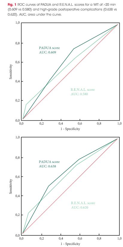
In this issue of BJUI Schiavina et al. [1] report on the RENAL and PADUA nephrometry scoring systems in predicting peri-operative outcomes, including warm ischaemia time and postoperative complications, in a multi-institutional cohort of patients undergoing robot-assisted partial nephrectomy. The authors showed that tumours classified as being of intermediate and high complexity on the PADUA score and high complexity on the RENAL score were associated with a nearly threefold higher risk of longer warm ischaemia times (>20 min). In addition, more complex tumours carried a higher risk of grade 3–4 postoperative complications (most commonly bleeding requiring angioembolization and urine leak requiring a ureteric stent). Notably, the two scoring systems were found to be similar predictors of these peri-operative outcomes on receiver-operating curve (ROC) analyses [1].

This represents the first large, multicentre study to evaluate the accuracy of these scoring systems in a cohort of patients who purely underwent robot-assisted surgery. A recent study by Borgmann et al. [2] found that, among the reported scoring systems, the RENAL nephrometry score correlated best with achieving negative surgical margins, shorter ischaemia times, and low postoperative complication rates; however, only 9% of patients underwent robot-assisted surgery. Another contemporary series showed concordance between the RENAL and PADUA scoring systems in predicting ischaemia times and complication rates, albeit in patients who only underwent open surgery [3].

Current guidelines recognize nephron-sparing approaches to small renal masses as the standard of care in well-selected patients, with the robot-assisted platform being predominantly adopted in clinical practice where available. Certainly, these nephrometry scores are valuable for urologists in counselling patients on the potential risk of complications specific to the surgical anatomy of the tumour. In addition, the RENAL and PADUA scores (and others) provide a quantitative, objective method for comparing data from different studies and different institutions.

As nephrometry scoring systems continue to be critically evaluated in the robotic surgery era, the question that naturally arises is: which system is best? With regard to this question, the data in the present study do not necessarily favour one or the other for the prediction of clinically relevant peri-operative outcomes. One must recognize, however, that several other anatomy-based scoring systems exist and were not examined in this manuscript [4-6]. While these are very valuable research and patient counselling tools, one must caution against using these nephrometry tools to make clinical decisions; for example, attempting to predict benign vs malignant histology (without a biopsy), attempting to predict high vs low grade tumours, or deciding on whether to perform a radical vs partial nephrectomy, or an open vs minimally invasive approach. After all, one must keep in mind that the area under the curve for these tools is in the range of 0.58–0.63 (0.50 being equivalent to toss of a coin).

It would have been interesting to include clinical size only in the present multivariate analysis (as was done for RENAL and PADUA scoring) and ROC analysis to compare this simple variable with the studied nephrometry scores. Future research should examine additional confounders that could potentially affect postoperative complication rates, such as BMI, adherent perinephric fat, experience of the surgeon actually performing the partial nephrectomy, technique of resection used (e.g. enucleation or resection) among others. This may help to distinguish a single system as the optimum model for use in research and in patient counselling regarding potential postoperative complications.

Matthew A. Meissner and Jose A. Karam


Department of Urology, University of Texas MD Andersonn Cancer Center, Houston, TX, USA


Read the full article


References


3 Kriegmair MC, Mandel P, Moses A et al. Dening Renal Masses: comprehensive Comparison of RENAL, PADUA, NePhRO, and C-Index Score. Clin Genitourin Cancer 2016; [Epub ahead of print]. doi: 10.1016/ j.clgc.2016.07.029.


5 Hakky TS, Baumgarten AS, Allen B, Lin HY, Ercole CE, Sexton WJSpiess PE et al. Zonal NePhRO scoring system: a superior renal tumor complexity classication model. Clin Genitourin Cancer 2014; 12: e138


6 Simmons MN, Ching CB, Samplaski MK, Park CH, Gill IS et al. Kidney tumor location measurement using the C index method. J Urol 2010; 183: 170813


Article of the Week: LCA – EuRECA study

Every week the Editor-in-Chief selects an Article of the Week from the current issue of BJUI. The abstract is reproduced below and you can click on the button to read the full article, which is freely available to all readers for at least 30 days from the time of this post.

In addition to the article itself, there is an accompanying editorial written by a prominent member of the urological community. This blog is intended to provoke comment and discussion and we invite you to use the comment tools at the bottom of each post to join the conversation.

If you only have time to read one article this week, it should be this one.

Oncological outcomes and complication rates after laparoscopic-assisted cryoablation: a European Registry for Renal Cryoablation (EuRECA) multi-institutional study

Tommy K. Nielsen*, Brunolf W. Lagerveld, Francis Keeley, Giovanni Lughezzani§Seshadri Sriprasad, Neil J. Barber**, Lars U. Hansen*,††, Nicole M. Buf§Giorgio Guazzoni§, Johan A. van der Zee, Mohamed Ismail, Khaled Farrag,Amr M. Emara**,‡‡, Lars Lund††,§§, Øyvind Østraat* and Michael Borre*


*Department of Urology, Aarhus University Hospital, Aarhus, Denmark, Department of Urology, Onze Lieve Vrouwe Gasthuis, Amsterdam, the Netherlands, Bristol Urological Institute, Bristol, UK, §Department of Urology, Istituto Clinico Humanitas IRCCS, Clinical and Research Hospital, Milano, Rozzano, Italy, Department of Urology, Darent Vally Hospital, Dartford, **Department of Urology, Frimley Park Hospital, Camberley, UK, ††Department of Urology, Odense University Hospital, Odense, Denmark, ‡‡Department of Urology, Ain Shams University, Cairo, Egypt, and §§Department of Urology, Viborg Regional Hospital, Viborg, Denmark


Read the full article

Abstract

Objective

To assess complication rates and intermediate oncological outcomes of laparoscopic-assisted cryoablation (LCA) in patients with small renal masses (SRMs).

Patients and Methods

A retrospective review of 808 patients treated with LCA for T1a SRMs from 2005 to 2015 at eight European institutions. Complications were analysed according to the Clavien–Dindo classification. Kaplan–Meier analyses were used to estimate 5- and 10-year disease-free survival (DFS) and overall survival (OS).

aotw-mar-2-2017

Results

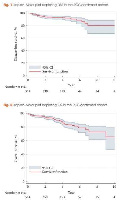
The median [interquartile (IQR)] age was 67 (58–74) years. The median (IQR) tumour size was 25 (19–30) mm. The transperitoneal approach was used in 77.7% of the patients. The median postoperative hospital stay was 2 days. In all, 514 patients with a biopsy-confirmed renal cell carcinoma (RCC) were available for survival analyses. The median (IQR) follow-up for the RCC-cohort was 36 (14–56) months. A total of 32 patients (6.2%) were diagnosed with treatment failure. The 5-/10-year DFS was 90.4%/80.0% and 5-/10-year OS was 83.2%/64.4%, respectively. A total of 134 postoperative complications (16.6%) were reported, with severe complications (grade ≥III) in 26 patients (3.2%). An American Society of Anesthesiologists score of 3 was associated with an increased risk of overall complications (odds ratio 2.85, 95% confidence interval 1.32–6.20; P = 0.005).

Conclusions

This large series of LCA demonstrates satisfactory long-term oncological outcomes for SRMs. However, although LCA is considered a minimally invasive procedure, risk of complications should be considered when counselling patients.

Editorial: Laparoscopic renal mass cryoablation: an operation in search of an indication

In this issue of BJUI, Nielsen et al. [1] report the oncological and surgical outcomes from a multi-institutional cohort of patients receiving laparoscopic cryoablation (LCA) as primary therapy for solitary renal masses <4 cm in size (cT1a). This work represents the latest addition to a growing body of literature in an important oncological space that lacks prospective/randomized evidence to guide practitioners counselling patients with kidney cancer. Although the article does not advance the discussion toward higher levels of evidence, the results are nonetheless provocative and several strengths and weaknesses deserve comment.

While nephron-sparing surgery has become the recognized standard of care for cT1a renal lesions [2, 3], the reality remains that certain patients carry unacceptable risk profiles for partial nephrectomy, making less invasive options preferable. Such indications might include being elderly or frail, having hereditary kidney cancer syndromes prone to metachronous renal tumours, or having a solitary kidney. For such patients, focal renal mass ablative techniques have emerged as a safe alternative to extirpation that avoids the permanent nephron loss associated with radical nephrectomy. From an oncological perspective, however, cryotherapy, radiofrequency ablation and microwave ablation (by any approach) all have yet to be studied against partial nephrectomy in a prospective fashion. Numerous retrospective analyses have attempted to fill the void [4], yet the general consensus among most academic kidney surgeons is that renal mass ablation offers acceptable but inferior cancer control compared with surgery [5].

In this retrospective analysis by Nielsen et al., 808 patients underwent LCA between 2005 and 2015, 514 (63.4%) of whom had pre-procedural biopsy-proven RCC. The principal findings described in the present study include not only 5- and 10-year disease-free and overall survival, but also morbidity and mortality outcomes after LCA. The authors should be commended for the structure of their design, which included a high proportion of patients with available preoperative biopsy data. Additionally, clear definitions of treatment success, ‘residual unablated tumour’ and ‘local tumour progression’ are provided, and consistent follow-up imaging protocols were employed by the institutions involved. In each of these ways, Nielsen et al. overcome many of the pitfalls that have clouded the interpretation of results from previous reports.

Nevertheless, the oncological outcomes reported in this study, which are on a par with those for partial nephrectomy as well as other ablative techniques, must be approached with a degree of skepticism. As there is no alternative treatment cohort included in the study, omission of anatomical complexity data (in the form of nephrometry scoring) prohibits any meaningful comparison with patients having undergone ablative procedures or partial nephrectomy from other series. Availability of these data is essential for the reader to gauge the influence of selection bias in the interpretation of the results.

From a morbidity and mortality standpoint, the reported 16% overall complication rate, 3% rate of severe complications (defined as Clavien III–V) and three deaths within 30 days of the procedure might have been strengthened by the missing nephrometry data, as the rate of complications would be expected to increase with the complexity of the renal mass [6]. Also noticeably absent from the analysis are granular comorbidity and previous surgery data, both of which intuitively predispose patients to complications when undergoing minimally invasive surgery.

With these limitations in mind, the experienced kidney surgeon is not likely to see LCA as an equally effective or safer alternative to minimally invasive partial nephrectomy, which can be performed with similar complication rates and length of hospital stay without sacrificing oncological efficacy in most patients. Similarly, the question of why the practitioner should assume the risks of LCA when percutaneous cryoablation is readily available at many contemporary kidney cancer centres is unanswered by the present study. Indeed, with increasingly complex renal masses being managed via minimally invasive nephron-sparing surgery, and active surveillance of small renal masses gaining traction in the appropriate patient population, cryoablation via a laparoscopic approach unfortunately may represent another urological application without a well-defined indication going forward. We hope that the results presented by Nielsen et al. in this issue of BJUI encourage investigators to enroll patients in prospective trials aimed at comparing available ablative techniques or partial nephrectomy in matched cohorts to identify the ideal patient population for this operation and further clarify the oncological and clinical outcomes compared with surgical excision.

Daniel C. Parker and Brian W. Cross


Department of Urologic Oncology, University of Oklahoma Stephenson Cancer Center, Oklahoma City, OK, USA


Read the full article


References


1 NielsenT, Lagerveld B, Keeley F et al. Oncologic outcomes and complication rates after laparoscopic-assisted cryoablation: a EuRECA multi-institutional study. BJU Int 2016. [Epub ahead of print].


2 Campbell SC, Novick AC, Belldegrun A et al. Guideline for management of the clinical T1 renal mass. J Urol 2009; 182: 12719


3 Ljungberg B, Bensalah K, Caneld S et al. EAU guidelines on renal cell carcinoma: 2014 update. Eur Urol 2015; 67: 91324


4 Wagstaff P, Ingels A, Zondervan P et al. Thermal ablation in renal cell carcinoma management: a comprehensive review. Curr Opin Urol 2014; 24: 47482


5 Kutikov A, Smaldone MC, Uzzo RG. Focal therapy for treatment of the small renal mass: dealers choice or a therapeutic gamble? Eur Urol 2015; 67: 2601


Article of the Week: Combining Nanotech Drug Delivery and Thermoablation in an in vivo mouse model of RCC

Every week the Editor-in-Chief selects an Article of the Week from the current issue of BJUI. The abstract is reproduced below and you can click on the button to read the full article, which is freely available to all readers for at least 30 days from the time of this post.

In addition to the article itself, there is an accompanying editorial written by a prominent member of the urological community. This blog is intended to provoke comment and discussion and we invite you to use the comment tools at the bottom of each post to join the conversation.

If you only have time to read one article this week, it should be this one.

Nanotechnology combined therapy: tyrosine kinase-bound gold nanorod and laser thermal ablation produce a synergistic higher treatment response of renal cell carcinoma in a murine model

James Liu*, Caleb Abshire*, Connor Carry*, Andrew B. Sholl, Sree Harsha Mandava*, Amrita Datta*, Manish Ranjan*, Cameron Callaghan*, Donna V. Peralta, Kristen S. Williams, Weil R. Lai*, Asim B. Abdel-Mageed*, Matthew Tarr and Benjamin R. Lee§


Departments of *Urology, Pathology, Tulane University School of Medicine, Department of Chemistry, University of New Orleans, New Orleans, LA, and §Division of Urology, University of Arizona College of Medicine, Tucson, AZ, USA


Read the full article

Abstract

Objectives

To investigate tyrosine kinase inhibitors (TKI) and gold nanorods (AuNRs) paired with photothermal ablation in a human metastatic clear cell renal cell carcinoma (RCC) mouse model. Nanoparticles have been successful as a platform for targeted drug delivery in the treatment of urological cancers. Likewise, the use of nanoparticles in photothermal tumour ablation, although early in its development, has provided promising results. Our previous in vitro studies of nanoparticles loaded with both TKI and AuNRs and activated with photothermal ablation have shown significant synergistic cell kill greater than each individual arm alone. This study is a translation of our initial findings to an in vivo model.

Materials and Methods

Immunologically naïve nude mice (athymic nude-Foxn1nu) were injected subcutaneously bilaterally in both flanks (n = 36) with 2.5 × 10⁶ cells of a human metastatic renal cell carcinoma cell line (RCC 786-O). Subcutaneous xenograft tumours developed into 1-cm palpable nodules. AuNRs encapsulated in human serum albumin protein (HSA) nanoparticles were synthesised with or without a TKI and injected directly into the tumour nodule. Irradiation was administered with an 808-nm light-emitting diode laser for 6 min. Mice were humanely killed 14 days after irradiation; tumours were excised, formalin fixed, paraffin embedded, and evaluated for size and the percentage of necrosis by a genitourinary pathologist. The untreated contralateral flank tumours were used as controls.

Results

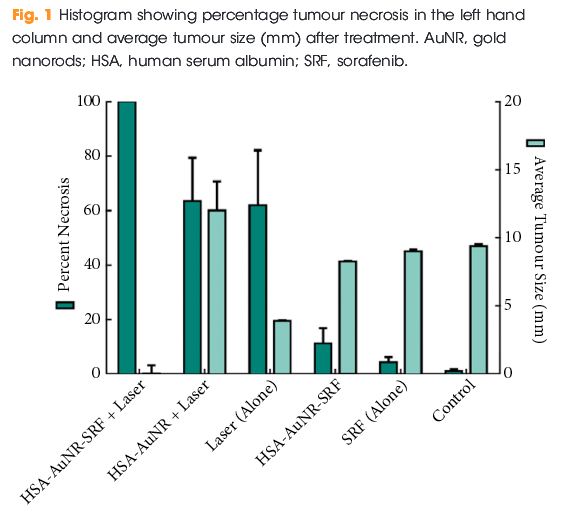
In mice that did not receive irradiation, TKI alone yielded 4.2% tumour necrosis on the injected side and administration of HSA-AuNR-TKI alone yielded 11.1% necrosis. In the laser-ablation models, laser ablation alone yielded 62% necrosis and when paired with HSA-AuNR there was 63.4% necrosis. The combination of laser irradiation and HSA-AuNR-TKI had cell kill rate of 100%.

aotw-22-2-17

Conclusions

In the absence of laser irradiation, TKI treatment alone or when delivered via nanoparticles produced moderate necrosis. Irradiation with and without gold particles alone also improves tumour necrosis. However, when irradiation is paired with gold particles and drug-loaded nanoparticles, the combined therapy showed the most significant and synergistic complete tumour necrosis of 100% (P < 0.05). This study illustrates the potential of combination nanotechnology as a new approach in the treatment of urological cancers.


Editorial: Synergistic effects in combinational drug delivery and thermal ablation using nanotechnology

Chemotherapy, the dominant therapeutic approach to the treatment of a wide variety of cancers, is intrinsically inefficient because its drug delivery is non-specific. This leads to a trade-off between undesirable cytotoxic effects in healthy cells and associated side effects and lower efficacy in killing cancerous cells at lower concentrations.

The study in the present issue of BJUI by Lui et al. [1], from the University of Arizona College of Medicine, attempts to circumvent these undesirable side effects by employing nanoparticles as drug delivery vectors, isolating chemotherapeutic agents from the systemic environment while allowing them to accumulate in solid tumours with leaky vasculature and impaired lymphatics, and improving cellular uptake both passively and through targeted therapy. In recent years, this therapeutic approach has been extended by combining targeted drug delivery via nanoparticles with temperature-based treatments. In the combined therapy, either the nanoparticles exhibit strong absorption in the human tissue transparency window in the near infra-red, enabling laser excitation, or alternatively, via the absorption of ultrasound. Synergy implies more than a simple linear addition of chemotherapeutic agents and high temperature ablation, and it has been suggested previously that the efficacy of chemotherapeutic agents is improved at elevated temperatures [2].

A wide variety of nanoparticle vectors has been investigated, but gold nanoparticles have been shown to be biocompatible and elementally stable, while possessing the ability to bind various compounds for immune system evasion, and are a malleable structure for further design considerations [3] as well as exhibiting a strong and wavelength-tunable absorption resonance for near infra-red laser excitation.

In genitourinary oncology, the use of nanotechnology as a carrier for drug delivery, laser ablation, gene therapy and imaging has grown rapidly in the past decade. The work reported in the present study follows one of the first studies on synergistic therapeutic treatments, which was conducted by Stern et al. [4] from the University of Texas Southwestern. They used a combination of gold and tyrosine kinase inhibitor (TKI) nanotechnology with laser ablation. Their work was performed in an in vivo metastatic RCC mouse model [4] and shows that gold nanoparticles improve heat delivery to cancer while both sparing local benign tissues and also significantly improving tumour necrosis.

A previous study by Lui’s group [5] has already demonstrated the efficacy of using gold nanorods loaded with human serum albumin (HSA) and a TKI (sorafenib) as effective drug delivery vectors, as well as gold nanorods (AuNR) for tumour ablation. The purpose of the study was to explore the synergistic effects when gold ablation is also paired with chemotherapeutics; therefore, in each individual experimental arm low amounts of HSA-AuNR and HSA-AuNR-TKI were used to further magnify any co-acting effect when combined with thermal ablation.

For that study, immunologically naïve nude mice (Athymic Nude-Foxn1nu) were injected bilaterally on the flanks (= 36) with 2.5 × 106 cells of a human metastatic RCC cell line (RCC 786-O). Subcutaneous xenograft tumours developed 1-cm palpable nodules. AuNR encapsulated in HSA protein nanoparticles were synthesized with or without a TKI and injected directly into the tumour nodule. Once tumours reached an appropriate size, they were directly injected with 0.1 mL of 10 mM sorafenib solution in PBS, 0.1 mL suspension of HSA-AuNR stock, or 0.1 mL of HSA-AuNR-TKI stock. In the treatment groups, laser ablation was performed 24 h later to allow the cellular uptake of the particle with irradiation, administered with an 808-nm LED diode laser for 6 min. The mice were killed 14 days after irradiation. The tumours were then excised, formalin-fixed, paraffin-embedded and evaluated for size and percent necrosis by a genitourinary pathologist. Untreated contralateral flank tumours were used as controls. The area of laser treatment was 1 cm in diameter, completely covering the tumours. The mice were anaesthetized during laser treatment with continuous isoflurane.

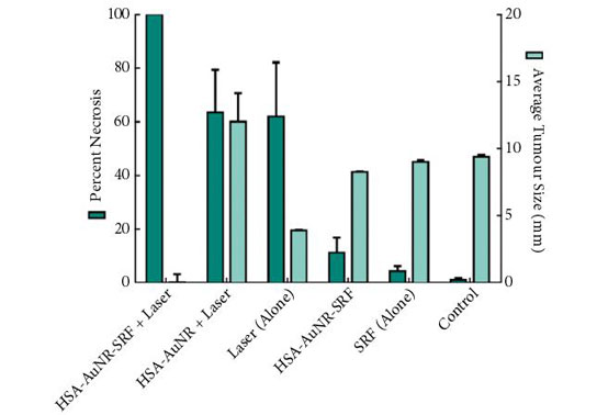
The results of the final percentage tumour necrosis and average tumour size are shown in Fig. 1. To be able to infer synergistic effects, a careful study design was used, such that neither HSA-AuNR vs sorafenib alone, nor HSA-AuNR vs laser treatment alone showed statistically significant differences, which showed that nanoparticles concentrations were insufficient in each individual experimental arm to result in any statistical improvement when compared with control settings (Fig. 1). In mice that did not receive irradiation, a TKI alone yielded 4.2% tumour necrosis on the injected side and administration of HSA-AuNR-TKI alone yielded 11.1% necrosis. In laser ablation models, laser ablation alone yielded 62% necrosis and, when paired with HSA-AuNR, yielded 63.4% necrosis; however, the combination of laser irradiation and HSA-AuNR-TKI had cell kill of 100%. The clear conclusion is that in the absence of laser irradiation, TKI treatment alone or when delivered via nanoparticle produced moderate necrosis. Irradiation with and without gold particles alone also improves tumour necrosis. When irradiation is paired with gold particle and drug-loaded nanoparticle treatment, however, the combination therapy showed the most significant and synergistic complete tumour necrosis of 100% (P < 0.05). The overwhelming tumour necrosis of combinational nanotechnology shows synergistic kill rather than simple additive effects of each treatment method.

aotw-ed-22-2-17

This significantly improved efficacy of combination nanotechnology can be explained by the complementary mechanisms of action for each individual arm. By combining AuNR and sorafenib in an albumin vehicle, better delivery of both chemotherapeutic drug and thermal ablative particle to the tumour site is possible. Likewise, when tumour cells are activated by laser there is not only cell lysis, attributable to rapid temperature increase, but also cells that do survive upregulate heat shock proteins, such as FasL which mediate apoptosis [6]. Studies have found that TKIs play a critical part in intracellular pathways that enhance this effect, possibly by upregulating FasR and thereby accelerating apoptosis [7]. A laser-induced temperature increase may disrupt albumin construction and facilitates intracellular drug delivery. The verification of synergistic tumour necrosis from combination nanotechnology is an exciting springboard for future experiments, while the translation of this effective in vitro model into a murine tumour model illustrates that nanotechnology is a reliable platform demanding future clinical evaluation.

The present study beautifully illustrates the enormous potential of combination nanotechnology as a new approach in the treatment of urological cancers. The next step in pursuing more effective combination nanotechnology is to better calibrate factors such as drug load, AuNR load and laser settings. In particular, narrow size distribution of the gold nanoparticles, fully optimizing their absorption resonance optimized at the irradiation wavelength will enable lower nanoparticle loading and either lower irradiation thresholds or deeper tissue activation. These studies will not only help find new ways to eradicate tumours but will also add to the precision of minimally invasive surgical technology.

Because of the hazardous nature of chemotherapeutics, a specialized CTSD pharmacy must handle, prepare, and dispense these kinds of medications.

Read the full article

Wayne Dickson
Department of Physics, King’s College London, London, UK

References


2 Zhang W, Guo Z, Huang D et al. Synergistic effect of chemo- photothermal therapy using PEGylated graphene oxide. Biomaterials 2011; 32: 855561


3 Stern JM, Staneld J, Lotan Y, Park S, Hsieh JT, Cadeddu JA. Efcacy of laseractivated gold nanoshells in ablating prostate cancer cells in vitro. J Endourol 2007; 8: 93


Article of the Week: Be Clear on Cancer – Blood in Pee

Every Week the Editor-in-Chief selects an Article of the Week from the current issue of BJUI. The abstract is reproduced below and you can click on the button to read the full article, which is freely available to all readers for at least 30 days from the time of this post.

In addition to the article itself, there is an accompanying editorial written by a prominent member of the urological community. This blog is intended to provoke comment and discussion and we invite you to use the comment tools at the bottom of each post to join the conversation.

Finally, the third post under the Article of the Week heading on the homepage will consist of additional material or media. This week we feature a video from Mr. Archie Hughes-Hallett,, discussing his paper.

If you only have time to read one article this week, it should be this one.

Assessing the impact of mass media public health campaigns. ‘Be Clear on Cancer: Blood in Pee’ a case in point

Archie Hughes-Hallett*, Daisy Browne, Elsie Mensah*, Justin Vale*† and Erik Mayer*†‡


*Department of Surgery and Cancer, Imperial College London, Department of Urology, Imperial College Healthcare Trust, and Institute of Global Health Innovation, Imperial College London, London, UK


Read the full article

Objectives

To assess the impact on suspected cancer referral burden and new cancer diagnosis of Public Health England’s recent Be Clear on Cancer ‘blood in pee’ mass media campaign.

Methods

A retrospective cohort study design was used. For two distinct time periods, August 2012 to May 2013 and August 2013 to May 2014, all referrals of patients deemed to be at risk of urological cancer by the referring primary healthcare physician to Imperial College NHS Healthcare Trust were screened. Data were collected on age and sex and whether the referral was for visible haematuria, non-visible haematuria or other suspected urological cancer. In addition to referral data, hospital episode data for all new renal cell (RCC) and upper and lower tract transitional cell carcinoma (TCC), as well as testicular and prostate cancer diagnoses for the same time periods were obtained.

Results

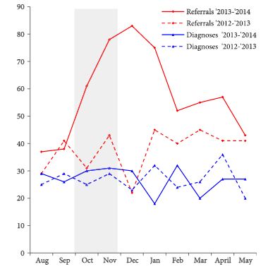
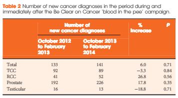
Over the campaign period and the subsequent 3 months, the number of haematuria referrals increased by 92% (P = 0.013) when compared with the same period a year earlier. This increase in referrals was not associated with a significant corresponding rise in cancer diagnosis; instead changes of 26.8% (P = 0.56) and −3.3% (P = 0.84) were seen in RCC and TCC, respectively.

AOTW2Apr


Conclusions

This study has shown that the Be Clear on Cancer ‘blood in pee’ mass media campaign significantly increased the number of new suspected cancer referrals, but there was no significant change in the diagnosis of target cancers across a large catchment. Mass media campaigns are expensive, require significant planning and appropriate implementation and, while the findings of this study do not challenge their fundamental objective, more work needs to be done to understand why no significant change in target cancers was observed. Further consideration should also be given to the increased referral burden that results from these campaigns, such that pre-emptive strategies, including educational and process mapping, across primary and secondary care can be implemented.

Editorial: Be better with public health campaigns (and taxpayers’ money)

In this month’s issue of the BJUI, Hughes-Hallette et al. [1] report on the impact of a mass media public health campaign for gross haematuria. The authors performed a retrospective analysis evaluating the effectiveness of the ‘Be Clear on Cancer: “Blood in the pee”’ campaign. Similar campaigns for colorectal cancer have shown increased referrals and cost, without increasing the number of cancer diagnoses [2, 3]. In the current study [1], cancer diagnosis similarly did not rise. The two questions that therefore needs to be asked are:

  1. Is gross i.e. visible haematuria a predictor for urological malignancy?
  2. Does a mass media public health campaign constitue an effective means of improving early diagnosis of cancer?

Recent data from large integrative datasets have shown that visible haematuria is a significant predictor for bladder cancer [4, 5]. If gross haematuria is a predictor for urological malignancy, however, why did the authors [1] fail to find an increase in diagnosis of urological malignancy in their study? While the authors indicate that the study may have been underpowered, and that the use of an unlinked dataset may have interfered with proper accounting of cancer incidence, one must also consider that mass media outreach may not be an effective method for cancer outreach.

The ‘Be Clear on Cancer’ campaign involved the use of television adverts, print media and ‘out of home’ advertisements. Using this method, patients who are at risk of renal and bladder cancer, i.e. men, those aged >50 years, and smokers, are targeted as frequently as non-smoking teenagers. This type of mass media outreach programme is analogous to traditional advertising, where the message is often diluted and ineffective. While the ‘Be Clear on Cancer’ campaign is a worthy endeavour, the data do not seem to support the use of taxpayers’ money given its ineffective nature. A novel approach to visible haematuria may be to encourage GPs to ask patients about visible, painless haematuria, much as they would ask about chest pain or blood pressure. This would create a more focussed and durable outreach programme that would reduce the number of non-oncological referrals. As stewards of taxpayers’ money, we must be careful of how public funds are spent. The next generation of mass media outreach may require a combination of traditional media in addition to, social media and targeted advertising [6]. Although the ‘Be Clear on Cancer’ campaign did not appear to achieve its intended goals at this time, we must continue to refine and create new interactive approaches to improve the diagnosis and treatment of urological malignancies.

Read the full article
Casey K. Ng
Department of Urology, Southern California Permanente Medical Group, Pasadena, and USC Institute of Urology, Keck School of Medicine, University of Southern California, Los Angeles, CA, USA


References


1 Hughes-Hallett A, Browne D, Mensah E, Vale J, Mayer E. Assessing the impact of mass media public health campaigns. Be Clear on Cancer blood in pee: a case in point. BJU Int 2016; 117: 57075


2 Peacock O, Clayton S, Atkinson F, Tierney GM, Lund JN. Be Clear on Cancer: the impact of the UK National Bowel Cancer Awareness Campaign. Colorectal Dis 2013; 15: 9637


3 Bethune R, Marshall MJ, Mitchell SJ et al. Did the Be Clear on Bowel Cancer public awareness campaign pilot result in a higher rate of cancer detection? Postgrad Med J 2013; 89: 3903


4 Jung H, Gleason JM, Loo RK, Patel HS, Slezak JM, Jacobsen SJAssociation of hematuria on microscopic urinalysis and risk of urinary tract cancer. J Urol 2011; 185: 1698703


5 Loo RK, Lieberman SF, Slezak JM et al. Stratifying risk of urinary tract malignant tumors in patients with asymptomatic microscopic hematuria. Mayo Clin Proc 2013; 88: 12938


© 2024 BJU International. All Rights Reserved.