Tag Archive for: renal cell carcinoma

Posts

Editorial: Does knowing the risk of relapse in localized renal cell carcinoma matter?

Shah et al. [1] report a retrospective analysis from the Mayo Clinic investigating the prognostic significance of different patterns of pathological T3a clear‐cell RCC in patients who underwent radical nephrectomy for localized disease. There was no difference in disease progression, cancer‐specific mortality or all‐cause mortality when comparing isolated perinephric fat invasion vs isolated renal sinus fat invasion vs isolated renal vein invasion. Multiple sites of extra‐renal extension compared with one site, however, was independently associated with an increased risk of disease progression (hazard ratio [HR] 1.31, P = 0.02), death from RCC (HR 1.64, P < 0.001) and all‐cause mortality (HR 1.32, P = 0.008) when adjusting for multiple key variables including age, tumour size, grade, presence of coagulative tumour necrosis and sarcomatoid differentiation. The authors incorporated multiple sites of extra‐renal extension vs one site into three RCC prognostic models: SSIGN score, UISS and MSKCC nomogram. After controlling for these three predictive tools independently, multiple sites of extra‐renal disease predicted progression, death from RCC and all‐cause death. These data suggest that risk stratification for pT3aN0MO clear‐cell RCC is improved by differentiating multiple vs one site of extra‐renal extension.

Does an improved ability to predict recurrence and mortality increase the likelihood of cure in high‐risk localized RCC patients in 2018? Unfortunately, the answer is no. Ideally, prognostic models would identify patients at sufficient risk to consider adjuvant therapy, which would increase cure rates by eradicating micro‐metastatic disease with an acceptable toxicity. Regrettably, in RCC management there are no well‐established post‐surgical therapies that improve cure rates. The deficiency of established adjuvant therapies is not attributable to a lack of investigative trials. In the era before vascular endothelial growth factor receptor (VEGFR) targeting, adjuvant vaccines, immunotherapies and other systemic therapies failed to demonstrate improved recurrence‐free (RFS) or overall survival (OS) [2]. The efficacy of VEGFR‐targeted therapies in the metastatic setting re‐energized the hope for adjuvant therapy in patients with high‐risk localized RCC after surgical resection in the past two decades. The results to date have been disappointing. To date, three trials (ASSURE, PROTECT and S‐TRAC) have been completed, comparing oral VEGFR tyrosine kinase inhibitors with placebo in high‐risk localized clear‐cell RCC, with disease‐free survival (DFS) as the primary endpoint [3,4,5]. ASSURE and PROTECT showed no difference in RFS or OS [3,4,5]. S‐TRACT demonstrated an improvement in DFS but not in OS [4]. A pooled analysis of these three trials also failed to demonstrate improved DFS or OS with adjuvant VEGFR‐targeted therapy [6]. Significant side effects with discontinuation of adjuvant therapy occurred in 28–45% of patients as a result of drug‐related toxicity [6]. Trials investigating immune checkpoint inhibitors have yet to be published and, with the established efficacy of these drugs in the metastatic setting, hope still remains for adjuvant therapy in resected high‐risk localized RCC.

If the current literature does not support adjuvant therapy for resected high‐risk RCC, does knowing the risk of relapse alter surveillance? National Comprehensive Cancer Network guidelines for resected stage III RCC recommend chest and abdominal imaging within 3–6 months, along with subsequent chest and abdominal imaging every 3–6 months for 3 years, and then annually up to 5 years. Although the ideal schedule for surveillance imaging is unknown, further characterizing of the risk of relapse in high‐risk localized RCC would not be likely to affect this schedule significantly.

Although knowing the risk of relapse in high‐risk localized RCC does not help management in 2018, there is still a value to enhancing our prognostic tools. For one, our prognostic tools help clinicians counsel patients appropriately about their risk of recurrence. In addition, enhanced prognostic tools will assist in selecting appropriate patients with high‐risk localized RCC for future clinical trials of adjuvant therapy and also help us understand the results when comparing cohorts within and between trials.

References

  1. Shah PH, Lyon TD, Lohse CM. Prognostic evaluation of perinephric fat, renal sinus fat, and renal vein invasion for patients with pathologic stage T3a clear cell renal cell carcinoma. BJU Int 2019; 123: 270–6
  2. Scherr AJO, Lima JPSN, Sasse EC et al. Adjuvant therapy for locally advanced renal cell cancer: a systematic review with meta‐analysis. BMC Cancer 2011; 11: 115–21
  3. Haas N, Manola J, Uzzo R et al. Adjuvant sunitinib or sorafenib for high‐risk, non‐metastatic renal‐cell carcinoma (ECOG‐ACRIN E2805): a double‐blind, placebo‐controlled, randomised, phase 3 trial. Lancet 2016; 387: 2008–16
  4. Ravaud A, Motzer RJ, Pandha HS et al. Adjuvant sunitinib in high‐ risk renal‐cell carcinoma after nephrectomy. N Engl J Med 2016; 375: 2246–54
  5. Motzer RJ, Haas NB, Donskov F et al. Randomized phase III trial of adjuvant pazopanib versus placebo after nephrectomy in patients with localized or locally advanced renal cell carcinoma. J Clin Oncol 2017; 35: 3916–23
  6. Sun M, Marconi L, Eisen T et al. Adjuvant vascular endothelial growth factore‐targeted therapy in renal cell carcinoma. Eur Urol 2018; 74: 611–20


Article of the Week: Impact of warm ischaemia time on postoperative renal function after partial nephrectomy for clinical T1 renal cell carcinoma

Every Week the Editor-in-Chief selects an Article of the Week from the current issue of BJUI. The abstract is reproduced below and you can click on the button to read the full article, which is freely available to all readers for at least 30 days from the time of this post.

In addition to the article itself, there is an accompanying editorial written by a prominent member of the urological community. This blog is intended to provoke comment and discussion and we invite you to use the comment tools at the bottom of each post to join the conversation.

If you only have time to read one article this week, it should be this one.

Impact of warm ischaemia time on postoperative renal function after partial nephrectomy for clinical T1 renal cell carcinoma: a propensity score-matched study

Hakmin Lee*, Byung D. Song*, Seok-Soo Byun*, Sang E. Lee* and Sung K. Hong*
*Department of Urology, Seoul National University Bundang Hospital, Seongnam, and Department of Urology, Seoul National University College of Medicine, Seoul, Korea


Read the full article

Objectives

To analyse the effect of prolonged warm ischaemia time (WIT) on long-term renal function after partial nephrectomy (PN), as controversy still exists as to whether prolonged WIT adversely affects the incidence of chronic kidney disease (CKD) after PN.

Patients and Methods

We reviewed data from 1816 patients who underwent PN for a clinical T1 renal tumour. The propensity scores for prolonged WIT were calculated with the shorter WIT group (<30 min) matched to the longer WIT group (≥30 min) in a 2:1 ratio. Multivariate analysis was used to determine independent predictors for occurrence of postoperative CKD [defined as an estimated glomerular filtration rate (eGFR) of <60 mL/min/1.73 m2] and major renal function deterioration (MRFD; defined as an eGFR decrease of ≥25% postoperatively).

Results

After propensity score matching, there was no significant difference in CKD-free survival between the two WIT groups (P = 0.787). Furthermore, longer WIT did not show any significant associations with postoperative CKD-free survival [hazard ratio (HR) 1.002, 95% confidence interval (CI) 0.989–1.015; P = 0.765) and MRFD-free survival (HR 1.014, 95% CI 1.000–1.028; P = 0.055). From further subgroup analyses using more specific WIT thresholds (≤20, 21–30, 31–40, 41–50, ≥50 min) and status of preoperative CKD, no significant differences were noted in CKD and MRFD-free survival amongst the subgroups (all P > 0.05).

Conclusions

Prolonged WIT was not associated with increased incidence of CKD or MRFD after PN.

Read more articles of the week

Editorial: Impact of warm ischaemia time during partial nephrectomy on renal function – is it really a matter of time?

In the latest edition of the BJUI, Lee et al. [1] have revisited the question of defining the ideal limit of warm ischaemia time (WIT) and its impact on postoperative renal function in patients undergoing partial nephrectomy (PN).

Partial nephrectomy has replaced radical nephrectomy as the preferred treatment for T1 renal masses. This publication challenges the theory that ischaemic nephropathy is inevitable if the renal vessels are clamped beyond 30 min, leading to a long-term decline in renal function.

The authors in this series are to be commended for analysing a prospectively collected database on 1 816 patients in two institutions who underwent PN for clinical T1 renal tumours. Their primary endpoint was to investigate the impact of prolonged WIT on long-term renal function focusing on two clinical endpoints; chronic kidney disease, as estimated by an estimated GFR of <60 mL/min/1.73 m2, and major renal function deterioration defined as an increase in creatinine of >25% of the preoperative value.

Warm ischaemia time using a threshold of 30 min created two comparative groups. Patients were followed for up to 40 months after surgery. In addition to this, patients were further sub-stratified into five subgroups, critiquing the effect of WIT up to 50 min. A key feature of this paper [1] is the use of propensity score matching to adjust for any potential preoperative confounders affecting postoperative renal function, a technique also allowing matching of the two groups.

The authors correctly emphasise the direct relationship between tumour size and duration of WIT, with larger tumours requiring excision of more renal parenchyma and adding to ‘on-clamp’ time. Tumour size and renal function are vital determinants of suitability for PN [2]. This publication [1] clearly demonstrates that although large tumour size equated with prolonged clamp time, this was not the sole determinant of impaired long-term renal function.

The importance of other independent variables such as preoperative renal function, patient age and preserved renal parenchyma have been highlighted here as potentially playing a greater role than was previously appreciated.

The second and possibly more remarkable finding from this paper is that ischaemic time was not an independent predictor of ultimate renal function after PN. This contrasts with most other reports to date. Although not recommending using a WIT of up to 50 min, the results here suggest this may not be relevant to future renal function. It appears that long-term renal function after PN is primarily determined by the quantity and quality of renal parenchyma preserved, although the type and duration of ischaemia remain the most important modifiable factors, and warrant further evaluation [3].

When discussing this topic, it is interesting to refer to the initial bench work on this issue. The current approach to WIT is extrapolated from data derived from the histological changes occurring in nephrons during operative stone cases. From the data presented in this and other studies, it seems more relevant than ever to conduct clinical trials to assess this appropriately. Traditionally the time threshold for WIT is taken as 30 min, an arbitrarily placed time-point based on the above laboratory data. Beyond this value in the setting of room temperature renal ischaemia creates an array of injury centred on cellular adaptations beginning ~20 min after clamping and persisting beyond 60 min. This indicates that the traditional 30-min limit of WIT is a somewhat subjective time point and was not based on clinical outcomes.

Previous evidence suggests a 5% increase in risk for acute renal failure for every additional minute of WIT [4]. It is hard to ignore such data in exchange for this a contemporary study when so much is at stake for patient longevity. Advocators of zero-ischaemia PN have shown that those who benefit most from a zero-ischaemia technique are those with the poorest baseline renal function [5]. Most of these studies have shown that the renal functional outcomes are either equivalent or superior in zero-ischaemia cases involving small renal tumours [6].

On balance, the authors are to be credited with tackling such a controversial matter and highlighting the lack of good quality laboratory data. Clearly, factors other than WIT contribute to postoperative renal function but for now we must conclude that every minute ‘on-clamp’ does count.

Eva M. Bolton and Thomas H. Lynch
St. Jamess Hospital, Dublin, Ireland

Read the full article

References

1 Lee H, Song BD, Byun SS, Lee SE, Hong SK. Impact of warm ischaemia time on postoperative renal function after partial nephrectomy for clinical T1 renal cell carcinoma: a propensity score-matched study. BJU Int 2018; 121: 4652


2 Volpe A, Blute ML, Ficarra V et al. Renal ischemia and function after partial nephrectomy: a collaborative review of the literature. Eur Urol 2015; 68: 6174


3 Lane BR, Russo P, Uzzo RG et al. Comparison of cold and warm ischemia during partial nephrectomy in 660 solitary kidneys reveals predominant role of nonmodiable factors in determining ultimate renal function. J Urol 2011; 185: 4217


4 Thompson RH, Lane BR, Lohse CM et al. Every minute counts when the renal hilum is clamped during partial nephrectomy. Eur Urol 2010;58: 3405


6 Salami SS, George AK, Rais-Bahrami S, Okhunov Z, Waingankar NKavoussi LR. Off-clamp laparoscopic partial nephrectomy for hilar tumors: oncologic and renal functional outcomes. J Endourol 2014; 28: 1915


Article of the Week: Management and Outcomes of RMC

Every Week the Editor-in-Chief selects an Article of the Week from the current issue of BJUI. The abstract is reproduced below and you can click on the button to read the full article, which is freely available to all readers for at least 30 days from the time of this post.

In addition to the article itself, there is an accompanying editorial written by a prominent member of the urological community. This blog is intended to provoke comment and discussion and we invite you to use the comment tools at the bottom of each post to join the conversation.

Finally, the third post under the Article of the Week heading on the homepage will consist of additional material or media. This week we feature a video discussing the paper.

If you only have time to read one article this week, it should be this one.

Management and outcomes of patients with renal medullary carcinoma: a multicentre collaborative study

Amishi Y. Shah*, Jose A. Karam*, Gabriel G. Malouf, Priya Rao*, Zita D. Lim*, Eric Jonasch*, Lianchun Xiao*, Jianjun Gao*, Ulka N. VaishampayanDaniel Y. Heng§, Elizabeth R. Plimack, Elizabeth A. Guancial**, Chunkit Fung**, Stefanie R. Lowas
††, Pheroze Tamboli*, Kanishka Sircar*, Surena F. Matin*, W. Kimryn Rathmell§§, Christopher G. Wood* and Nizar M. Tannir*


*MD Anderson Cancer Center, Houston, TX, USA, Groupe Hospitalier Pitie-Salpetriere, University Pierre and Marie Curie,
Paris, France, Karmanos Cancer Center, Detroit, MI, USA, §Tom Baker Cancer Center, Calgary, Canada, Fox Chase Cancer Center, Philadelphia, PA, **University of Rochester, Rochester, NY, ††University of Nebraska Medical Center and
Childrens Hospital and Medical Center, Omaha, NE, and §§Vanderbilt-Ingram Cancer Center, Nashville, TN, US


Read the full article

Abstract

Objective

To describe the management strategies and outcomes of patients with renal medullary carcinoma (RMC) and characterise predictors of overall survival (OS).

Patients and Methods

RMC is a rare and aggressive malignancy that afflicts young patients with sickle cell trait; there are limited data on management to date. This is a study of patients with RMC who were treated in 2000–2015 at eight academic institutions in North America and France. The Kaplan–Meier method was used to estimate OS, measured from initial RMC diagnosis to date of death. Cox regression analysis was used to determine predictors of OS.

Results

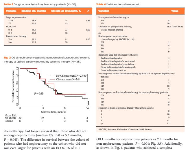
In all, 52 patients (37 males) were identified. The median (range) age at diagnosis was 28 (9–48) years and 49 patients (94%) had stage III/IV. The median OS for all patients was 13.0 months and 38 patients (75%) had nephrectomy. Patients who underwent nephrectomy had superior OS compared to patients who were treated with systemic therapy only (median OS 16.4 vs 7.0 months, P < 0.001). In all, 45 patients received chemotherapy and 13 (29%) had an objective response; 28 patients received targeted therapies, with 8-week median therapy duration and no objective responses. Only seven patients (13%) survived for >24 months.

Conclusions

RMC carries a poor prognosis. Chemotherapy provides palliation and remains the mainstay of therapy, but <20% of patients survive for >24 months, underscoring the need to develop more effective therapy for this rare tumour. In this study, nephrectomy was associated with improved OS.

Read more articles of the week

Editorial: New Strategies for Treating RMC

In the current issue of BJUI, Shah et al. [1] present a multi-institutional study of 52 patients with renal medullary carcinoma (RMC) collected over a 15-year period. This notoriously lethal and rare form of kidney cancer, associated with sickle cell trait and disease, usually affects young adults. In the study, the median age was 28 years, 94% of the patients presented with stage 3 or 4 disease, and the median overall survival was only 13 months. Nephrectomy, performed in 75% of patients, as opposed to systemic therapy alone, was associated with longer survival (16.4 vs 7.0 months). Of the 45 patients who received platinum- or carboplatinum-based systemic chemotherapy, 13 (29%) had an objective response, while there was no objective response in 28 patients treated with vascular endothelial growth factor-targeted agents, which are highly effective in conventional clear-cell carcinoma of the kidney. Only seven patients survived >2 years, with two long-term survivors at 5 and 9 years after nephrectomy and various combinations of systemic therapy.

Recent reports suggest new insight into this lethal form of kidney cancer, raising hope about the development of effective systemic agents. Calderaro et al. [2] used gene expression profiling, array genomic hybridization, and RNA and whole-exome sequencing to study frozen tissue in five patients with RMC. They reported an interchromosomal balanced translocation that disrupts the SMARCB1 gene, a tumour suppressor on chromosome 22 encoding BAF47 protein, which impairs the SWI/SNF complex regulating chromatin remodelling, which, in turn, leads to increased cyclin D transcription and downstream over-expression of the transcriptional regulator EZH2. EZH2 is the enzymatic subunit of the PRC2 complex and its histone methylation function. EZH2 also has a PRC2-independent role in transcriptional activation and can methylate a number of non-histone proteins. Over-expression of EZH2 can lead to cancer by changing expression of tumour suppressor (pRB) and DNA-damage repair genes. EZH2 over-expression and loss of function mutations are associated with a diverse group of cancers including liver, breast, prostate, endometrial, melanoma, bladder and lymphoma, none of which are as rare as RMC but in which targetable agents can be tested for their ability to disrupt this pathway [3]. Interestingly, SMARCB1 gene truncating and or deletion mutations have been reported in the equally rare and lethal paediatric rhabdoid tumour of the kidney [4] and loss of immunoexpression of SMARCB1 reported in the clinically aggressive collecting duct renal cancer, which is morphologically similar to and often difficult to distinguish from RMC [5].

A number of small-molecule compounds able to target both EZH2 and PRC2 complex are currently undergoing preclinical testing (i.e. DZNEP, E11, EP2005687), phase I trials (GSK126), and phase II trials (EPZ-648, Tazemetostat) [3]. A phase II multicentre study of tazemetostat, a selective small-molecule inhibitor of EZH2, is underway and accruing patients with rare tumours with abnormalities in this pathway, including synovial-cell sarcoma, RMC and rhabdoid tumour of the kidney, for which there are no standard therapies [6]. Contemporary genomic research has great potential to identify such critical oncogenic pathways, shared in both rare and more common malignancies, with the potential for effective drugs to be designed to improve the grave prognosis of RMC and related cancers.

Paul Russo
Weill Cornell School of Medicine Memorial Sloan Kettering Cancer Center New York NY USA


Read the full article


References


1 Shah AYKaram JAMalouf GG et al. Management and outcomes of patients with renal medullary carcinoma: a multicentre collaborative study. BJU Int2017; 120: 78292.


2 Calderaro JMasliah-Planchon JRicher W et al. Balanced translocations disrupting SMARCB1 are hallmark recurrent genetic alterations in renal medullary carcinomas. Eur Urol2016; 69: 105561.


3 Kim KHRoberts CWM. Targeting EZH2 in cancer. Nat Med 2016; 22: 128– 34


4 Versteege I, Sevenet NLange J et al. Truncating mutations of hSNF5/INI1 in aggressive paediatric cancer. Nature 1998; 394:2036


5 Elwood H1Chaux ASchultz L et al. Immunohistochemical analysis of SMARCB1/INI-1 expression in collecting duct carcinoma. Urology 2011;78: 474


Video: Management and Outcomes of RMC

Management and outcomes of patients with renal medullary carcinoma: a multicentre collaborative study

Amishi Y. Shah*, Jose A. Karam*, Gabriel G. Malouf, Priya Rao*, Zita D. Lim*, Eric Jonasch*, Lianchun Xiao*, Jianjun Gao*, Ulka N. VaishampayanDaniel Y. Heng§, Elizabeth R. Plimack, Elizabeth A. Guancial**, Chunkit Fung**, Stefanie R. Lowas
††, Pheroze Tamboli*, Kanishka Sircar*, Surena F. Matin*, W. Kimryn Rathmell§§, Christopher G. Wood* and Nizar M. Tannir*


*MD Anderson Cancer Center, Houston, TX, USA, Groupe Hospitalier Pitie-Salpetriere, University Pierre and Marie Curie,
Paris, France, Karmanos Cancer Center, Detroit, MI, USA, §Tom Baker Cancer Center, Calgary, Canada, Fox Chase Cancer Center, Philadelphia, PA, **University of Rochester, Rochester, NY, ††University of Nebraska Medical Center and
Childrens Hospital and Medical Center, Omaha, NE, and §§Vanderbilt-Ingram Cancer Center, Nashville, TN, US


Read the full article

Abstract

Objective

To describe the management strategies and outcomes of patients with renal medullary carcinoma (RMC) and characterise predictors of overall survival (OS).

Patients and Methods

RMC is a rare and aggressive malignancy that afflicts young patients with sickle cell trait; there are limited data on management to date. This is a study of patients with RMC who were treated in 2000–2015 at eight academic institutions in North America and France. The Kaplan–Meier method was used to estimate OS, measured from initial RMC diagnosis to date of death. Cox regression analysis was used to determine predictors of OS.

Results

In all, 52 patients (37 males) were identified. The median (range) age at diagnosis was 28 (9–48) years and 49 patients (94%) had stage III/IV. The median OS for all patients was 13.0 months and 38 patients (75%) had nephrectomy. Patients who underwent nephrectomy had superior OS compared to patients who were treated with systemic therapy only (median OS 16.4 vs 7.0 months, P < 0.001). In all, 45 patients received chemotherapy and 13 (29%) had an objective response; 28 patients received targeted therapies, with 8-week median therapy duration and no objective responses. Only seven patients (13%) survived for >24 months.

Conclusions

RMC carries a poor prognosis. Chemotherapy provides palliation and remains the mainstay of therapy, but <20% of patients survive for >24 months, underscoring the need to develop more effective therapy for this rare tumour. In this study, nephrectomy was associated with improved OS.

Read more articles of the week

Video: Stereotactic ablative body radiotherapy for inoperable primary kidney cancer

Stereotactic ablative body radiotherapy for inoperable primary kidney cancer

Read the full article

Abstract

Objective

To assess the feasibility and safety of stereotactic ablative body radiotherapy (SABR) for renal cell carcinoma (RCC) in patients unsuitable for surgery. Secondary objectives were to assess oncological and functional outcomes.

Materials and Methods

This was a prospective interventional clinical trial with institutional ethics board approval. Inoperable patients were enrolled, after multidisciplinary consensus, for intervention with informed consent. Tumour response was defined using Response Evaluation Criteria In Solid Tumors v1.1. Toxicities were recorded using Common Terminology Criteria for Adverse Events v4.0. Time-to-event outcomes were described using the Kaplan–Meier method, and associations of baseline variables with tumour shrinkage was assessed using linear regression. Patients received either single fraction of 26 Gy or three fractions of 14 Gy, dependent on tumour size.

Results

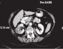
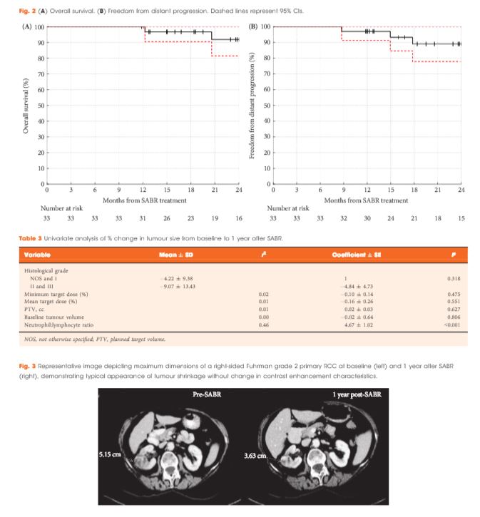
Of 37 patients (median age 78 years), 62% had T1b, 35% had T1a and 3% had T2a disease. One patient presented with bilateral primaries. Histology was confirmed in 92%. In total, 33 patients and 34 kidneys received all prescribed SABR fractions (89% feasibility). The median follow-up was 24 months. Treatment-related grade 1–2 toxicities occurred in 26 patients (78%) and grade 3 toxicity in one patient (3%). No grade 4–5 toxicities were recorded and six patients (18%) reported no toxicity. Freedom from local progression, distant progression and overall survival rates at 2 years were 100%, 89% and 92%, respectively. The mean baseline glomerular filtration rate was 55 mL/min, which decreased to 44 mL/min at 1 and 2 years (P < 0.001). Neutrophil:lymphocyte ratio correlated to % change in tumour size at 1 year, r2 = 0.45 (P < 0.001).

Conclusion

The study results show that SABR for primary RCC was feasible and well tolerated. We observed encouraging cancer control, functional preservation and early survival outcomes in an inoperable cohort. Baseline neutrophil:lymphocyte ratio may be predictive of immune-mediated response and warrants further investigation.

View more videos

Editorial: Stereotactic radiotherapy for primary renal cell carcinoma: time for larger-scale prospective studies

A number of important trends in kidney cancer diagnosis have emerged in recent decades, including the increasing detection of renal tumours in older patients with more comorbidities. In the UK in 2012–2014, 50% of new cases were diagnosed in people aged 70 years and over. Whilst many of these lesions are incidental small renal masses suitable for active surveillance, the dilemma of how to manage the higher-risk lesion (rapid growth kinetics, larger size, symptomatic lesion) is increasingly encountered. Surgical management may pose an unacceptable risk of morbidity, mortality or dialysis, yet these patients may live long enough to experience the consequences of disease progression. Thermal ablation is an option for small cortical tumours (≤3 cm), but there are limitations for larger or centrally located tumours.

In this issue of BJUI, Siva and colleagues [1] report promising early efficacy and toxicity data using stereotactic ablative body radiotherapy (SABR) for the treatment of primary RCC in this difficult cohort. SABR is a non-invasive treatment that delivers very high doses of radiation over one to five outpatient sessions. It uses advanced motion management, radiation planning and image guidance techniques to ensure delivery of an ablative dose with millimetre precision. Survival benefits with stereotactic radiosurgery have been demonstrated in patients with solitary brain metastases [2], and SABR is now an accepted standard of care for patients with medically inoperable early-stage lung cancer [3]. Randomized phase III trials are currently under way, testing SABR against standard of care in primary prostate (clinicaltrials.gov ID NCT01584258) and liver cancer (NCT01730937) and in the oligometastatic setting (NCT02759783). Historically considered radio-resistant, both pre-clinical and clinical data now support the sensitivity of RCC to high-dose per fraction radiotherapy, as used in SABR [4].

The study by Siva and colleagues is one of the largest, early-phase, prospective studies of SABR for primary RCC to date, accruing 37 patients with cT1a–cT2a RCC not suitable for other therapies. Importantly, this was not a cohort of incidentally detected small renal masses. The majority (65%) of tumours were >4 cm (median 4.8 cm), were growing on surveillance or symptomatic, and were biopsy-proven. The inclusion of enlarging T1a tumours is not unreasonable. A recent analysis of patients with localized T1a kidney cancer from the Surveillance, Epidemiology and End Results (SEER) Medicare data reported an excess of kidney cancer deaths for non-surgically managed patients aged >75 years, highlighting how difficult it can be to find the right balance between active and expectant management in this group [5]. Indeed 11% of patients in the present study by Siva et al. developed distant metastases by 2 years.

In the present study, tumours <5 cm received a single 26-Gy fraction of SABR, whilst tumours >5 cm received 42 Gy over three fractions. Whilst acknowledging a number of uncertainties in modelling, this should equate to an equivalent biological dose in excess of 100 Gy. The authors found that delivering this SABR regimen was feasible and well tolerated with one grade 3 toxicity (transient fatigue) and no grade 4–5 toxicities. Most patients sustained only transient minor side effects (78%) or no treatment-related side effects (18%). The mean baseline estimated GFR was 55 mL/min, which decreased to 44 mL/min at 1 year, and was maintained for those with 2 years follow-up.

Similarly, short-term efficacy appears promising. With a median follow-up of 24 months, freedom from local progression at 2 years was 100%, with one patient subsequently progressing locally with concurrent distant metastases 28 months after treatment. Local progression was defined using Response Evaluation Criteria In Solid Tumors (RECIST) v1.1. This is a pragmatic definition and takes into account the challenges in interpreting standard imaging after SABR and the difficulties in obtaining and interpreting repeat biopsies in this cohort. Similarly to the study by Sun et al. [6] it appears that stable or partial radiological responses will predominate in the early years after SABR and, unlike thermal ablation, changes in enhancement patterns can be very slow to evolve.

Longer-term follow-up is required to confirm these promising tumour control and nephron preservation rates, in addition to evaluating longer-term late effects. To that end, the present study has provided a platform for the authors to launch an international phase II clinical trial under the auspices of the TransTasman Radiation Oncology Group (TROG 15.03 FASTRACK, clinicaltrials.gov ID NCT02613819). Larger-scale prospective studies are essential to confirm the efficacy and safety of this non-invasive, nephron-sparing, ablative technique and provide further information to help refine patient selection and develop better biomarkers of response.

Read the full article

David I. Pryor *† and Simon Wood†‡


*Department of Radiation Oncology, Princess Alexandra Hospital, Wooloongabba, School of Medicine, University of Queensland, Brisbane, and Department of Urology, Princess Alexandra Hospital, Wooloongabba, Qld, Australia


References


1 Siva S. Stereotactic ablative body radiotherapy for inoperable primary kidney cancer: a prospective clinical trial. BJU Int 2017; 120: 62330


2 Andrews DW, Scott CB , Sperduto PW et al. Whole brain radiation therapy with or without stereotactic radiosurgery boost for patients with  one to three brain metastases: phase III results of the RTOG 9508 randomised trial. Lancet (London, England) 2004; 363: 166572


4 De Meerleer G, Khoo V, Escudier B et al. Radiotherapy for renal-cell carcinoma. Lancet Oncol 2014; 15: e1707


6 Sun MR, Brook A, Powell MF et al. Effect of stereotactic body radiotherapy on the growth kinetics and enhancement pattern of primary renal tumors. AJR Am J Roentgenol 2016; 206: 54453


Article of the Week: Stereotactic ablative body radiotherapy for inoperable primary kidney cancer: a prospective clinical trial

Every week the Editor-in-Chief selects an Article of the Week from the current issue of BJUI. The abstract is reproduced below and you can click on the button to read the full article, which is freely available to all readers for at least 30 days from the time of this post.

In addition to the article itself, there is an accompanying editorial written by a prominent member of the urological community. This blog is intended to provoke comment and discussion and we invite you to use the comment tools at the bottom of each post to join the conversation.

If you only have time to read one article this week, it should be this one.

Stereotactic ablative body radiotherapy for inoperable primary kidney cancer: a prospective clinical trial


Shankar Siva*,, Daniel Pham*, Tomas Kron*,, Mathias Bressel*, Jacqueline Lam*, Teng Han Tan*, Brent Chesson*, Mark Shaw*, Sarat Chander*, Suki Gill*,Nicholas R. Brook§, Nathan Lawrentschuk*,, Declan G. Murphy*,† and Farshad Foroudi*,


*Peter MacCallum Cancer Centre, Melbourne, Vic., Australia, † Sir Peter MacCallum Department of Oncology, University of Melbourne, Melbourne, Vic. Australia, Sir Charles Gairdner Hospital, Nedlands Perth, WA, Australia, §Royal Adelaide Hospital, Adelaide, SA, Australia, and Olivia Newton John Cancer Centre, Heidelberg, Vic., Australia


Read the full article

Abstract

Objective

To assess the feasibility and safety of stereotactic ablative body radiotherapy (SABR) for renal cell carcinoma (RCC) in patients unsuitable for surgery. Secondary objectives were to assess oncological and functional outcomes.

Materials and Methods

This was a prospective interventional clinical trial with institutional ethics board approval. Inoperable patients were enrolled, after multidisciplinary consensus, for intervention with informed consent. Tumour response was defined using Response Evaluation Criteria In Solid Tumors v1.1. Toxicities were recorded using Common Terminology Criteria for Adverse Events v4.0. Time-to-event outcomes were described using the Kaplan–Meier method, and associations of baseline variables with tumour shrinkage was assessed using linear regression. Patients received either single fraction of 26 Gy or three fractions of 14 Gy, dependent on tumour size.

Results

Of 37 patients (median age 78 years), 62% had T1b, 35% had T1a and 3% had T2a disease. One patient presented with bilateral primaries. Histology was confirmed in 92%. In total, 33 patients and 34 kidneys received all prescribed SABR fractions (89% feasibility). The median follow-up was 24 months. Treatment-related grade 1–2 toxicities occurred in 26 patients (78%) and grade 3 toxicity in one patient (3%). No grade 4–5 toxicities were recorded and six patients (18%) reported no toxicity. Freedom from local progression, distant progression and overall survival rates at 2 years were 100%, 89% and 92%, respectively. The mean baseline glomerular filtration rate was 55 mL/min, which decreased to 44 mL/min at 1 and 2 years (P < 0.001). Neutrophil:lymphocyte ratio correlated to % change in tumour size at 1 year, r2 = 0.45 (P < 0.001).

Conclusion

The study results show that SABR for primary RCC was feasible and well tolerated. We observed encouraging cancer control, functional preservation and early survival outcomes in an inoperable cohort. Baseline neutrophil:lymphocyte ratio may be predictive of immune-mediated response and warrants further investigation.

Read more articles of the week

Highlights from the Urological Association of Asia Annual Congress 2017

Having trained and worked in London throughout my urology career, I have recently relocated and joined the exciting, dynamic urology community of my birthplace, Hong Kong. Coincidentally, it so happens to be this year’s host of the Urological Association of Asia (UAA) annual congress #UAA2017.

1-1

The beautiful and mesmerising night view of the Victoria Harbour of Hong Kong.

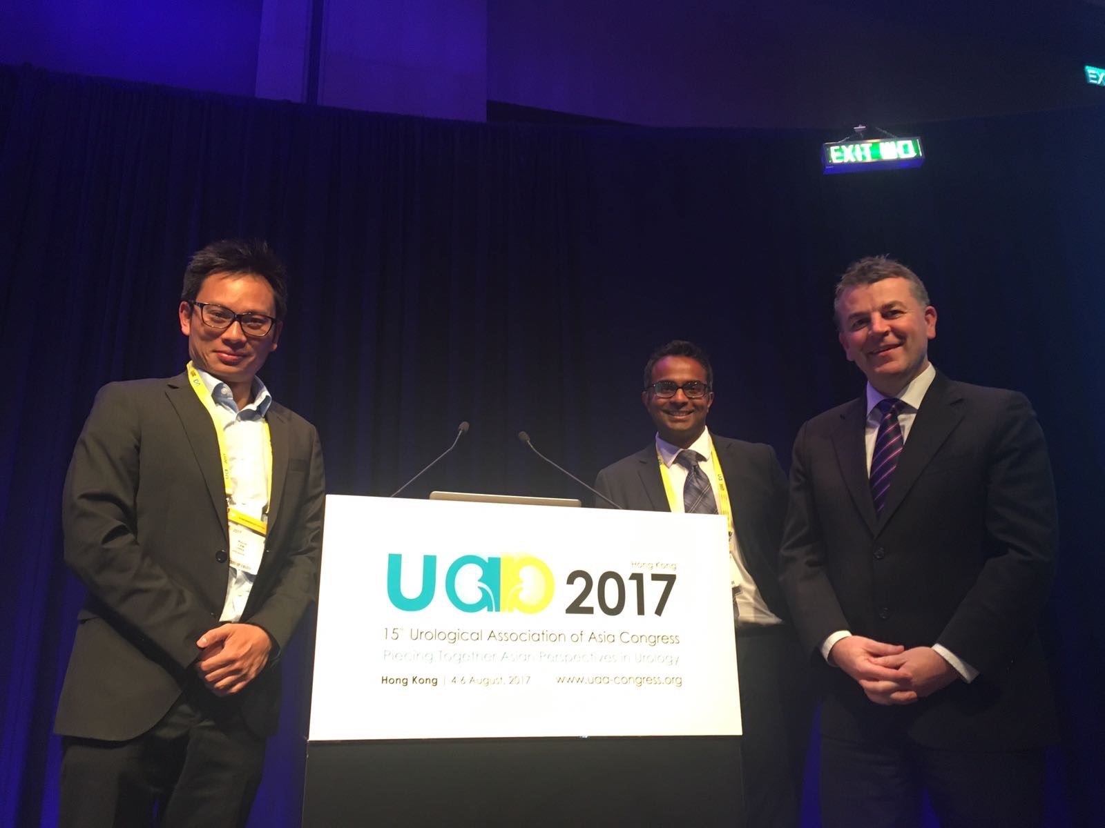
Established in 1990 in Fukuoka, Japan, the #UAA currently has 25 urological associations as members or affiliated members across Asia and Australasia with and over 25,000 members. This was my attendance at the #UAA and it most certainly did not disappoint. What made the conference even more special was the chance to meet up with my good friend and ex-colleague from Guy’s Hospital @nairajesh – both of us were honoured to speak at the meeting. With over 1600 delegates attending the meeting and over 500 scientific abstracts presented, the congress served as an excellent platform for knowledge exchange and the establishment of professional links with many urological greats in Asia and beyond.


Pre #UAA2017 Congress Activities

#UAA2017 started off with a pre-congress ‘wet-lab’ 3D laparoscopic skills and endourology workshop hosted by @HKUniversity and the European School of Urology @UrowebESU. Both transperitoneal laparoscopic and retroperitoneoscopic techniques were taught by eminent leaders and pioneers in minimally invasive urological surgery by faculties from Europe, India and China, including Professor Jens Rassweiller, Professor Christian Schwentner (@Schwenti1977), Dr Domenico Veneziano (@d_veneziano), Professor Janak Desai (@drjanajddesai), and Professor Zhang Shudong.

1-2

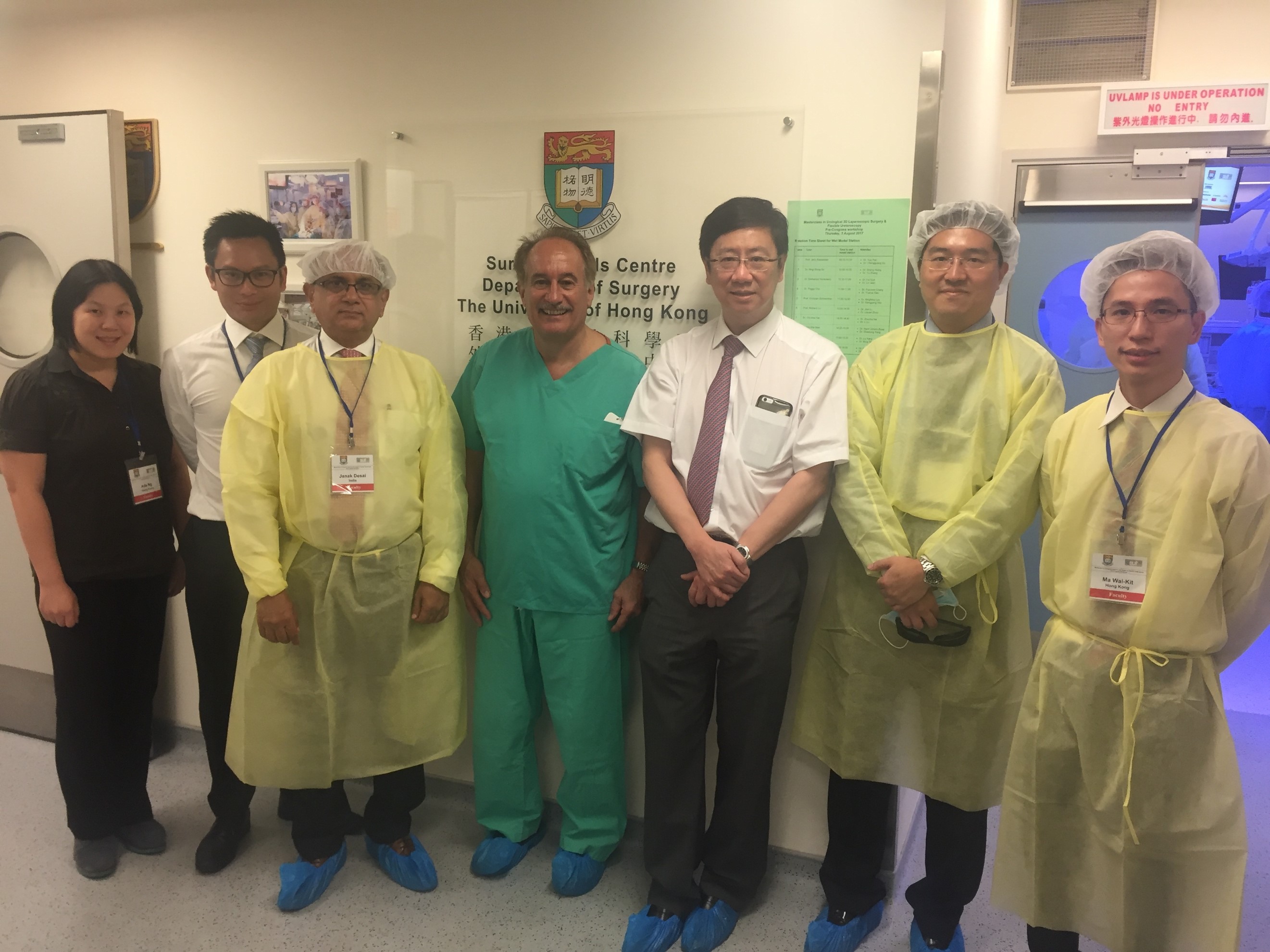
Joint UAA-ESU 3D laparoscopic and endourological skills course faculties. Left to right: Dr Ada Ng (Hong Kong), Dr Wayne Lam @WayneLam_Urol (Hong Kong), Professor Janek Desai @drjanajddesai (India), Professor Jens Rassweiller (Germany), Professor MK Yiu (Hong Kong), Dr James Tsu (Hong Kong), Dr WK Ma (Hong Kong).

1-3

1-4

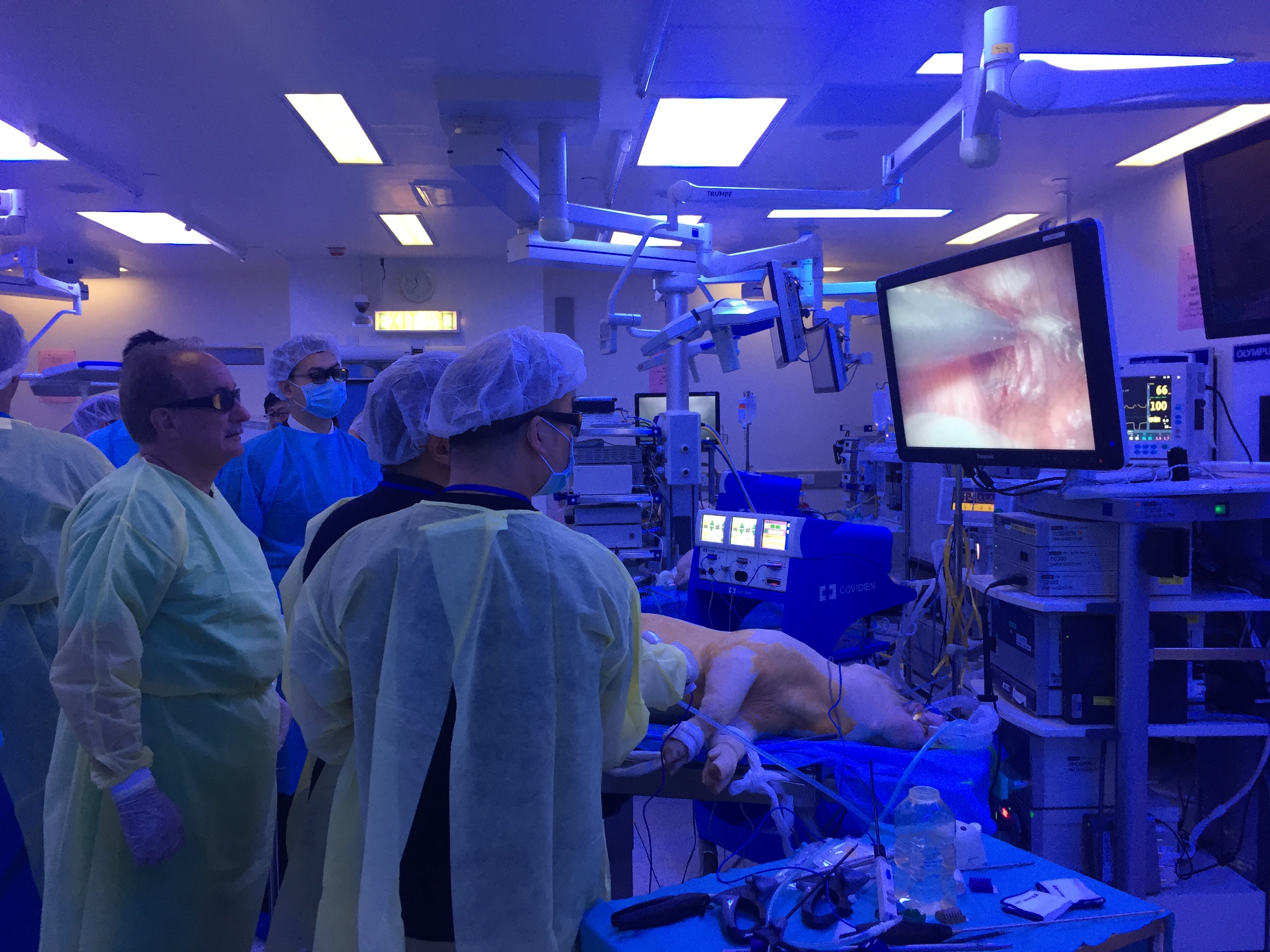
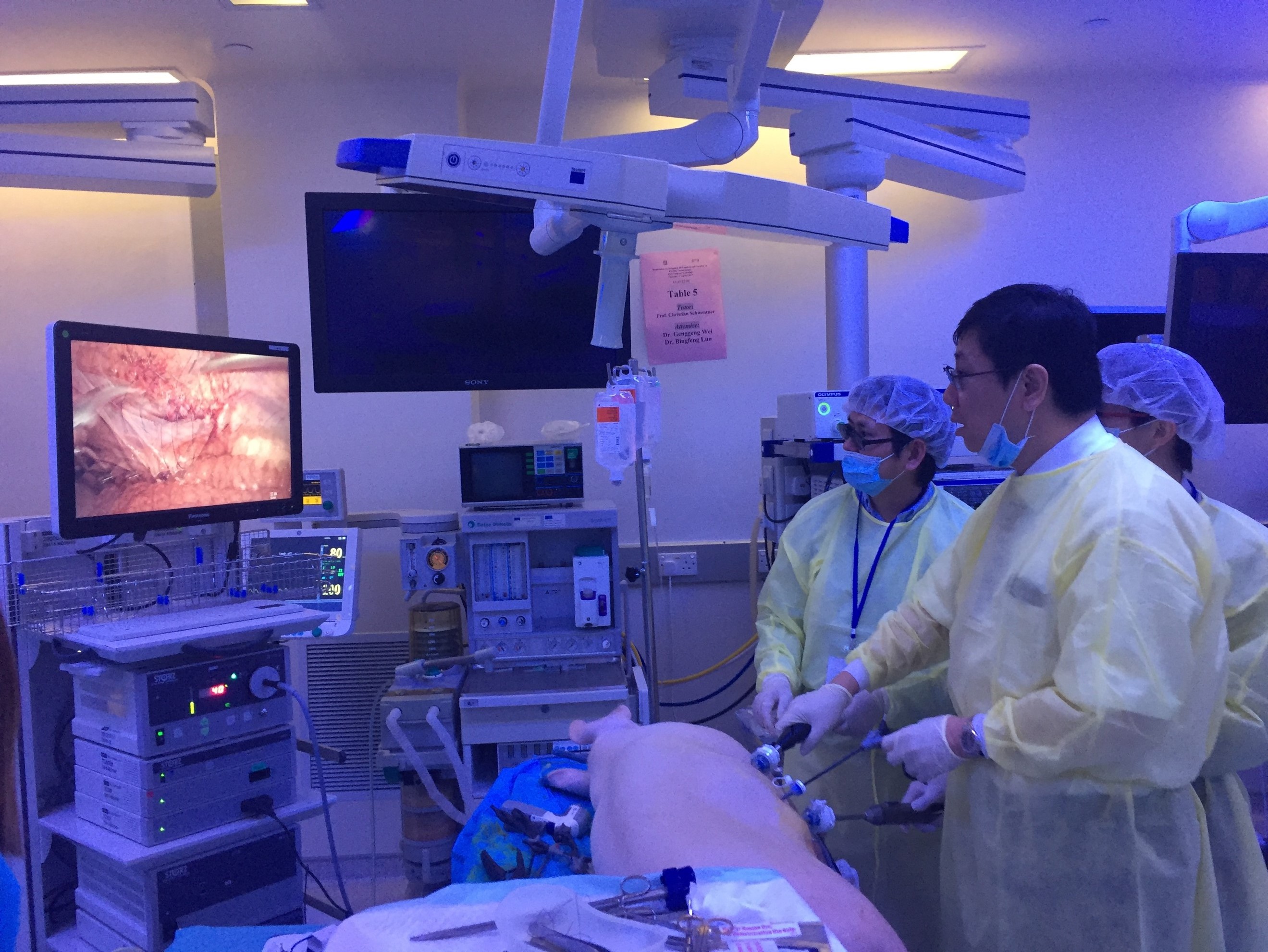
Joint UAA-ESU 3D Laparoscopic skills workshop – Above: Professor Rassweiller (Germany) supervising overseas delegates. Below: Professor MK Yiu (Hong Kong) demonstrating techniques of laparoscopic suturing to delegates from China.

The renal cell carcinoma #RCC masterclass was a particular highlight. A whole day of excellent lectures and speakers entertained both local and international delegates, and was particularly popular with trainees. There were talks examining the role of percutaneous biopsy of renal tumours presented by Alessandro Volpe (@foxal72), an update of current trends and techniques in robotic and laparoscopic partial nephrectomy by Dr. Joseph Wong (Hong Kong) and Dr. Shuo Wang (China) and a fantastic discussion examining the role of non-clamping partial nephrectomy by Dr. Ringo Chu (Hong Kong). The afternoon session kicked off with @nairajesh giving a comprehensive review on the surgical management of advanced #RCC. Professor Axel Bex (Netherlands) continued with an examination of neoadjuvant and adjuvant systemic therapy in #RCC and the emerging role of #immunotherapy. Professor Alessandro Volpe (@foxal72) discussed the current #EAU guidelines and recent updates.

1-5

Masterclass in #RCC: @foxal72 , @nairajesh , Professor Axel Bex (Netherlands), with moderators Dr Ringo Chu (Hong Kong) and Dr Joseph Wong (Hong Kong).


Day 1 of #UAA2017

The plenary session on day 1 of #UAA started off with Professor Zengnan Mo from China on the epidemiology of prostate cancer in Asia. There is, not surprisingly, a very diverse range of incidence of #prostatecancer rate across the largest continent in the world, inevitably effected by the presence of #PSA screening in countries such as Japan and Korea, ethnicity (East Asia vs Middle East), genetics (Israeli Jewish population), and their local healthcare system and policies. Arguably the currently available #prostatecancer screening trials may not be applicable to the Asian populations, and various on-going studies in Japan and China are going to address these issues. One in particular is an ongoing population-based study funded by the Chinese Ministry of Science and Technology, in which over 50,000 men will be recruited into the screening, early detection, localised, and advanced #prostatecancer cohorts and to be followed up with time. Obviously, we will not expect to see the results of the trial anytime soon, but will surely answers to address the behaviour of prostate cancer in the Asian population in the future.

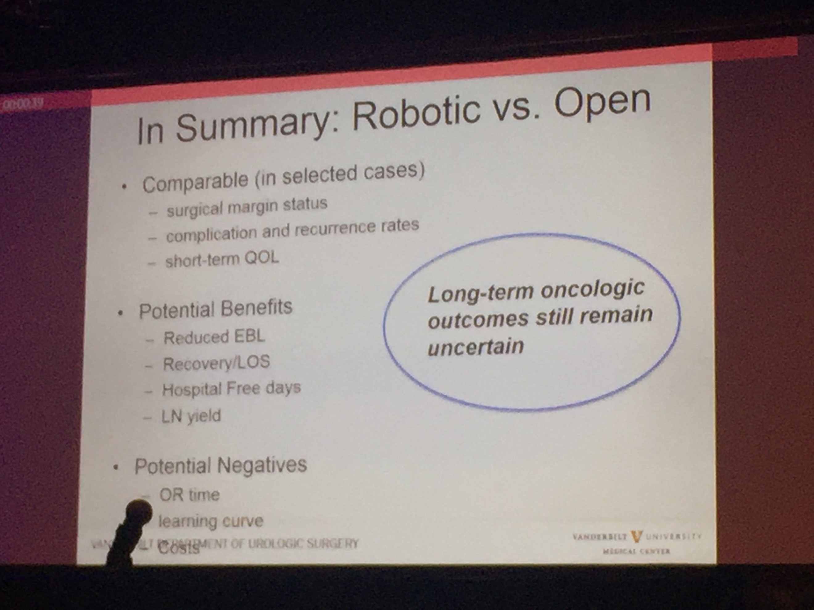
Professor Sam Cheng (@UroCancerMD) from Vanderbilt University then gave a comprehensive review on the current status of #cystectomy. Robotic cystectomy appears to have the benefit of reduced blood loss and length of stay. However, long-term oncological outcome still remains uncertain, and certainly, patient reported outcome measures (PROMs) is lacking.

1-6


Day 2 of #UAA2017

After early showers on day 2 of UAA, Hong Kong was heating up with temperatures over 33 degrees Celsius. So were the discussions in the plenary session in the morning. Professor Freddie Hamdy (@Freddie_Hamdy) gave the #EAU lecture on #activesurveillance for #prostatecancer. This was followed on nicely with the current status of #prostatecancer management in Hong Kong by Dr. Yau-Tung Chan and Dr. Gerhardt Attard (UK) enlightened the audience with a concise update in the management of hormone sensitive prostate cancer, an area where the landscape is ever-changing.

The advanced oncology session started off with a heated debate in the use of mass clamping (Dr Ringo Chu, Hong Kong) versus selective artery clamping in partial nephrectomy (Dr Tae-Gyun Kwon, Korea). Both speakers presented with very valid arguments and perhaps it was fair to say it ended up with all square. Professor Krishna Sethia (UK) gave a fascinating summary of the current local management of #penilecancer at centralised penile cancer centres in the UK, after which I was honoured to provide an update on current nodal management in #penilecancer from my recent experience at St George’s University Hospitals @StGeorgesTrust in the UK. It was exciting to see the centralisation of services in #penilecancer in the UK has given great opportunities to understand and optimise management of patients with such rare disease.

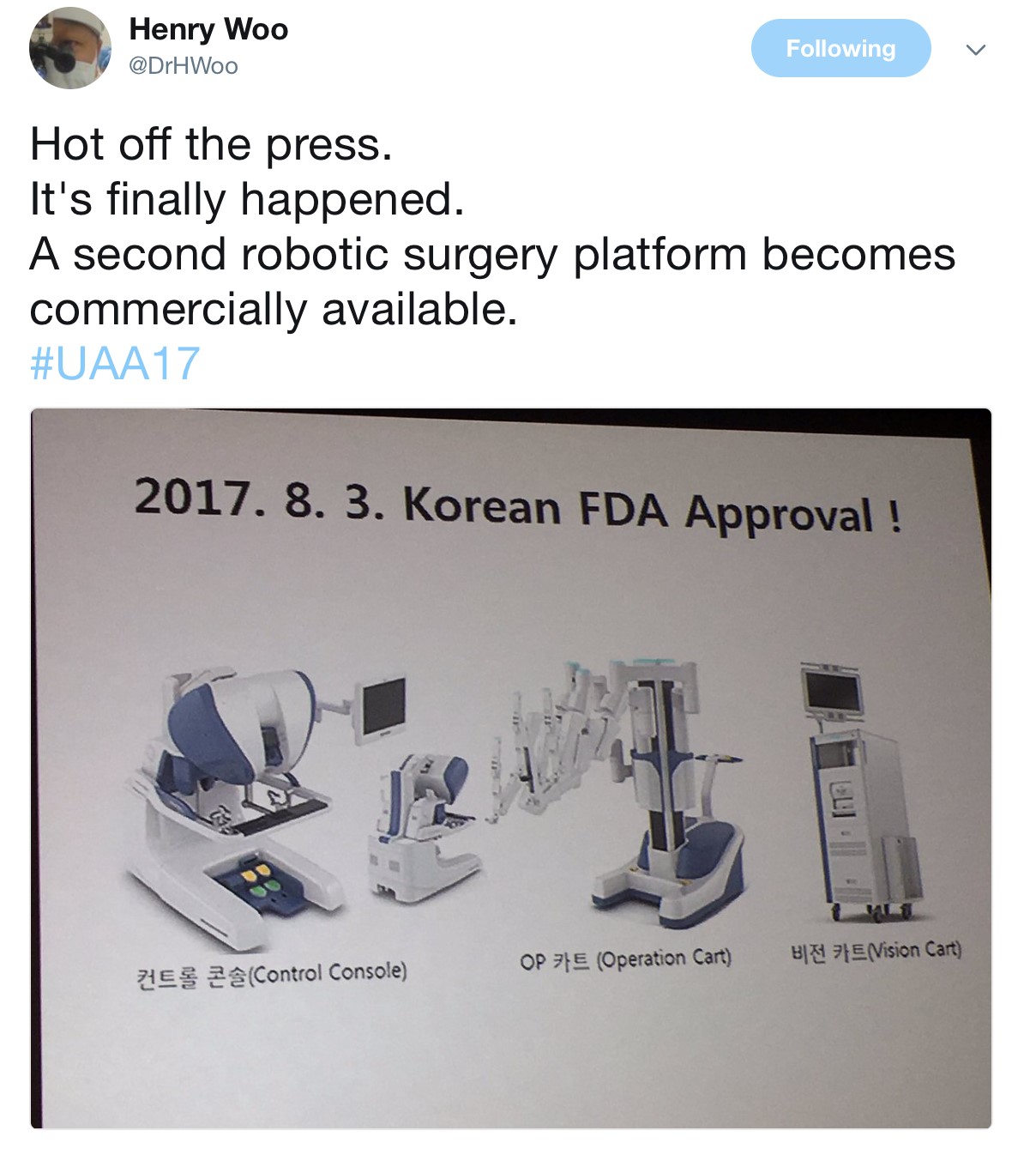
The Semi-live sessions entertained the audience on both days of the conference. Excellent videos were presented throughout. Professor Koon Rha of Yonsei University in South Korea gave a fantastic semi-live talk on his tricks and techniques of Retzius-space sparing Robot-assisted radical prostatectomy. Perhaps what’s even more exciting to know is that a Korean company has produced a new robot for surgery which has been well tested by Professor Rha’s group, which has just literally been licenced and approved in Korea just days before #UAA2017. Will this finally drive the cost of robotic surgery down? Time will tell.

1-7

Associate Professor Declan Murphy (@declanmurphy) and Mr. Rajesh Nair (@nairajesh) both contributed with a beautiful video showcasing techniques in total pelvic exenteration and long-term outcomes of urinary diversion and reconstruction in this cohort of patients.

The Gala dinner in the evening was full of fun and entertainment. Following the performance of a soprano quartet formed by local Hong Kong urologists (who sang the classic My Way with a twist on prostate examination!), the rock stars of urology – Professor Jens Rassweiller, Dr Samuel Yee from Hong Kong, and Dr Domenico Veneziano (@d_veneziano) provided an energetic and electrifying live performance of some rock classics!

1-8

Urology Rock N’ Roll! Left to right: Professor Jens Rassweiller (Germany), Dr Domenico Veneziano (Italy), Dr Samuel Yee (Hong Kong).

However, perhaps the highlight and the most touching moment of the evening the performance of a song written and sung by a young local former patient with a history of #ketaminebladder , who was successfully treated by the urology team lead by Professor Anthony Ng at the Prince of Wales Hospital in Hong Kong. His surgery and treatment has transformed his life – he is now enjoying a career as both a singer-songwriter of a rock band and as a footballer!


Day 3 of #UAA2017

The morning plenary session also saw the evergreen Dr Peggy Chu of Hong Kong, renown for her discovery of ketamine-associated uropathy and pioneered the management of this challenging 21st century urological disease.

1-9

Left to right: Dr CW Man (Hong Kong), Congress President of #UAA2017, and Dr Peggy Chu (Hong Kong).

She delivered a very interesting talk on revisiting the role of #gastrocystoplasty. Interestingly, the operation was first described and carried out in human by the honourable Professor CH Leong at my current institution, Queen Mary Hospital @HKUniversity , in the 1970s following a successful animal study at the same institution. Its use has been limited due to its associated metabolic disturbances, but arguably it is still a weapon that can be used when tackling patients with tuberculosis-associated severely contracted bladder, in particular those who have already been rendered to have a single solitary kidney due to the disease. Another situation when #gastrocystoplasty can still be considered are those patients with #ketamine uropathy. Although patients are usually required to be completely abstinence from #ketamine abuse for a certain lengthy period of time before they are eligible for surgical treatment, many fear the avalanche effect of ileal re-absorption of the drug if an ileo-cystoplasty has been carried out in these patients, if they happen to resume ketamine use in the future. Hence, #gastrocystoplasty may be a better substitution tissue for cystoplasty in the management of such patients.

The meeting also provided an opportunity to catch up with fellow Guy’s Hospital urology graduates @nairajesh and @declanmurphy over a cold pint of Hong Kong locally made #MoonzenBeer, when the temperature outside the conference centre was hitting 34 degrees Celsius!

1-10

Left to right: Dr Wayne Lam (Hong Kong), Mr Rajesh Nair (Australia/United Kingdom), A/Prof Declan Murphy (Australia).

All credits to #UAA and the local organisers’ immense effort and hard work, making this congress a valuable learning experience for everyone who participated. We very much look forward to #UAA2018. Bring on Kyoto, Japan!


Wayne Lam

Assistant Professor in Urology, Queen Mary Hospital, University of Hong Kong

Twitter: @WayneLam_Urol

1-11


Rajesh Nair

Fellow in Robotic Surgery and Uro-Oncology

1-12


© 2024 BJU International. All Rights Reserved.