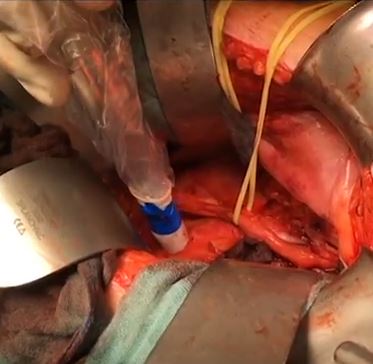
Tag Archive for: radioguided surgery

Posts

Article of the Week: Value of 111In-PSMA-RGS for salvage lymphadenectomy in recurrent PCa

Every Week the Editor-in-Chief selects an Article of the Week from the current issue of BJUI. The abstract is reproduced below and you can click on the button to read the full article, which is freely available to all readers for at least 30 days from the time of this post.

In addition to the article itself, there is an accompanying editorial written by a prominent member of the urological community. This blog is intended to provoke comment and discussion and we invite you to use the comment tools at the bottom of each post to join the conversation.

Finally, the third post under the Article of the Week heading on the homepage will consist of additional material or media. This week we feature a video discussing the paper.

If you only have time to read one article this week, it should be this one.

Value of 111In-prostate-specific membrane antigen (PSMA)-radioguided surgery for salvage lymphadenectomy in recurrent prostate cancer: correlation with histopathology and clinical follow-up

Isabel Rauscher*, Charlotte Duwel, Martina Wirtz, Margret SchotteliusHans-Jurgen Wester, Kristina Schwamborn§, Bernhard Haller, Markus Schwaiger*, Jurgen E. Gschwend, Matthias Eiber* and Tobias Maurer


*Departments of Nuclear Medicine, Urology, Technical University of Munich, Klinikum rechts der Isar, Munich, Institute of Pharmaceutical Radiochemistry, Technical University of Munich, Garching, §Department of Pathology, and Institute of Medical Statistics and Epidemiology, Technical University of Munich, Klinikum rechts der Isar, Munich, Germany


How to Cite

Rauscher, I., Düwel, C., Wirtz, M., Schottelius, M., Wester, H.-J., Schwamborn, K., Haller, B., Schwaiger, M., Gschwend, J. E., Eiber, M. and Maurer, T. (2017), Value of 111In-prostate-specific membrane antigen (PSMA)-radioguided surgery for salvage lymphadenectomy in recurrent prostate cancer: correlation with histopathology and clinical follow-up. BJU International, 120: 40–47. doi: 10.1111/bju.13713

Abstract

Objectives

To evaluate the use of 111In-labelled prostate-specific membrane antigen (PSMA)-I&T-based radioguided surgery (111In-PSMA-RGS) for salvage surgery in recurrent prostate cancer (PCa) using comparison of intra-operative gamma probe measurements with histopathological results of dissected specimens. In addition, to determine the success of 111In-PSMA-RGS with regard to postoperative prostate-specific antigen (PSA) responses, PCa-specific treatment-free survival rates and postoperative complication rates.

Patients and Methods

A total of 31 consecutive patients with localized recurrent PCa undergoing salvage surgery with PSMA-targeted radioguided surgery using a 111In-labelled PSMA ligand between April 2014 and July 2015 were retrospectively included in this study. The preoperative (interquartile range; range) median PSA level was 1.3 (0.57–2.53 ng/mL; 0.2–13.9 ng/mL). Results of ex vivo radioactivity rating (positive vs negative) of resected tissue specimens were compared with findings of postoperative histological analysis. Best PSA response without additional treatment was determined after 111In-PSMA-RGS, and salvage-surgery-related postoperative complications and PCa-specific additional treatments were recorded.

aotw-jul-2017-3-results

Results

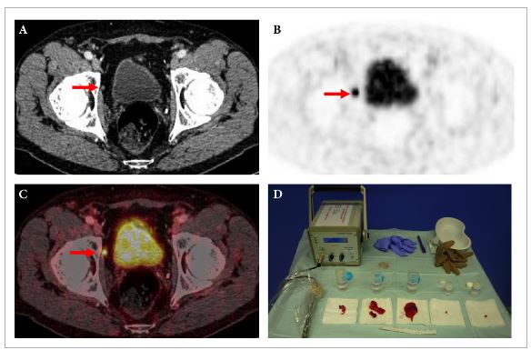
In 30/31 patients, 111In-PSMA-RGS allowed intra-operative identification of metastatic lesions. In total, 145 surgical specimens were removed and 51 showed metastatic involvement at histological analysis. According to 111In-PSMA-RGS ex vivo measurements, 48 specimens were correctly classified as metastatic and 87 as cancer-free, four were false-negative and six were false-positive compared with histological evaluation. Follow-up information was available for 30/31 patients. PSA declines of >50% and >90% were observed in 23/30 patients and in 16/30 patients, respectively. In 18/30 patients, a PSA decline to <0.2 ng/mL was observed. In 10/30 patients further PCa-specific treatment was given after a median (range) of 125 (48–454) days post-111In-PSMA-RGS. The remaining 20 patients remained treatment-free at a median (range) follow-up of 337 (81–591) days. Of 30 patients, 10 presented with surgery-related complications (Clavien–Dindo grade 1, n = 6, Clavien–Dindo grade 3b, n = 4).

Conclusion

111In-PSMA-RGS proved to be of high value for intra-operative detection of even small metastatic lesions in patients with PCa scheduled for salvage lymphadenectomy. It allows the exact localization and resection of metastatic tissue during 111In-PSMA-RGS and is therefore anticipated to have a beneficial influence on further disease progression; however, identification of suitable patients on the basis of PSMA-positron-emission tomography imaging as well as clinical variables is essential for satisfactory results to be obtained.

Editorial: PSMA-RS – a promising utility

There is no doubt that, in the field of prostate cancer, few recent topics have been the subject of as much captivation and discussion as prostate-specific membrane antigen (PSMA) positron-emission tomography (PET) imaging. The body of literature on this imaging technique, the majority on the use of 68Ga-PSMA-HBED-CC as a radiotracer, is growing unceasingly [1], and includes data to support the superior accuracy of PSMA PET/CT for lymph node staging in prostate cancer [2] and in identifying patients unlikely to benefit from radiotherapy after radical prostatectomy [3].

In the present paper, Rauscher et al. [4] present their data on the use of an 111In-PSMA-I&T tracer during salvage lymphadenectomy for recurrent prostate cancer. In a previous study by the same research group, 111In-PSMA-I&T has already proven to be a high-affinity radiotracer, with enhanced internalization efficiency compared with other molecules and significant accumulation in prostate cancer tissue [5].

In the present pilot study, salvage lymphadenectomy was performed in 31 patients with recurrent prostate cancer after primary treatment. Using intra-operative γ-probe measurements and comparing these with the histopathological results of the specimens, the authors found that these correlated well, resulting in a sensitivity of 92.3%, a specificity of 93.5% and an accuracy of 93.1%, with a positive predictive value of 88.9% and a negative predictive value of 95.6%.

These findings also translated well into PSA response after surgery. Postoperative PSA reductions of >50% were observed in 76.7% of patients and of >90% in 53.3% of patients. Further cancer-specific treatment was given to 33% of patients at a median of 125 days after surgery. The remaining patients remained free of treatment at a median follow-up of 337 days.

The study sample was relatively small and the analysis was conducted retrospectively. These are obvious drawbacks; nonetheless the intra- and postoperative results presented are promising. However, as the authors of the present paper point out, careful patient selection is important, and the follow-up period for these patients is still quite short. We will need to wait several years to compare the outcomes of this series with those reported in the literature with regard to salvage lymphadenectomy without prior PSMA PET CT. These data were recently published in a review by Heidenreich et al. [6]. They reported 5-year biochemical recurrence-free survival of 19–25% after salvage lymphadenectomy without the use of PSMA PET, and a median time to systemic treatment of 20–30 months [6].

As physicians, of course, our primary goal is to do the best for our patients. Certainly, we would like to believe that aggressively treating every single lesion made visible with this new imaging technique would be to the patient’s benefit. It is certainly tempting to chase these colourful lesions now demonstrated so nicely by PSMA PET/CT, but we owe it to ourselves as scientists to gather the facts and the evidence to determine whether or not our current course of action makes sense. PSMA PET radio-guided surgery is no exception to the rule, and only the evidence will tell what exact role this new technology is to have in the treatment of prostate cancer. A number of questions need to be addressed. Can we justify putting patients through surgical procedures with the morbidity associated with them? Do we not need to define oligometastatic disease in the molecular imaging era? Should we then start using PSMA PET in primary staging of prostate cancer patients? What of those tumours that do not express PSMA? Can this approach be offered laparoscopically or robotically?

It seems the introduction of PSMA PET has, instead of giving us all the answers, given rise to even more questions.

Nicolas Geurts,*Alastair D. Lamb,*† Nathan Lawrentschuk*†‡ and Declan G. Murphy*§¶


*Division of Cancer Surgery, University of Melbourne, Peter MacCallum Cancer Centre, Melbourne, Department of Surgery, Austin Hospital, University of Melbourne, Heidelberg§ Australian Prostate Cancer Research Centre, Epworth Healthcare, and
Sir Peter MacCallum Department of Oncology, University of Melbourne, Melbourne, Vic., Australia


How to Cite

Geurts, N., Lamb, A. D., Lawrentschuk, N. and Murphy, D. G. (2017), Prostate-specific membrane antigen radioguided surgery: a promising utility. BJU International, 120: 5–6. doi: 10.1111/bju.13838

References


Video: Value of 111In-PSMA-RGS for salvage lymphadenectomy in recurrent PCa

Value of 111In-prostate-specific membrane antigen (PSMA)-radioguided surgery for salvage lymphadenectomy in recurrent prostate cancer: correlation with histopathology and clinical follow-up


Abstract

Objectives

To evaluate the use of 111In-labelled prostate-specific membrane antigen (PSMA)-I&T-based radioguided surgery (111In-PSMA-RGS) for salvage surgery in recurrent prostate cancer (PCa) using comparison of intra-operative gamma probe measurements with histopathological results of dissected specimens. In addition, to determine the success of 111In-PSMA-RGS with regard to postoperative prostate-specific antigen (PSA) responses, PCa-specific treatment-free survival rates and postoperative complication rates.

Patients and Methods

A total of 31 consecutive patients with localized recurrent PCa undergoing salvage surgery with PSMA-targeted radioguided surgery using a 111In-labelled PSMA ligand between April 2014 and July 2015 were retrospectively included in this study. The preoperative (interquartile range; range) median PSA level was 1.3 (0.57–2.53 ng/mL; 0.2–13.9 ng/mL). Results of ex vivo radioactivity rating (positive vs negative) of resected tissue specimens were compared with findings of postoperative histological analysis. Best PSA response without additional treatment was determined after 111In-PSMA-RGS, and salvage-surgery-related postoperative complications and PCa-specific additional treatments were recorded.

Results

In 30/31 patients, 111In-PSMA-RGS allowed intra-operative identification of metastatic lesions. In total, 145 surgical specimens were removed and 51 showed metastatic involvement at histological analysis. According to 111In-PSMA-RGS ex vivo measurements, 48 specimens were correctly classified as metastatic and 87 as cancer-free, four were false-negative and six were false-positive compared with histological evaluation. Follow-up information was available for 30/31 patients. PSA declines of >50% and >90% were observed in 23/30 patients and in 16/30 patients, respectively. In 18/30 patients, a PSA decline to <0.2 ng/mL was observed. In 10/30 patients further PCa-specific treatment was given after a median (range) of 125 (48–454) days post-111In-PSMA-RGS. The remaining 20 patients remained treatment-free at a median (range) follow-up of 337 (81–591) days. Of 30 patients, 10 presented with surgery-related complications (Clavien–Dindo grade 1, n = 6, Clavien–Dindo grade 3b, n = 4).

Conclusion

111In-PSMA-RGS proved to be of high value for intra-operative detection of even small metastatic lesions in patients with PCa scheduled for salvage lymphadenectomy. It allows the exact localization and resection of metastatic tissue during 111In-PSMA-RGS and is therefore anticipated to have a beneficial influence on further disease progression; however, identification of suitable patients on the basis of PSMA-positron-emission tomography imaging as well as clinical variables is essential for satisfactory results to be obtained.

View more videos
© 2024 BJU International. All Rights Reserved.