Tag Archive for: prostatic neoplasms

Posts

Editorial: Nerve wrapping with biomaterials during radical prostatectomy to improve potency recovery

Radical prostatectomy is one of the standard treatment options for localized prostate cancer. The functional outcomes of radical prostatectomy are steadily improving along with better understanding of the surgical anatomy involved. Technological and technical advancements have helped improve continence outcomes significantly. High‐volume centres have consistently reported continence rates of >95% 1; however, potency recovery is the major limiting factor in achieving trifecta, even with full nerve‐sparing. Neuropraxia secondary to surgical dissection is one of the factors delaying potency recovery. We were the first to introduce the concept of protecting the neurovascular bundle using a wrap. In 2015, we first published our work on dehydrated human amnion‐chorion membrane (dHACM) nerve wrapping, a potential means of improving functional outcomes after radical prostatectomy 2.

Clinical applications of biomaterials are increasingly being explored. Their biological and physiochemical properties influence their role in peripheral nervous system regenerative therapy 3. Amniotic membrane graft has multiple growth factors including epidermal growth factor, vascular endothelial growth factor and anti‐inflammatory chemokines and cytokines including interleukin (IL)‐1, IL‐10 and IL‐1ra. In vitro and in vivo studies have reported that dHACM minimizes the surgical trauma‐induced inflammation and peri‐neural adhesions. These membranes are commercially available in various sizes for clinical use.

Porpiglia et al. 4 have reported their work on chitosan membrane application on the prostatic neurovascular bundle. Their phase II study is a step towards finding an ideal biomaterial favouring peripheral nerve healing. Chitosan is another potential biomaterial made of glucosamine and N‐acetyl glucosamine polymer which are natural components of mammalian tissues 5. Chitosan is hypoallergenic and only transiently stimulates the immune system and ultimately becomes bio‐tolerated and metabolized. It is not possible to develop specific antibodies against it because there are no proteins and lipids in its structure. Chitosan has inherent antimicrobial activity as its positive loads destabilize the membrane integrity of microorganisms. The inherent haemostatic and antimicrobial action of chitosan favour its application in wound healing. Chitosan has been extensively researched as a carrier molecule for biologically active particles and a scaffold in tissue engineering. Porpiglia et al. 4 have reported the safety and feasibility of its application for neurovascular bundle wrap during radical prostatectomy. In their non‐comparative study, they observed 96.4% continence and 68.6% potency recovery within 6 months. Comparative clinical trials are recommended to study its advantages in both partial and full nerve‐sparing settings. Membranes were manufactured from chitosan solution and sterilized for the purposes of the study. Pending approval by the regulators, study in other centres using chitosan membrane may be challenging.

The urological community has long been searching for ways to optimize functional outcomes after radical prostatectomy. Even for an ideal candidate with full nerve‐sparing, potency recovery is not assured. Several technical and technological modifications are being explored to address this concern. Bio-materials hold potential, and further exploration is warranted in the form of multicentre and randomized trials.

Hariharan Palayapalayam GanapathiFikret OnolTravis Rogers and Vipul Patel
Global Robotics Institute at Florida Hospital, University of Central Florida College of Medicine, Celebration, FL, USA


References


  • Patel VR, Abdul‐Muhsin HM, Schatloff O et al.Critical review of ‘pentafecta’ outcomes after robot‐assisted laparoscopic prostatectomy in high‐volume centresBJU Int2011108: 1007–17


  • Patel VR, Samavedi S, Bates AS et al.Dehydrated Human Amnion/Chorion membrane allograft nerve wrap around the prostatic neurovascular bundle accelerates early return to continence and potency following robot‐assisted radical prostatectomy: propensity score‐matched analysisEur Urol201567: 977–80


  • Dalamagkas K, Tsintou M, Seifalian A. Advances in peripheral nervous system regenerative therapeutic strategies: a biomaterials approachMater Sci Eng C Mater Biol Appl201665: 425–32


  • Porpiglia F, Bertolo R, Fiori C, Manfredi M, De Cillis S, Geuna S. Chitosan membranes applied on the prostatic neurovascular bundles after nerve‐sparing robot‐assisted radical prostatectomy: a phase II studyBJU Int2018121: 473–9


  • Rodríguez‐Vázquez M, Vega‐Ruiz B, Ramos‐Zúñiga R, Saldaña‐Koppel DA, Quiñones‐Olvera LF. Chitosan and its potential use as a scaffold for tissue engineering in regenerative medicineBiomed Res Int20152015: 821279


Video: Chitosan membranes applied on the prostatic neurovascular bundles after nerve‐sparing robot‐assisted radical prostatectomy: a phase II study

Chitosan membranes applied on the prostatic neurovascular bundles after nerve‐sparing robot‐assisted radical prostatectomy: a phase II study


Abstract

Objective

To evaluate the feasibility and the safety of applying chitosan membrane (ChiMe) on the neurovascular bundles (NVBs) after nerve‐sparing robot‐assisted radical prostatectomy (NS‐RARP). The secondary aim of the study was to report preliminary data and in particular potency recovery data.

Patients and Methods

This was a single‐centre, single‐arm prospective study, enrolling all patients with localised prostate cancer scheduled for RARP with five‐item version of the International Index of Erectile Function scores of >17, from July 2015 to September 2016. All patients underwent NS‐RARP with ChiMe applied on the NVBs. The demographics, perioperative, postoperative and complications data were evaluated. Potency recovery data were evaluated in particular and any sign/symptom of local allergy/intolerance to the ChiMe was recorded and evaluated.

Results

In all, 140 patients underwent NS‐RARP with ChiMe applied on the NVBs. Applying the ChiMe was easy in almost all the cases, and did not compromise the safety of the procedure. None of the patients reported signs of intolerance/allergy attributable to the ChiMe and potency recovery data were encouraging.

Conclusion

In our experience, ChiMe applied on the NVBs after NS‐RARP was feasible and safe, without compromising the duration, difficulty or complication rate of the ‘standard’ procedure. No patients had signs of intolerance/allergy attributable to the ChiMe and potency recovery data were encouraging. A comparative cohort would have added value to the study. The present paper was performed before Conformité Européene (CE)‐mark achievement.

View more videos


Article of the month: MRI and active surveillance for prostate cancer

Every week the Editor-in-Chief selects the Article of the Week from the current issue of BJUI. The abstract is reproduced below and you can click on the button to read the full article, which is freely available to all readers for at least 30 days from the time of this post.

In addition to the article itself, there is an accompanying editorial written by a prominent member of the urological community. This blog is intended to provoke comment and discussion and we invite you to use the comment tools at the bottom of each post to join the conversation.

Finally, the third post under the Article of the Week heading on the homepage will consist of additional material or media. This week we feature a video from Dr. Hyun M. Lee discussing his paper.

If you only have time to read one article this week, it should be this one.

Role of multiparametric 3.0-Tesla magnetic resonance imaging in patients with prostate cancer eligible for active surveillance

Bong H. Park, Hwang G. Jeon, Seol H. Choo, Byong C. Jeong, Seong I. Seo, Seong S. Jeon, Han Y. Choi and Hyun M. Lee

Department of Urology, Samsung Medical Center, Sungkyunkwan University School of Medicine, Seoul, Korea
Current address: Bong H. Park, Department of Urology, Incheon St. Mary’s Hospital, The Catholic University of Korea College of Medicine, Seoul, Korea

Read the full article
OBJECTIVE

• To evaluate predictors of more aggressive disease and the role of multiparametric 3.0-T magnetic resonance imaging (MRI) in selecting patients with prostate cancer for active surveillance (AS).

PATIENTS AND METHODS

• We retrospectively assessed 298 patients with prostate cancer who met the Prostate Cancer Research International: Active Surveillance (PRIAS) criteria, defined as T1c/T2, PSA level of ≤10 ng/mL, PSA density (PSAD) of <0.2 ng/mL2, Gleason score <7, and one or two positive biopsy cores.

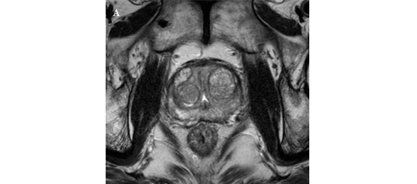
• All patients underwent preoperative MRI, including T2-weighted, diffusion-weighted, and dynamic contrast-enhanced imaging, as well as radical prostatectomy (RP) between June 2005 and December 2011.

• Imaging results were correlated with pathological findings to evaluate the ability of MRI to select patients for AS.

RESULTS

• In 35 (11.7%) patients, no discrete cancer was visible on MRI, while in the remaining 263 (88.3%) patients, a discrete cancer was visible.

• Pathological examination of RP specimens resulted in upstaging (>T2) in 21 (7%) patients, upgrading (Gleason score >6) in 136 (45.6%), and a diagnosis of unfavourable disease in 142 (47.7%) patients.

• The 263 patients (88.3%) with visible cancer on imaging were more likely to have their cancer status upgraded (49.8% vs 14.3%) and be diagnosed with unfavourable disease (52.1% vs 14.3%) than the 35 patients (11.7%) with no cancer visible upon imaging, and these differences were statistically significant (P < 0.001 for all).

• A visible cancer lesion on MRI, PSAD, and patient age were found to be predictors of unfavourable disease in multivariate analysis.

CONCLUSION

• MRI can predict adverse pathological features and be used to assess the eligibility of patients with prostate cancer for AS.

Read more articles of the week


Editorial: Multiparametric MRI and active surveillance for prostate cancer: future directions

A growing body of data exists suggesting an important role of MRI in selecting men with prostate cancer for active surveillance (AS). In the present study, Park et al. [1] show that a suspicious lesion on MRI was independently predictive of adverse pathology after radical prostatectomy (RP). This finding supports existing data suggesting that suspicious lesions on MRI confer an increased risk of disease reclassification among men enrolled in AS [2]. Indeed, in our institutional AS experience we found that men with a suspicious lesion on MRI were more likely to have biopsy reclassification with extended follow-up [3].While these data are provocative, much work remains to be done before the adoption of MRI as a standard screening tool for entry into AS for men with very low-risk prostate cancer.

Introduction of functional sequence imaging into multiparametric MRI protocols has resulted in improved detection and characterisation of clinically localised prostate cancer. However, before widespread implementation into AS protocols can occur, increased rigor and standardisation in image interpretation is needed. As in the present study, 5-point Likert scales have become an increasingly popular method of quantifying a lesions likelihood of representing cancer [1]. Still other authors have quantified a lesions level of suspicion using both weighted and non-weighted scoring systems based on the number of positive MRI sequences [3,4]. While useful for statistical analysis, these reporting methods are fraught with concerns of inter-observer variability and generalizability. Additionally, a recent report by Lee et al. [5] found that a simple measurement of lesion diameter on diffusion-weighted MRI was predictive of insignificant disease after RP. Combining the plethora of functional and morphological data obtained by multiparametric MRI into a standardised, reproducible tool will greatly facilitate implementation of MRI into current AS screening protocols.

As a step in the right direction, Stamatakis et al. [4] recently generated a nomogram for predicting biopsy reclassification in men on AS after taking into consideration both functional and morphological characteristics of MRI lesions. Adding an additional layer of complexity, they also assessed the utility of calculated values, e.g. lesion density (lesion volume/prostate volume), in predicting biopsy reclassification. Briefly, their analysis showed that the number of lesions, lesion suspicion, and lesion density were predictive of biopsy reclassification. While nomogram validation and testing of its predictive value on pathological outcomes is needed, this represents a major advance in the standardised application of MRI to AS cohorts.

Despite great strides in the application of multiparametric MRI to AS cohorts, a significant concern about the false-negative rate exists. Considering the present report, of the 35 men with no visible lesion on MRI, 14.3% men had unfavourable pathology after RP [1]. This is similar to previous studies reporting disease reclassification rates of <18% [2,6]. These men with normal imaging and high-grade cancer highlight the importance of incorporating imaging and clinical data when selecting men for AS. Better defining the false-negative rate of multiparametric MRI, and effectively identifying men with a normal MRI and high-grade disease remain major challenges.

Considering all of the available data, it is becoming increasingly clear that MRI has the potential for improving the identification of patients for whom AS would be safe. It is currently the practice at our institution to refer eligible men for multiparametric MRI before enrolment in AS. Our future scholarly efforts should be directed at the standardisation of reporting MRI data and the development of user-friendly AS criteria that synthesise MRI results with clinicopathological data.

Jeffrey K. Mullins and H. Ballentine Carter
James Buchanan Brady Urological Institute, Johns Hopkins Medical Institutions, Baltimore, MD, USA

Read the full article

References

  1. Park BH, Jeon HG, Choo SH et al. Role of multiparametric 3.0-Tesla magnetic resonance imaging in patients with prostate cancer eligible for active surveillance. BJU Int 2014; 113: 864–70
  2. Margel D, Yap SA, Lawrentschuk N et al. Impact of multiparametric endorectal coil prostate magnetic resonance imaging on disease reclassification among active surveillance candidates: a prospective cohort study. J Urol 2012; 187: 1247–52
  3. Mullins JK, Bonekamp D, Landis P et al. Multiparametric magnetic resonance imaging findings in men with low-risk prostate cancer followed using active surveillance. BJU Int 2013; 111: 1037–45
  4. Stamatakis L, Siddiqui MM, Nix JW et al. Accuracy of multiparametric magnetic resonance imaging in confirming eligibility for active surveillance for men with prostate cancer. Cancer 2013; 119: 3359–66
  5. Lee DH, Koo KC, Lee SH et al. Tumor lesion diameter on diffusion weighted magnetic resonance imaging could help predict insignificant prostate cancer in patients eligible for active surveillance: preliminary analysis. J Urol 2013; 190: 1213–7
  6. Guzzo TJ, Resnick MJ, Canter DJ et al. Endorectal T2-weighted MRI does not differentiate between favorable and adverse pathologic features in men with prostate cancer who would qualify for active surveillance. Urol Oncol 2012; 30: 301–5


Video: MRI can assess the eligibility of patients with prostate cancer for AS

Role of multiparametric 3.0-Tesla magnetic resonance imaging in patients with prostate cancer eligible for active surveillance

Bong H. Park, Hwang G. Jeon, Seol H. Choo, Byong C. Jeong, Seong I. Seo, Seong S. Jeon, Han Y. Choi and Hyun M. Lee

Department of Urology, Samsung Medical Center, Sungkyunkwan University School of Medicine, Seoul, Korea
Current address: Bong H. Park, Department of Urology, Incheon St. Mary’s Hospital, The Catholic University of Korea College of Medicine, Seoul, Korea

Read the full article
OBJECTIVE

• To evaluate predictors of more aggressive disease and the role of multiparametric 3.0-T magnetic resonance imaging (MRI) in selecting patients with prostate cancer for active surveillance (AS).

PATIENTS AND METHODS

• We retrospectively assessed 298 patients with prostate cancer who met the Prostate Cancer Research International: Active Surveillance (PRIAS) criteria, defined as T1c/T2, PSA level of ≤10 ng/mL, PSA density (PSAD) of <0.2 ng/mL2, Gleason score <7, and one or two positive biopsy cores.

• All patients underwent preoperative MRI, including T2-weighted, diffusion-weighted, and dynamic contrast-enhanced imaging, as well as radical prostatectomy (RP) between June 2005 and December 2011.

• Imaging results were correlated with pathological findings to evaluate the ability of MRI to select patients for AS.

RESULTS

• In 35 (11.7%) patients, no discrete cancer was visible on MRI, while in the remaining 263 (88.3%) patients, a discrete cancer was visible.

• Pathological examination of RP specimens resulted in upstaging (>T2) in 21 (7%) patients, upgrading (Gleason score >6) in 136 (45.6%), and a diagnosis of unfavourable disease in 142 (47.7%) patients.

• The 263 patients (88.3%) with visible cancer on imaging were more likely to have their cancer status upgraded (49.8% vs 14.3%) and be diagnosed with unfavourable disease (52.1% vs 14.3%) than the 35 patients (11.7%) with no cancer visible upon imaging, and these differences were statistically significant (P < 0.001 for all).

• A visible cancer lesion on MRI, PSAD, and patient age were found to be predictors of unfavourable disease in multivariate analysis.

CONCLUSION

• MRI can predict adverse pathological features and be used to assess the eligibility of patients with prostate cancer for AS.

Read more articles of the week


Article of the week: Out of the COLD: cryoablation for locally advanced PCa

Every week the Editor-in-Chief selects the Article of the Week from the current issue of BJUI. The abstract is reproduced below and you can click on the button to read the full article, which is freely available to all readers for at least 30 days from the time of this post.

In addition to the article itself, there is an accompanying editorial written by a prominent member of the urological community. This blog is intended to provoke comment and discussion and we invite you to use the comment tools at the bottom of each post to join the conversation.

If you only have time to read one article this week, it should be this one

Cryoablation for locally advanced clinical stage T3 prostate cancer: a report from the Cryo-On-Line Database (COLD) Registry

John F. Ward, Christopher J. DiBlasio*, Christopher Williams, Robert Given and J. Stephen Jones

§The University of Texas MD Anderson Cancer Center, Houston, TX, *Urology, Mount Sinai School of Medicine, Huntington, NY, Urology, University of Florida and Shands Medical Center, Jacksonville, FL, Urology, Eastern Virginia Medical School, Norfolk, VA, and §Urology, Cleveland Clinic Lerner College of Medicine, Cleveland, OH, USA

Read the full article
OBJECTIVE

• To assess the oncological and functional outcomes of primary prostate cryoablation for men with clinical stage T3 (cT3) prostate cancer, as although radical prostatectomy (RP) or external beam radiotherapy (EBRT) are the standard treatments for locally advanced cT3 prostate cancer some patients opt for nonextirpative prostate cryoablation instead.

PATIENTS AND METHODS

• The Cryo-On-Line Database (COLD) Registry was queried to identify patients with cT3 prostate cancer treated with whole-gland cryoablation (366 patients).

• We assessed biochemical disease-free survival (bDFS) using the Phoenix definition and determined reported rates of urinary incontinence and retention, sexual activity, and rectourethral fistulisation after treatment.

• Patients were subsequently assessed according to whether they were administered neoadjuvant androgen-deprivation therapy or not (ADT; 115 patients, 31.4%).

RESULTS

• For the entire cohort, the 36- and 60-month bDFS rates were 65.3% and 51.9%, respectively.

• Patients who received neoadjuvant ADT had statistically nonsignificantly higher 36- and 60-month bDFS rates (68.0% and 55.4%, respectively) than patients who did not receive neoadjuvant ADT (55.3% and 36.9%, respectively).

• The after treatment urinary incontinence rate was 2.6%; urinary retention rate, 6.0%; sexual activity rate, 30.4%; and rectourethral fistulisation rate, 1.1%.

CONCLUSIONS

• Cryoablation for patients with cT3 prostate cancer leads to less favourable bDFS than that after RP or RT for the same group of men.

• The after treatment rectourethral fistulisation rates for patients with cT3 disease are higher than in those with organ-confined prostate cancer treated with cryoablation; however, urinary dysfunction and sexual activity rates are similar for men with cT3 to those reported from this same registry in men with cT2 disease.

• The addition of neoadjuvant ADT (though not studied prospectively here) should be strongly considered if a patient with cT3 prostate cancer is to be treated with cryoablation.

Read more articles of the week


Editorial: Cryosurgery for clinical T3 prostate cancer

There are limited data available on the outcomes of cryosurgery for clinical T3 prostate cancer, and as such, the role of cryosurgery for clinical T3 disease is currently undetermined [1]. Modern cryosurgery of the prostate, utilizing gas-based third-generation technology, a real-time monitoring system with ultrasonography and thermocouples, is associated with a low complication rate [7], although comparative outcomes of the different treatment modalities and long-term follow-up data remain to be seen.

Several aspects of cryosurgery can make it difficult to adequately control locally advanced prostate cancer. First, cryosurgery for clinical T3 cancer requires unique surgical expertise to control local disease while minimizing side-effects. Secondly, staging of locally advanced prostate cancer is challenging – it is difficult to accurately identify the extent of extracapsular extension, seminal vesicle involvement and/or lymph node metastasis. Thirdly, challenges in managing clinical T3 disease include the requirement of a more extensive ablation technique to appropriately target the extraprostatic disease and seminal vesicle involvement as well as treatment for possible microscopic metastasis, which might not be clinically detectable.

Two recent randomized trials compared outcomes of external beam radiation therapy with those of cryosurgery (including cT3 diseases with use of neo-adjuvant androgen deprivation therapy [ADT]), with contrasting results [2, 3]. Chin et al. [2] reported superiority of biochemical disease-free survival favouring external beam radiation therapy in relatively more advanced (bulky) disease, while Donnelly et al. [3] reported significantly fewer positive biopsy rates favouring cryosurgery in the relatively less advanced disease. These findings could suggest that more advanced bulky cases that require wider local control of bulky extraprostatic diseases are not suitable for cryosurgery, while in appropriately selected cases with fewer extraprostatic diseases, cryosurgery is an acceptable option (when combined with neo-adjuvant ADT). Although appropriately extended cryo-lesions that achieve lethal temperatures can control extraprostatic disease, there is a certain limitation in the extension of cryo-lesions without injury to vital peri-prostate organs, such as the urinary sphincter, rectal wall, bladder wall and ureters.

Evolving accuracy of preoperative diagnostic imaging to assess extraprostatic disease can enhance outcomes, and staging tissue sampling from suspected extraprostatic disease could also identify actual microscopic extension of the extraprostatic disease [4]. A recently updated nomogram predicting lymph node invasion [5] suggests that the probability of lymph node invasion in patients with cT3, PSA level >10 ng/mL, and biopsy primary Gleason grade 4 is 20% or greater. Clearly, the preoperative risk assessment of lymph node involvement using such a modern calculator is pertinent for appropriate patient selection. Finally, management decision should be made by a multidisciplinary team.

When combined with radiotherapy, neo-adjuvant ADT for high-risk and locally advanced prostate cancer has been associated with clinical benefit; however, when combining neo-adjuvant ADT with prostatectomy, there is pathological down-staging and reduction in the surgical positive margin but minimal improvement in overall or disease-free survival [6]. The role of neo-adjuvant and adjuvant ADT when combined with cryosurgery is still unknown. Clearly, a prospective study is needed to determine the optimal duration and method of ADT (whether to use LHRH analogue or combined blockade) and to analyse the side-effects, the quality of life and the cost-effectiveness of a combination of cryosurgery with ADT for cT3a and cT3b prostate cancer.

Osamu Ukimura, Andre Luis de Castro Abreu, Andrew J. Hung and Inderbir S. Gill
USC Institute of Urology, Keck School of Medicine, University of Southern California, Los Angeles, CA, USA

Read the full article

References

  1. Babaian RJ, Donnelly B, Bahn D et al. Best practice statement on cryosurgery for the treatment of localized prostate cancer. J Urol 2008; 180: 1993–2004
  2. Chin JL, Al-Zahrani AA, Autran-Gomez AM, Williams AK, Bauman G. Extended followup oncologic outcome of randomized trial between cryoablation and external beam therapy for locally advanced prostate cancer (T2c-T3b). J Urol 2012; 188: 1170–1175
  3. Donnelly BJ, Saliken JC, Brasher PM et al. A randomized trial of external beam radiotherapy versus cryoablation in patients with localized prostate cancer. Cancer 2010; 116: 323–330
  4. Ukimura O, Coleman JA, de la Taille A et al. Contemporary role of systematic prostate biopsies: indications, techniques, and implications for patient care. Eur Urol 2013; 63: 214–230
  5. Briganti A, Larcher A, Abdollah F et al. Updated nomogram predicting lymph node invasion in patients with prostate cancer undergoing extended pelvic lymph node dissection: the essential importance of percentage of positive cores. Eur Urol 2012; 61: 480–487
  6. Shelley MD, Kumar S, Wilt T, Staffurth J, Coles B, Mason MD. A systematic review and meta-analysis of randomised trials of neo-adjuvant hormone therapy for localised and locally advanced prostate carcinoma. Cancer Treat Rev 2009; 35: 9–17
  7. Ward JF, DiBlasio CJ, Williams C, Given R, Jones JS. Cryoablation for locally advanced clinical stage T3 prostate cancer: a report from the Cryo-On-Line Database (COLD) Registry. BJU Int 2014; 113: 714–718


Article of the week: Rethinking inflammation in PCa

Every week the Editor-in-Chief selects the Article of the Week from the current issue of BJUI. The abstract is reproduced below and you can click on the button to read the full article, which is freely available to all readers for at least 30 days from the time of this post.

In addition to the article itself, there is an accompanying editorial written by a prominent member of the urological community. This blog is intended to provoke comment and discussion and we invite you to use the comment tools at the bottom of each post to join the conversation.

If you only have time to read one article this week, it should be this one.

Histological inflammation and risk of subsequent prostate cancer among men with initially elevated serum prostate-specific antigen (PSA) concentration in the Finnish prostate cancer screening trial

Tytti H. Yli-Hemminki*, Marita Laurila*, Anssi Auvinen, Liisa Määttänen§, Heini Huhtala, Teuvo L.J. Tammela and Paula M. Kujala*

*Department of Pathology, Fimlab Laboratories, Tampere University Hospital, Tampere, Department of Pathology, Seinäjoki Central Hospital, Seinäjoki, School of Health Sciences, University of Tampere, Tampere, §Finnish Cancer Registry, Helsinki, and Department of Urology, Tampere University Hospital and University of Tampere, Tampere, Finland

T. H. Y.-H. and M. L. contributed equally to this study.

Read the full article

OBJECTIVE

• To assess whether histological signs of inflammation are associated with an increased risk of subsequent prostate cancer (PCa) in men with elevated serum prostate-specific antigen (PSA) concentrations and benign initial biopsy.

MATERIALS AND METHODS

• Study subjects were men aged 54–67 years with an elevated PSA (≥4 ng/mL or 3–4 ng/mL and free to total PSA ratio ≤0.16 or positive digital rectal examination), but a benign biopsy result within the Finnish population-based randomised screening trial for PCa, which started in 1996.

• A total of 293 prostate biopsies without PCa or suspicion of malignancy from the first screening round in the Tampere centre were re-evaluated by a uropathologist to assess histological inflammation.

• Results of the subsequent screening rounds were obtained from the trial database and PCa diagnoses made outside the screening were obtained from the Finnish Cancer Registry.

• The median length of follow-up was 10.5 years.

• Cox regression analysis was used to assess PCa risk after the initial benign biopsy.

RESULTS

• Histological inflammation was found in 66% of the biopsies.

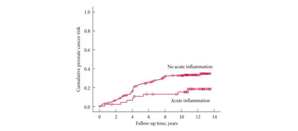
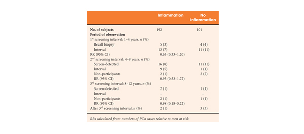
• Subjects with inflammation at the biopsy had a slightly lower PCa risk in the second screening round (18 vs 27%, rate ratio 0.69, 95% confidence interval [CI] 0.35–1.34) relative to men without inflammation. In further follow-up, the PCa risk remained nonsignificantly lower (hazard ratio [HR] 0.71, CI 0.46–1.10; P = 0.13). The risk was not appreciably affected by adjustment for age, PSA, prostate volume and family history of PCa (HR 0.67, CI 0.42–1.07; P = 0.092).

CONCLUSIONS

• Histological inflammation in a prostate biopsy among men with an initial false-positive screening test was not associated with an increased risk of subsequent PCa, but instead with a decreased risk which was of borderline significance.

• Inflammation in prostate biopsy is not a useful risk indicator in PCa screening.


Read Previous Articles of the Week


Editorial: Does inflammation reduce the risk of prostate cancer?

Chronic inflammation is thought to play an aetiological role in tumorigenesis in several cancers including bladder, oesophagus and liver [1]. Molecular studies show that it plays a critical role in several stages of the carcinogenic process including tumour initiation, promotion, metastases and response to therapy. However the role in prostate cancer is less clear and to date, clinical studies are inconclusive.

The article in this issue of BJUI by Yli-Hemminki et al. [2] appears to show an inverse association with histological inflammation and the risk of prostate cancer. Using data from the Finnish subgroup of the European Randomised Study of Prostate Cancer Screening (ERSPC) study they examined 293 patients with previous negative biopsies over a 10.5-year period and reported an 18% risk of prostate cancer in men with inflammation on initial biopsy (34 of 101 men) as opposed to 27% in those without inflammation (51 of 192 men). Perhaps somewhat surprisingly, histological inflammation did not appear to be significantly associated with PSA concentration, although it did appear that the free/total PSA ratio was higher in men with inflammation.

One potential confounding factor is that inflammation may also play a role in the pathogenesis of BPH. A large scale study showed that the odds ratio for BPH was 8.0 with a history of prostatitis [3]. Furthermore, the Medical Therapy of Prostatic Symptoms (MTOPS) study showed that men with inflammation had a significantly higher risk of BPH progression and acute urinary retention. These factors may impact on PSA levels and the chance of a subsequent prostate biopsy. However, the authors report that the inverse association of prostate cancer and inflammation did not alter when corrected for prostate volume, PSA level and age.

Significantly, those patients who screened positive at first biopsy were already excluded as were those with a suspicion of prostate cancer, such as a small atypical focus. Despite this, the study group probably represents high-risk patients, as they had all previously met the criteria for the first round of biopsies. This is borne out by the fact that the risk of prostate cancer was significantly increased when the men with inflammation on biopsy were compared with the initially screened negative men (hazard ratio 4.3). Unfortunately, we do not know what the rate of inflammation was in the control arm as they were screened negative and hence no biopsies were taken.

The significance of PSA level or prostatic intraepithelial neoplasia was not specifically investigated, although the reported rates were 32.1% and 7.5% respectively. Overall the incidence of inflammation was reported as 65%, but this included both acute and chronic inflammation. A smaller number of patients were given grade 2/3 chronic inflammation (80 and 20 patients, respectively) and only 14 patients had grade 2/3 acute, indicating most had milder degrees of inflammation. Whether this is a representative sample is not entirely clear. As the authors comment, published rates of histological inflammation do vary significantly from 8 to 99% and this does appear to vary according to detection method.

Several case-control studies and a meta-analysis [4] have shown that there is a significant increase in the relative risk of prostate cancer in men with prostatitis; however, these epidemiological studies all suffer from selection bias, in that men with clinical symptoms are more likely present and to be investigated and followed up by a Urologist. This study [2] only examines the role of histological inflammation and, at least partially, removes the selection bias associated with clinical symptoms, although it could still be argued that men with clinical prostatitis may be more likely to present for screening. This study represents a highly selected group of patients that are high risk for prostate cancer and no firm conclusions can be drawn on the general population. As inflammation is so common in prostate specimens, further high-quality large-scale studies are needed with similar long-term follow-up.

Miles A. Goldstraw and Roger S. Kirby
The Prostate Centre, London, UK

Read the full article

References

  1. Coussens LM, Werb Z. Inflammation and cancer. Nature 2002; 420: 860–867
  2. Yli-Hemminski T, Laurila M, Auvinen A et al. Histological inflammation and risk of subsequent prostate cancer among men with initially elevated serum prostate-specific antigen (PSA) concentration in the Finnish prostate cancer screening trial. BJU Int 2013; 112: 735–741
  3. Alcarez A, Hammerer P, Tubaro A, Schroder FH, Castro R. Is there evidence of a relationship between benign prostatic hyperplasia and prostate cancer? Findings of a literature review. Eur Urol 2009; 55: 864–875
  4. Dennis LK, Lynch CF, Torner JC. Epidemiologic association between prostatitis and prostate cancer. Urology 2002; 60: 78–83
© 2024 BJU International. All Rights Reserved.