Tag Archive for: prostate

Posts

Video: Prostatic urethral lift for the treatment of LUTS

Multicentre prospective crossover study of the ‘prostatic urethral lift’ for the treatment of lower urinary tract symptoms secondary to benign prostatic hyperplasia

Anthony L. Cantwell, William K. Bogache*, Steven F. Richardson, Ronald F. Tutrone, Jack Barkin§, James E. Fagelson, Peter T. Chin†† and Henry H. Woo

‡‡Atlantic Urological Associates, Daytona Beach, FL, *Carolina Urological Research Center, Myrtle Beach, SC, Western Urological Clinic, Salt Lake City, UT, Chesapeake Urology, Baltimore, MD, USA, §University of Toronto, Toronto, ON, Canada, Urology Associates of Denver, Denver, CO, USA, ††Figtree Private Hospital, Figtree, and ‡‡Sydney Adventist Hospital Clinical School, University of Sydney, Sydney, NSW, Australia

OBJECTIVE

• To assess the clinical effect of the ‘prostatic urethral lift’ (PUL) on lower urinary tract symptoms (LUTS) associated with benign prostatic hyperplasia (BPH) through a crossover design study.

PATIENTS AND METHODS

• Men aged ≥50 years with an International Prostate Symptom Score of ≥13, a maximum urinary flow rate (Qmax) of ≤12 mL/s, and a prostate of 30–80 mL were enrolled into a crossover study after completing a prospective, randomised, controlled, ‘blinded’ pivotal study in which they were control subjects receiving a sham procedure.

• Patients were followed for 1 year after crossover PUL at 19 centres in the USA, Canada and Australia. The sham procedure involved rigid cystoscopy with simulated active treatment sounds.

• PUL involved placing permanent UroLift® (NeoTract, Inc., Pleasanton, CA, USA) implants into the lateral lobes of the prostate to enlarge the urethral lumen.

• Urinary symptom relief, health-related quality of life (HRQL) impact, urinary flow parameters, sexual function, and adverse events were assessed and compared between the sham and PUL using paired statistical analysis.

RESULTS

• Symptom, flow, HRQL and sexual function assessments showed response improvements from baseline results, similar to results from other published studies, and most parameters were markedly improved after PUL vs the sham procedure in the same patients.

• Symptom, flow, and HRQL improvements were durable over the 12 months of the study.

• Adverse events associated with the procedure were typically transient and mild to moderate; one patient (2%) required re-intervention with transurethral resection of the prostate in the first year.

• There were no occurrences of de novo, sustained ejaculatory or erectile dysfunction.

CONCLUSION

• The PUL can be performed under local anaesthesia, causes minimal associated perioperative complications, allows patients to quickly return to normal activity, provides rapid and durable improvement in symptoms, and preserves sexual function.


Article of the week: Getting to the core of the matter with PIRADS scoring

Every week the Editor-in-Chief selects the Article of the Week from the current issue of BJUI. The abstract is reproduced below and you can click on the button to read the full article, which is freely available to all readers for at least 30 days from the time of this post.

In addition to the article itself, there is an accompanying editorial written by prominent members of the urological community. This blog is intended to provoke comment and discussion and we invite you to use the comment tools at the bottom of each post to join the conversation.

If you only have time to read one article this week, it should be this one.

Histology core-specific evaluation of the European Society of Urogenital Radiology (ESUR) standardised scoring system of multiparametric magnetic resonance imaging (mpMRI) of the prostate

Timur H. Kuru*, Matthias C. Roethke, Philip Rieker*, Wilfried Roth, Michael Fenchel, Markus Hohenfellner*, Heinz-Peter Schlemmer and Boris A. Hadaschik*

*Department of Urology, University Hospital Heidelberg, Department of Radiology, German Cancer Research Center (DKFZ), and Institute of Pathology, University of Heidelberg, Heidelberg, Germany

Read the full article

Link to Video: MRI-Navigated Stereotactic Prostate Biopsy

OBJECTIVES

• To evaluate the Prostate Imaging Reporting and Data System (PIRADS) in multiparametric magnetic resonance imaging (mpMRI) based on single cores and single-core histology.

• To calculate positive (PPV) and negative predictive values (NPV) of different modalities of mpMRI.

PATIENTS AND METHODS

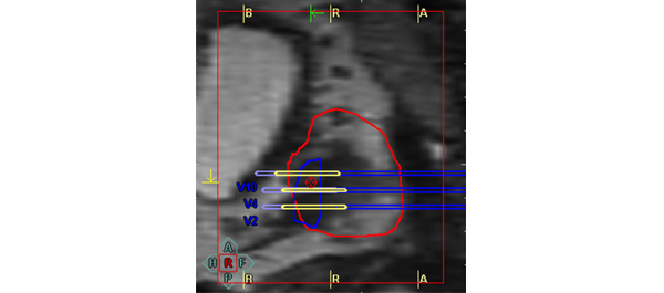
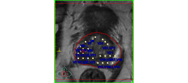
• We performed MRI-targeted transrectal ultrasound-guided perineal prostate biopsies on 50 patients (mean age 66 years, mean PSA level of 9.9 ng/mL) with suspicion of prostate cancer. The biopsy trajectories of every core taken were documented in three dimensions (3D) in a 3D-prostate model.

• Every core was evaluated separately for prostate cancer and the performed biopsy trajectories were projected on mpMRI images.

• PIRADS scores of 1177 cores were then assessed by a histology ‘blinded’ uro-radiologist in T2-weighted (T2W), dynamic contrast-enhanced (DCE), diffusion-weighted imaging (DWI) and magnetic resonance spectroscopy (MRS).

RESULTS

• The PIRADS score was significantly higher in cores positive for cancer than in negative cores.

• There was a significant correlation between the PIRADS score and histopathology for every modality.

• Receiver operating characteristic (ROC) analysis showed excellent specificity for T2W (90% peripheral zone/97% transition zone) and DWI (98%/97%) images regardless of the prostate region observed. These numbers decreased for DCE (80%/93%) and MRS (76%/83%).

• All modalities had NPVs of 99%, if a PIRADS score threshold of 2 (for T2W, DCE, and MRS) or 3 (for DWI) was used. However, PPVs were low.

CONCLUSIONS

• Our results show that PIRADS scoring is feasible for clinical routine and allows standardised reporting.

• PIRADS can be used as a decision-support system for targeting of suspicious lesions.

• mpMRI has a high NPV for prostate cancer and, thus, might be a valuable tool in the initial diagnostic evaluation.


Read Previous Articles of the Week


Editorial: Too many men still undergo needless prostate biopsy

Multiple studies have shown that only one in three or four men with a raised PSA level prove to have prostate cancer and many men suffer potentially life-threatening complications from transrectal prostate biopsy. There is an urgent need for better risk stratification of men with elevated PSA levels. Any such test should have a high negative predicative value (NPV; small number of significant cancers missed) but also a high positive predictive value (PPV; i.e. the yield would be high and there would be very few false positives) to diminish the number of unnecessary biopsies. Multiparametric MRI (mpMRI) of the prostate, especially with a stronger 3 T magnet, has been advocated for this purpose. The parameters refer to the separate MRI sequences used, typically at least three. Sequences can not only study the anatomy of the gland (standard T2-weighted MRI), but there is also a measure of the tissue cellularity (diffusion-weighted MRI), vascularity (dynamic contrast-enhanced MRI) or biochemistry (magnetic resonance spectroscopy). Initial data have shown promise but the changes seen on these various sequences can be subtle and interpretation is subjective. Naturally observer experience plays a large part but a standardised scoring system, the so called Prostate Imaging Reporting and Data System (PIRADS) system, has been proposed to improve reporting performance [1]. Each parameter is scored on a scale of 1–5 according to the likelihood of cancer. Scoring systems are always a compromise between the NPV and PPV, and so far there is no agreement where the threshold for each parameter should be set. In the original document, the authors proposed that a score of 4 or 5 signifies a high likelihood or almost certainty of cancer, whilst scores of 1 or 2 denote a high likelihood of benign tissue. A score of 3 is evens. The paper by Kuru et al. [2] shows a high NPV only when the threshold was set at the low level of 2 for each parameter. Predictably, at this threshold the PPV was extremely low, and therefore many men would still undergo unnecessary biopsy. Another similar paper advocated a mean threshold of 3, but even then the PPV was 38% with a NPV of 95% [3]. Both these papers are retrospective studies, in particular the MRI readings were done retrospectively. Nevertheless, the low PPV is disappointing. The results of prospective studies with multiple readers are keenly awaited and I hope that that these will find a higher PPV for mpMRI, and we can to move to an era when fewer men undergo needless prostate biopsy.

Uday Patel
St George’s Hospital, London, UK

Read the full article

References

  1. Barentsz JO, Richenberg J, Clements R et al. ESUR prostate MR guidelines 2012. Eur Radiol 2012; 22: 746–757
  2. Kuru T, Roethke M, Rieker P et al. Histology core-specific evaluation of the European Society of Urogenital Radiology (ESUR) standardised scoring system of multiparametric magnetic resonance imaging (mpMRI) of the prostate. BJU Int 2013; 112:1080–1087
  3. Portalez D, Mozer P, Cornud F et al. Validation of the European Society of Urogenital Radiology scoring system for prostate cancer diagnosis on multiparametric magnetic resonance imaging in a cohort of repeat biopsy patients. Eur Urol 2012; 62: 986–996

Patient with De Novo Adenosquamous Carcinoma of the Prostate

We report a case of ASC arising spontaneously in a 54 year old male with no previous risk factors.

Authors: Love, Matthew; Storey, Barckley; Alatassi, Houda; Tonkin, Jeremy
Corresponding Author: Love, Matthew


Abstract
Primary adenosquamous cell carcinoma of the prostate (ASC) is an exceedingly rare and aggressive form of prostate cancer, making up <1% of all diagnoses. Since its initial description by Thompson in 1942, there have been fewer than 30 cases reported in the literature. Recent reports of age-adjusted incidence rates of ASC have been shown to be around 0.03 cases per million people per year, making it less prevalent than pure squamous cell carcinoma, an exceedingly rare subtype in itself. While the majority of these tend to arise subsequent to endocrine or radiation treatment with squamous differentiation, approximately one-third of cases have arisen in a de novo setting. We report a case of ASC arising spontaneously in a 54 year old male with no previous risk factors.

Introduction
Primary adenosquamous cell carcinoma of the prostate (ASC) is an exceedingly rare and aggressive form of prostate cancer. Since its initial description by Thompson in 1942, there have been fewer than 30 cases reported in the literature [1]. While the majority of these tend to arise subsequent to endocrine or radiation treatment with squamous differentiation, approximately one-third of cases have arisen in a de novo setting [2]. We report a case of ASC arising spontaneously in a 54 year old African American male with no previous risk factors.

Case Report
A 54 year old African American male was referred to the University of Louisville Department of Urology following detection of an elevated PSA of 20 ng/mL on annual screening. He denied hematuria, dysuria, ejaculatory issues, or lower urinary tract symptoms at the time of presentation. Past medical history was noncontributory and there was no history of prostate cancer or any other malignancies in his family. On digital rectal examination the patient was noted to have a 40 gram prostate that was firm, non-tender, and with no discernible nodules. Repeat PSA was obtained and was found to be 46.7 ng/mL and he was subsequently scheduled for an ultrasound guided prostate biopsy.

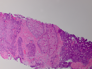
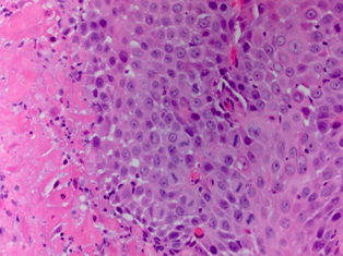
Standard 12 core template prostate biopsy revealed adenosquamous carcinoma of the prostate, Gleason 4+4=8 in 7/12 cores and involving more than 50% of each core (See Figure 1 and Figure 2). As a result, an extensive metastatic workup was performed. Bone scan revealed multiple metastatic sites including the patient’s right rib, pubic symphysis, and iliac crest. CT abdomen/pelvis was obtained showing extensive retroperitoneal lymphadenopathy. Chest X-ray was negative for disease.

Due to the extensive metastatic nature of the disease, the patient opted for hormone deprivation therapy and an elective orchiectomy was performed. He was then referred to medical oncology for chemotherapeutic intervention.

Discussion/Conclusion
ASC of the prostate is among one of the rarest and most aggressive subtypes of prostate cancer making up <1% of all diagnoses [3]. Recent reports of age-adjusted incidence rates of ASC have been shown to be around 0.03 cases per million people per year, making it less prevalent than pure squamous cell carcinoma, an exceedingly rare subtype in itself [4].

The underlying mechanism for the progression and development of ASC is unknown and debated; however, most theories contend that differentiation is triggered by the various treatment modalities rather than de novo development [5]. While over two thirds of the cases of ASC originate in patients with previously diagnosed adenocarcinoma of the prostate who have been treated with additional endocrine/radiation therapy, it is extremely rare to find a primary case of this particular subtype[5]. While some believe that hormonal treatment and radiotherapy induce squamous metaplasia in the glandular cells of adenomatous prostate cancer, others contend that ASC develops de novo from divergent differentiation from epithelial stem cell lines within the prostatic cells [6] [7]. Due to the infrequent occurrence of this disease there are very few available studies to examine the mechanism behind this metaplastic process and research is currently undergoing.

ASC is defined by the presence of both glandular and squamous components on histological examination. The squamous elements of ASC constitute on average of 40% of the tumor volume but can range anywhere from 5-95% [8]. This wide range of cell types is reflected in ASC variability through immunohistochemical staining. Stains such as PSA, PSAP, and low molecular weight keratin (CAM5.3) are commonly found only in the glandular components of ASC and therefore patients with a large squamous fraction can have normal serum levels of PSA, possibly further delaying diagnosis [3]. Similarly, the squamous portion can stain positive for high molecular weight keratin (AE3), but this can vary depending on the amount of tissue that has squamous components involved (See Table 1) [9]. Additionally, glandular components have a tendency to be more high grade with an average Gleason score of >6, however, this is also a controversial point as some have suggested that Gleason scoring should not be applied to this subtype [8].

ASC tends to follow the traditional metastatic pathways similar to adenocarcinoma of the prostate, starting with local invasion and then spreading to bone and other distant soft tissue sites. However, one notable difference is that unlike standard prostatic adenocarcinoma, bone metastatic sites are characteristically osteolytic rather than osteoblastic in nature [3].

ASC is an extremely aggressive subtype of prostate cancer and in most cases is found to have widely metastasised at time of diagnosis, suggesting that this disease has a tendency to disseminate early. Most cases reported in the literature presented in individuals who were found to be in urinary retention and the pathological diagnosis was made on TURP specimens [8]. The disease is highly resistant to radiation and chemotherapy and in those individuals fortunate enough to be diagnosed early with localized/regional disease, prostatectomy shows some survival advantage [2]. It is not clear whether hormone ablation is an effective treatment modality, as some authors suggest an early response while others have noted that patients are refractory to hormone deprivation. Long term survival is extremely poor. Wang et al, evaluating SEER data and isolating for patients with an ASC diagnosis, found that the median cancer specific survival was 16 months [2]. For patients who presented with distant disease, the 6 month survival rate was only 20% with all dying within one year of diagnosis.

While literature on the subject of ASC is limited, it appears that the best initial treatment for this particular type of cancer is aggressive surgical intervention in patients with regionally restricted disease. However, due to the highly aggressive nature of this disease in most cases, such as this one, patients present with widely disseminated disease and are relegated to a regimen of hormone ablation and chemotherapy. Currently there are no recommended chemotherapeutic regimens targeted at ASC.

087Figure1


Fig. 1

087Figure2


Fig. 2

References
1. Thompson GJ. Transurethral resection of malignant lesions of the prostate gland. JAMA, 1942;120: 1105-9.
2. Wang J, Wang FW, LaGrange CA, et al. Clinical features and outcomes of 25 patients with primary adenosquamous cell carcinoma of the prostate. Rare Tumors, 2010; 2(3): e47.
3. Humphrey PA. Histological variants of prostatic carcinoma and their significance. Histopathology, 2012; 60(1): 59-74.
4. Marcus DM, Goodman M, Jani AB, et al. A comprehensive review of incidence and survival in patients with rare histological variants of prostate cancer in the United States from 1973 to 2008. Prostate Cancer and Prostatic Disease, 2012. doi: 10.1038/pcan.2012.4.
5. Mazzucchelli R, Lopez-Beltran A, Cheng L, et al. Rare and unusual histological variants of prostatic carcinoma: clinical significance. BJU International, 2008; 102: 1369–1374.
6. Baydar DE, Kosemehmetoglu K, Akdogan B, et al. Prostatic Adenosquamous Carcinoma Metastasizing to Testis. The Scientific World Journal, 2006; 6: 2491–2494.
7. Egilmez T, Bal N, Guvel S, et al. Adenosquamous carcinoma of the prostate. Int J Urol, 2005; 12(3): 319-21.
8. Parwani AV, Kronz JD, Genega EM, et al. Prostate carcinoma with squamous differentiation: an analysis of 33 cases. Am J Surg Pathol, 2004; 28(5): 651-7.
9. Gattuso P, Carson HJ, Candel A, et al. Adenosquamous carcinoma of the prostate. Hum Pathol, 1995; 26(1): 123-6.


Table 1

Capture


Immunohistochemicalstaining patterns of adenocarcinomas and squamous cell carcinomas of the prostate.


Date added to bjui.org: 14/12/2012

DOI: 10.1002/BJUIw-2012-087-web


Inadvertent injury to an incidental ectopic ureter in a completely duplicated collecting system during open radical perineal prostatectomy

We present the diagnosis and management of a man with clinically localised prostate cancer and a complete duplication of the right collecting system associated with an asymptomatic upper pole ectopic ureter, that was inadvertently injured during open RPP.

Authors: Miguel Suhady (MS) Cabalag1, Henry Han-I (HH) Yao1, Gideon Adam (GA) Blecher1, Antonio (A) DeSousa1, Diana (D) Tran2

1 Department of Urology, Ballarat Base Hospital, Ballarat, Victoria, Australia
2 Department of Radiology, Royal Melbourne Hospital, Melbourne, Victoria, Australia

Corresponding Author: Richard (R) McMullin,  Consultant Urologist, Ballarat Base Hospital, Drummond Street North  Ballarat VIC 3350, Ballarat, Victoria, Australia E-mail: richard.mcmullin@ballaraturology


Abstract
 
It is rare for embryologic abnormalities of the urinary tract to present in adulthood, especially as an incidental finding post radical perineal prostatectomy (RPP). We present the incidental diagnosis and subsequent management of a 68 year old man with clinically localised prostate cancer and a complete duplication of the right collecting system associated with an asymptomatic upper pole ectopic ureter, that was inadvertently injured during open RPP.


Introduction
 
With the earlier detection of clinically localised prostate cancer using prostate specific antigen (PSA) testing, it has become routine not to perform extensive staging investigations to avoid unnecessary tests. This approach may lead to rare unforeseen post-operative complications in patients with asymptomatic congenital anomalies of the urinary tract. We report the case of a 68 year old man who presented with persistent urinary extravasation from his surgical wound following RPP. Subsequent CT intravenous urogram demonstrated an incidental complete duplication of the right collecting system and an injured right upper pole ectopic ureter. To our knowledge, there are only four cases reported in the literature of ectopic ureters found incidentally in men undergoing radical prostatectomy for prostate cancer (1-4), and we describe the first case reported during RPP.


Case Report
 
A 68 year old asymptomatic man initially presented with an incidental elevated PSA of 4.8 ng/ml (12% free) detected on routine testing. The patient otherwise had no significant past medical history. On digital rectal examination, the prostate was mildly enlarged and firm but with no palpable nodules bilaterally. Trans-rectal ultrasound (TRUS) of the prostate with concurrent 12 core guided biopsies, revealed no suspicious areas and an estimated prostate volume of 60 cm3. Histopathology showed a 2mm focus of Gleason 6 (3+3) prostate adenocarcinoma, which was only detected following further immunohistochemical staining. An active surveillance approach with six monthly PSA tests was initially adopted given the very low volume of malignancy. Over the next 18 months, the PSA gradually rose to 8.2 ng/ml whereby a repeat TRUS guided biopsy revealed prostate adenocarcinoma with Gleason 6 (3+3) in 33% of 1 core obtained from the right lateral prostate (clinical stage T1c).
Following patient counselling, a radical nerve-sparing perineal prostatectomy was performed using a previously described technique (5). A routine pre-operative rigid cystoscopy revealed a normal urethra and bladder with two appropriately located ureteric orifices. Surgery went as planned and a 16Fr two-way silicone indwelling catheter was inserted into the bladder intra-operatively. The urethral anastomosis was subsequently checked to ensure a watertight seal and a tube drain was also inserted through a separate stab incision into the perineum. Pelvic lymphadenectomy was not performed due to the patient’s low risk pathology. The final pathology demonstrated a Gleason 7 (3+4) adenocarcinoma, localised predominantly to the right lower zone involving approximately 15% of total prostatic volume. Lymphovascular invasion was absent and the surgical margins were clear.
Early post-operative recovery was uneventful and the drain tube was removed on post-operative day (POD) 2. A routine trial of void 2 weeks post surgery resulted in a persistent urine leak from the apex of his perineal wound, which was exacerbated by urination. Of note, the patient remained pain free and afebrile throughout the post-operative period. This was initially suggestive of an anastomotic leak and therefore the indwelling urinary catheter was re-inserted. However, a cystogram did not show any extravasation of contrast (Figure 1) and the leak persisted through the apex of the wound, so the re-introduced urinary catheter was removed.
Figure 1. Post-operative retrograde cystogram showing no anastomotic leakage


Subsequent computed tomography (CT) intravenous urogram demonstrated a complete duplication of the right collecting system with an ectopic right upper pole ureter (Figure 2a and 2b), the distal end of which could not be properly visualised.


Figure 2a.  Coronal view of the CT intravenous urogram showing the right lower pole ureter (LPU) and the right upper pole ectopic ureter (UPEU).


Figure 2b. 3D reconstruction of the coronal CT-intravenous urogram showing the orthotopic insertion of the right lower pole ureter (LPU) into the bladder (solid arrow), the tortuous right upper pole ectopic ureter (UPEU, dotted arrow). Note an indwelling urinary catheter is in situ.


There was also a fistulous tract originating from the ectopic ureter, draining into the perineum (Figure 3). The patient subsequently developed right-sided epididymo-orchitis, which was successfully treated with intravenous gentamicin and amoxicillin.


Figure 3. Coronal view of the CT intravenous urogram demonstrating the right upper and lower pole renal moieties, as well as a fistulous tract (solid arrow) originating from the ectopic ureter into the perineum on the right side (dotted arrows). 


Management
 
The patient subsequently underwent a rigid cystoscopy and a right retrograde pyelogram, which demonstrated a non-dilated right ureter draining into a lower renal moiety, producing a ‘drooping lily’ type image of the right kidney (Figure 4).


Figure 4. Right retrograde pyelogram demonstrating the characteristic ‘drooping lily’ image of the lower renal moiety. 


After patient counselling, the decision was then made to perform an open right ureteric reimplantation. Intra-operatively, two ureters were identified in the retroperitoneum adjacent to the bifurcation of the right common iliac vessel. The lateral ureter was entered via a small stab incision and dye was injected. This dye was noted to be draining out of the indwelling catheter and therefore this was thought to be the normal ureter that was draining into the bladder. The medial ureter was also pierced and dye was injected, but the dye was not detected in the bladder. Thus, this was thought to be the ectopic ureter, most likely to have been injured during the RPP. Given the close proximity of the bladder to the ectopic ureter, a uretero-vesical anastomosis was performed. The distal ectopic ureter was spatulated and a cystotomy was made at a corresponding site. A 6Fr 26 cm ureteric stent was passed through the ectopic ureter over a guide wire up to the right kidney, and the distal end was placed through the cystotomy. Using four, 4-zero vicryl sutures in an interrupted fashion, the ureter was securely implanted onto the bladder with good mucosal apposition. A pelvic drain tube was placed adjacent to the newly formed uretero-vesical anastomosis. There were no peri-operative complications, the drain was removed on POD 4 and the patient was discharged home on POD 6. A flexible cystoscopy was performed approximately 4 weeks after to remove the ureteric stent. Repeat CT intravenous urogram performed 2 months post re-implantation demonstrated complete duplication of the right collecting system with double ureters in which contrast drained normally into the bladder. Importantly, it did not show any hydronephrosis bilaterally, no perineal collection nor any leak from the right renal tract (Figure 5).


Figure 5. Coronal view of the CT intravenous urogram post uretero-vesical anastomosis of the right upper pole ectopic ureter, showing resolution of the fistulous tract to the perineum.


Discussion
 
We report the case of a patient who was incidentally found to have a complete duplication of the right collecting system with an asymptomatic ectopic upper pole ureter that was inadvertently injured during a RPP. To our knowledge, this is the first case reported in the literature associated with such surgery.
A duplex system refers to a kidney with two pelvicalyceal systems within a single renal parenchyma, which may have either a single or bifid ureter (partial duplication), or two discrete ureters (complete duplication) (6).  Renal duplication is a relatively common congenital anomaly, with a reported prevalence of 0.3-6% (7). The majority of cases are detected during childhood, but up to 20% of patients remain asymptomatic into adulthood (8). In contrast, ectopic ureters are rare, with a reported incidence of 1 in 1900 (9). Ectopic ureters are more common in females and are usually associated with a duplicated collecting system, whereas they are typically associated with a single collecting system in men (10).The anatomic site of insertion of an ectopic ureter in a duplicated collecting system follows the Weigert-Meyer rule, whereby the upper pole ureter is more commonly ectopic and the lower pole ureter typically inserts into the trigone, or laterally and cranially to this structure. In 50-60% of patients, renal duplication is associated with vesicoureteric reflux, which may affect one or both ureters. Reflux is more common in the lower pole kidney (97%), while ureteroceles and ectopic ureters are commoner in the upper pole kidney (11).
The presentation of ectopic ureters depends on their site of insertion. Ectopic ureters inserting into the prostatic urethra typically present with urinary tract infections or lower urinary tract symptoms of urgency and frequency. Ectopic ureters inserting into a seminal vesicle, vas deferens or epididymis may present with epididymitis, chronic prostatitis, abdominal or pelvic pain, discomfort during ejaculation, constipation, or a large abdominal mass secondary to obstruction and hydronephrosis. Males with an ectopic ureter may also present with infertility (3).
In males, the majority of ectopic ureters (50%) insert into the prostatic urethra, and 33% into a seminal vesicle. The least common sites include the prostatic utricle and the vas deferens (12). Ectopic ureters in males almost never insert distal to the external sphincter, where it would present as urinary incontinence. One such case has been reported in the literature (13). Of note, this patient had no lower urinary tract symptoms prior to the RPP. It is likely that in this case, the upper pole ectopic ureter was transected during the bladder neck dissection at the level of the prostatic urethra.
There are multiple treatment options for the pathological ectopic ureter associated with a duplicated system, including ureteropyelostomy, heminphrectomy, ipsilateral ureteroureterostomy and common sheath reimplantation (14). An ureterovesical anastomosis was performed in this case due to the upper pole ectopic ureter being a distinct entity from the lower pole ureter, and because of its close proximity to the bladder.
Current guidelines do not recommend imaging of the prostate with CT or MRI for low risk disease prior to radical prostatectomy. Consequently, any asymptomatic congenital anomalies of the urinary tract will not be detected preoperatively. However, due to the rarity of such anomalies, as well as the expense and potential risk to patients, routine preoperative imaging to detect congenital abnormalities is not justifiable in this setting. In the retrospective study conducted by Costa et al, only 3 out of 254 (1.1%) surgical descriptions of nephroureterectomy samples showed anatomical variations of the collecting system, with all cases demonstrating ureteric duplications (15).
Of interest, given the established genetic association of duplex systems, the patient’s twin brother subsequently underwent a CT intravenous urogram, which did not demonstrate any evidence of a duplicated system or ectopic ureter.


Conclusion
 
Preoperative imaging may help detect asymptomatic congenital abnormalities of the urinary tract, enabling appropriate intraoperative management. However, given the rarity of such anomalies and the expense of imaging, extensive investigations prior to radical prostatectomy for the sole purpose of screening for congenital abnormalities is not justifiable. However, this case highlights the need to consider congenital anomalies of the urinary tract as a possible differential in peri-operative complications post radical prostatectomy.


References
 
1. Funahashi Y, Kamihira O, Kasugai S, Kimura K, Fukatsu A, Matsuura O. [Radical prostatectomy for prostate carcinoma with ectopic ureter ; a case report]. Nihon Hinyokika Gakkai zasshi The japanese journal of urology. 2007 2007;98(3):580-2.
2. Ghazi A, Zimmermann R, Janetschek G. Delayed detection of injury to an ectopic ureter of a duplicated collecting system following laparoscopic radical prostatectomy for early organ-confined prostate cancer. Urologia internationalis.  2011 Feb (Epub 2010 Nov;86(1):121-4.
3. Marien TP, Shapiro E, Melamed J, Taouli B, Stifelman MD, Lepor H. Management of localized prostate cancer and an incidental ureteral duplication with upper pole ectopic ureter inserting into the prostatic urethra. Reviews in urology. 2008 2008;10(4):297-303.
4. Nakai Y, Tanaka M, Yoshikawa M, Tanaka N, Hirayama A, Fujimoto K, et al. [Prostate cancer and left ectopic ureter opening to seminal vesicle with left renal agenesis: a case report]. Hinyokika kiyo Acta urologica Japonica. 2009 2009;55(1):47-50.
5. Weiss JP, Schlecker BA, Wein AJ, Hanno PM. Preservation of periprostatic autonomic nerves during total perineal prostatectomy by intrafascial dissection. Urology. 1985 1985;26(2):160-3.
6. Glassberg KI, Braren V, Duckett JW, Jacobs EC, King LR, Lebowitz RL, et al. Suggested terminology for duplex systems, ectopic ureters and ureteroceles. The Journal of urology. 1984 1984;132(6):1153-4.
7. Hartman GW, Hodson CJ. The duplex kidney and related abnormalities. Clinical radiology. 1969;20(4):387-400.
8. Privett JT, Jeans WD, Roylance J. The incidence and importance of renal duplication. Clinical radiology. 1976 1976;27(4):521-30.
9. Campbell M, Harrison J. Anomalies of the Ureter. In: Campbell M, editor. Urology. 3rd ed. Philadelphia: WB Saunders; 1970.
10. Schlussel R, Retik A. Ectopic ureter, ureterocele, and other anomalies of the ureter. In: Wein A, Kavoussi L, Novick A, editors. Campbell-Walsh Urology. 9th ed. Philadelphia: Saunders Elsevier; 2008. p. 3383-422.
11. Jelloul L, Valayer J. Ureteroureteral anastomosis in the treatment of reflux associated with ureteral duplication. The Journal of urology. 1997 1997;157(5):1863-5.
12. Ellerker AG. The extravesical ectopic ureter. The British journal of surgery. 1958 1958;45(192):344-53.
13. Ejaz T, Malone PS. Male duplex urinary incontinence. The Journal of urology. 1995 1995;153(2):470-1.
14. Chacko JK, Koyle MA, Mingin GC, Furness PD, 3rd. Ipsilateral ureteroureterostomy in the surgical management of the severely dilated ureter in ureteral duplication. The Journal of urology. 2007 2007 Oct (Epub 2007 Aug;178(4 Pt 2):1689-92.
15. Costa HC, Moreira RJ, Fukunaga P, Fernandes RC, Boni RC, Matos AC. Anatomic variations in vascular and collecting systems of kidneys from deceased donors. Transplantation proceedings. 2011 2011;43(1):61-3.


Date added to bjui.org: 23/05/2012
DOI: 10.1002/BJUIw-2011-145-web


Fatal gas embolism during transurethral enucleation of the prostate with bipolar electrodes

In this report, we describe a case of gas embolism occurring during TUEB, which might have been induced by gas generated from the electrode.


Authors: Inokuchi, Go; Yajima, Daisuke; Hayakawa, Mutsumi; Sakuma, Ayaka; Makino, Yohsuke; Iwase, Hirotaro
Corresponding Author: Go Inokuchi, Department of Legal Medicine, Graduate School of Medicine, Chiba University, Chiba, Japan.   Email: [email protected]


Introduction 
Transurethral resection of the prostate (TURP) is the most common surgical treatment for benign prostatic hypertrophy. In recent years, however, new transurethral resection techniques, including transurethral resection of the prostate in saline (TURis) and transurethral enucleation with bipolar (TUEB) electrodes, have been developed to minimise surgical complications such as TURP syndrome, obturator nerve reflex and haemorrhage. Application of the TUEB procedure to clinical cases has only recently started (1).
During prostate surgery, gas embolism can occur as a complication of medical intervention and may lead to death (2). Consequently, it is vital to understand whether the specific cause of the gas embolism can be elucidated and meaningful feedback given to the medical community. In this report, we describe a case of gas embolism occurring during TUEB, which might have been induced by gas generated from the electrode.


Case report


Clinical course
A 72 year old man (72kg, 165cm) was scheduled to undergo TUEB for the treatment of benign prostate enlargement. Preoperative tests were unremarkable. The procedure was performed with the patient in the reverse Trendelenburg lithotomy position under spinal anaesthesia, and no problems with the induction of anaesthesia were noted. The patient was not wearing anti-embolic stockings. Thirteen minutes into the operation, the patient complained of epigastric discomfort and blood pressure gradually dropped to 73/48mmHg from 131/97 mmHg, but heart rate remained unchanged at 60-70 bpm.
After administration of a plasma substitute was started, blood pressure returned to between 80 and 100mmHg, and the operation proceeded without major bleeding from the bladder or prostate. There were no gas bubbles other than the gas generated from the electrodes seen in the surgical field on the surgical monitor during the procedure. Around 90 min later, enucleation of the prostate was completed. The surgeon then used a tissue morcellator to cut the resected prostate into small pieces. A few minutes after inserting the morcellator into the bladder, the patient lost consciousness and the electrocardiogram was flat.
Cardiopulmonary resuscitation was performed and the patient was taken to the medical care center, but subsequently died. Immediate postmortem computed tomography (CT) showed a large volume of gas in the heart (both left and right ventricles and left and right atria), pulmonary artery, cerebral vessels, vessels around bladder and bladder. (Figs.1, 2)


Figure 1.


Figure 2.

 
Autopsy findings
 
On external examination, there were no remarkable findings. On internal examination, left and right atrial puncture clearly revealed the presence of gas which had been confirmed by CT prior to autopsy. The foramen ovale was closed and the heart showed no organic problem. In the bladder, the urethral orifice was enlarged and the residual prostate gland remained (weight 40g). Furthermore, there was no major blood vessel injury into which gas might have entered. Histological examination showed congestion of the heart, liver and kidney, but there were no specific findings. We concluded that the cause of death was air embolism.


Verification experiment
On autopsy, we resected part of the prostate taken from the cadaver using the resection apparatus used clinically in 10 L saline at room temperature. Resection  took2 min and the gas generated was collected with a syringe in order to measure the amount. In our measurements, gas was generated at approximately 15 mL/min in resection mode (Fig. 3). No gas was generated in coagulation mode.


Figure 3. 
 

Discussion
It is well known that gas embolism can occur during invasive diagnostic or therapeutic intervention (2).  Gas embolism during TURP, however, is exceedingly rare and there are only a few case reports in the anesthesia and urology literature (3–8).
The mechanisms of gas generation discussed in previous reports are as follows.
・ Incorrect assembly of the bladder irrigation-resectoscope-drainage system: gas flows into the bladder due to reverse connection of the inflow and the outflow lines (6, 8)
・ Infusion of gas into the irrigation fluid used to rinse the bladder at the end of the procedure (Ellik’s evacuator or three-way Foley catheter) (3, 4)
・ Infusion of gas into the irrigation bag (5)
・ Inclusion of gas when the internal cylinder is inserted and removed (5)
・ Gas generation by an electrosurgical knife (5)
In the present case, neither the drain system nor Ellik’s evacuator had been employed during the procedure. In addition, it had been strictly confirmed that no gas was present in the irrigation bag. Tsou et al. considered the possibility that air embolism can be induced by infusion of gas generated from irrigation fluid that is vaporized by the electrical discharge of an electrode and by the infusion of gas into the bladder when the internal cylinder is inserted and removed (5). They did conclude, however, that the probability of a lethal volume of gas generated by such procedures is very low.
An electrosurgical knife resects tissue by arc discharge which is produced though the high-voltage potential formed at the part with the highest resistance value in the electrical circuit. While monopolar electrodes produce this arc discharge at the site of contact in the human body, bipolar electrodes (loop electrodes) generate a high voltage potential with gas bubble produced all around the electrodes, because normal saline has lower resistance than human tissue. For this reason, bipolar electrodes can generate more gas than when monopolar electrodes are used for conventional TURP (9).
In the present case, the ventral mucosa of the prostate adenoma was resected at an early stage. Therefore, it was thought that a vascular stump may have been exposed to gas over a clinically significant time period, so that avolume of gas could have entered and accumulated in the veins. However, Tsou et al. considered it doubtful that a lethal volume of gas could have been generated from an electrode. To address this question we conducted an experiment to elucidate the volume of gas that can be produced when resecting in saline with bipolar electrodes, observing that gas was generated at a rate of approximately 15mL/min. It has been reported that the lethal volume of air is approximately 200mL(10). This volume would be generated in 14 min in our observations; if a large proportion  of the generated gas is taken up by the vascular system, sufficient volume may be accumulated intravascularly to cause death.
The patient’s complaint and the reduction of blood pressure shortly before cardiopulmonary arrest might have indicated that gas embolism had already developed at this point. Although gas embolism caused by the loop electrode may have been the decisive event, it might have been overlooked because the patient exhibited only minor symptoms and fatal cases are rare. Taken together, urologists should be aware of the possibility of gas embolism when a patient complains of discomfort or when there is a reduction in blood pressure during surgery.


References
1. Fagerström T, Nyman CR, Hahn RG. Bipolar transurethral resection of the prostate causes less bleeding than the monopolar technique: a single-centre randomized trial of 202 patients. BJU Int. 2010 Jun; 105(11):1560-4.
2. Mirski MA, Lele AV, Fitzsimmons L, Toung TJ. Diagnosis and treatment of vascular air embolism. Anesthesiology. 2007 Jan; 106(1):164-77.
3. Hofsess DW. Fatal air embolism during transurethral resection. J Urol. 1984 Feb; 131(2):355.
4. Vacanti CA, Lodhia KL. Fatal massive air embolism during transurethral resection of the prostate. Anesthesiology. 1991 Jan; 74(1):186-7.
5. Tsou MY, Teng YH, Chow LH, Ho CM, Tsai SK. Fatal air embolism during transurethral incision of the bladder neck under spinal anesthesia. Anesth Analg. 2003 Dec; 97(6):1833-4
6. Frasco PE, Caswell RE, Novocki D. Venous air embolism during transurethral resection of the prostate. Anesth Analg. 2004 Dec; 99(6):1864-6.
7. Fukano N, Sasaki J, Iida R, Ichihara Y, Suzuki T, Ogawa S. Pulmonary embolism induced by evacuator during transurethral resection of the prostate. Masui. 2007 Feb; 56(2):178-80.
8. Matsuno D, Cho S, Isshiki S, Kojima S, Sato N, Suzuki F, Furuya Y. A case of venous air embolism during transurethral resection of the prostate. Hinyokikakiyo. 2007 Jun; 53(6):409-11.
 9. Ioritani N. The characteristics of TURis system and the techniques in TURBT. Jpn J
Urol Surg. 2008 Jun; 21(6):789-94.
10. Toung TJ, Rossberg MI, Hutchine GM. Volume of air in a lethal venous air embolism.Anesthesiology. 2001 Feb; 94(2):360-1.


Date added to bjui.org: 06/06/2011 


DOI: 10.1002/BJUIw-2011-012-web


Radiotherapy for Leukaemic infiltration of Prostate

We present an 81 year old gentleman with haematuria and LUTS who had TURP for his outflow symptoms. Radiotherapy is an excellent option for local symptom control as evidenced in this patient.


Authors: Venugopal S, Das.S,  Hamid BN, Doyle.G, Leggat H, Powell CS. Countess of Chester Hospital NHS Foundation Trust
 
Corresponding Author: Suresh Venugopal, Countess of Chester Hospital NHS Foundation Trust Email: [email protected]

Abstract
Haemopoietic malignant infiltration of the prostate is unusual. When present, it brings about the dilemma on the best approach to manage it. Usually, chronic lymphocytic leukaemia is an indolent process that waxes and wanes in its course and when present with lymphadenopathy and clinical symptoms would warrant treatment with chemotherapy. When organ confined, it has a more favourable course and would be amenable to local treatments.
We present an 81 year old gentleman with haematuria and LUTS who had TURP for his outflow symptoms. The histology had confirmed a malignant lymphocytic infiltration of the prostate and he had elevated lymphocytic count and peripheral blood marker study confirming chronic lymphocytic leukaemia. He was treated with radiotherapy of his prostate. His presenting WBC count was 19.2 and PSA was 4.41 and presently his white count is 10.3 and PSA is 1.77.  He was treated with 24 Gray external beam radiotherapy to the prostate.
As we encounter an aging population, we will be seeing more of these cancers with higher incidence in the elderly. Radiotherapy is an excellent option for local symptom control as evidenced in this patient.


Case Report
 
Presentation
 
An 81 year old gentleman had simultaneous referral to the haematologist for low haemoglobin and raised white count and the urologist for visible haematuria and LUTS. A digital rectal examination revealed a smooth, benign-feeling prostate with a normal age-specific PSA. A CT scan done to evaluate his haematuria had picked up a large mass in the pelvis. It was difficult to differentiate whether it was of bladder or prostate origin (Figure-1).


Figure 1: CT scan showing pre treatment pelvic mass (left) and post treatment pelvic mass (right).


Trans-urethral resection of the mass showed diffuse infiltration of the prostate gland by a malignant B-cell type lymphoid infiltrate. His peripheral blood markers for chronic lymphocytic leukaemia were positive. He did not have generalised lymphadenopathy, splenomegaly or B type symptoms of leukaemia.


Pathology
 
The trans-urethral resection chippings of prostate showed large foci of basal cell hyperplasia and extensive infiltration with monotonous population of small lymphoid cells (Figure-2).


Figure 2: Prostate tissue infiltrated by lymphocytes staining strongly for CD20 (left) and H&E staining showing sheets of monotonous lymphocytic infiltration of prostate (right). 

The abnormal lymphoid infiltrate stained positively for CD5, CD20, CD79a but was negative for CD3, CD10, BCL2 and BCL6. This is keeping in with a picture of Chronic Lymphocytic Leukaemia of Prostate. There was no evidence of high grade PIN or adenocarcinoma of prostate.


Radiotherapy
 
In view of his symptomatic haematuria and potential for the lesion to cause local symptoms of persistence of haematuria and possible obstruction of adjacent structures, he was considered for radiotherapy after due discussion at the multidisciplinary team meeting. The pros and cons of the treatment was discussed with the patient and he opted to have the treatment. He received standard 24 Gray in 12 fractions for low grade lymphoma of the prostate (1).


Follow-up
 
He has since been regularly followed up at the urology clinic for his lower urinary tract symptoms with a flow rate and rectal examination on the six monthly visits as well as a yearly PSA test. He does not have any lower urinary tract symptoms. His six monthly follow-up in the haematology clinic for his Chronic Lymphocytic Leukaemia involves assessing the presence or absence of B symptoms of leukaemia as well as a peripheral blood smear assessment. At the end of three years a re-biopsy of the prostate revealed the presence of a residual malignant lymphomoid infiltrate (Figure 3).


Figure3: Core biopsy specimen showing lymphocytes staining strongly for CD 20 (left) and H&E staining showing monotonous lymphocytic infiltration of the core biopsy specimen of prostate (right).


A rescan showed the persistence of the pelvic mass but without increase in size and without any accompanying symptomatic haematuria or obstructive features.


Discussion
Infiltration of prostate by Chronic Lymphocytic Leukaemia is not an uncommon finding in patients with CLL. Autopsy studies have revealed CLL as the commonest secondary tumour of the prostate. Leukemic infiltration is symptomatic in only 1% of them (2). They usually present with outflow obstructive features. Chronic lymphocytic leukaemia has an indolent course with waxing and waning of the white cell count. As a haemopoietic malignancy involving the prostate, it cannot be cured by local therapies, though this has been claimed on short term follow-up in literature (3,4,5).
The prostatic component of the disease is best dealt with by radiotherapy if local problems are anticipated. Chemotherapy is reserved for patients who have systemic disease progression. Outflow obstruction is best relieved by Trans-urethral resection of the prostate. Though various doses have been cited for curative treatment of this disease, this claim is to be disputed. We currently recommend a dose of 24 Gray in 12 fractions for palliative control of symptoms as per the recommendation for a low-grade non Hodgkin’s lymphoma.
Urological follow-up of these patients is as that of other patients with outflow symptoms, who generally get a flow rate and rectal examination assessment. In addition they need to have continuous follow-up at a haematology clinic for monitoring their systemic symptoms, which may dictate the need for further intervention.
 

Lesson learnt:
• Leukaemic infiltration is not uncommon.
• It usually presents as bladder outflow obstruction requiring TURP.
• Radiotherapy is indicated only if local complications are anticipated and is not always curative.


References
1) https://www.rcr.ac.uk/docs/oncology/pdf/DoseFract_49_Lymphoma.pdf
2) E. H. Eddes et al, Urinary symptoms due to leukemic infiltration of the prostate A case report, Ann Haematol 1993, 66:323 – 324.
3) Mitch Jr et al, Leukemic infiltration of the prostate: A reversible form of urinary obstruction, Cancer 1970, 26: 1361-1365.
4) Belis JA, Lizza EF, Kim JC, Raich PC, Acute leukemic infiltration of the prostate. Successful treatment with radiation, Cancer 1983, 51: 2164–2167.
5) Belhiba H et al, Prostatic involvement in leukemia. Report of a case Progrès en Urologie 1992, Aug-Sep;2(4):650-2.


Date added to bjui.org: 15/12/2010


DOI: 10.1002/BJUIw-2010-063-web


© 2024 BJU International. All Rights Reserved.