Tag Archive for: prostate specific antigen

Posts

Editorial: PSA persistence after radical prostatectomy needs more than standard therapeutic options to improve outcomes

In their retrospective study, Bartkowiak et al. [1] report the therapeutic outcomes of salvage radiation therapy (sRT) after radical prostatectomy (RP) for lymph‐node‐negative prostate cancer in 422 and 133 patients with biochemical relapse or persistently detectable PSA, respectively. In the total cohort, patients with persistent PSA serum levels ≥0.1 ng/mL postoperatively had significantly shorter progression‐free survival as compared to patients with undetectable PSA levels (P < 0.001). After risk‐matched analysis, PSA persistence was not a risk factor associated with poor outcome and only a PSA serum concentration ≥0.5 ng/mL at time of sRT was associated with early relapse in both patients with detectable and those with undetectable PSA levels postoperatively.

Although this retrospective study adds some additional evidence to support the already well‐known recommendation to initiate sRT as early as possible [2], there are various issues that need to be considered when it comes to the interpretation of sRT results in patients with PSA persistence. The patient cohort is heterogeneous since the men underwent surgery between the years 1989 and 2012 and sRT between the years 1997 and 2012. The treatment strategies and techniques used with regard to surgery and sRT are outdated and no longer reflect current practice. No patient underwent modern imaging studies to identify extent and anatomical distribution of relapsing lesions, and neither was a risk‐adapted approach realized using nomograms or molecular markers in order to stratify treatment dependent on the biological aggressiveness of the disease.

PSA persistence is associated with an increased risk of metastases and impaired cancer‐specific survival as compared to undetectable PSA levels after RP for patients with negative and positive lymph nodes [3,4,5]. In fact, the majority of patients with persisting PSA serum levels postoperatively have locally advanced prostate cancer, positive lymph nodes, positive surgical margins and high Gleason scores. In almost all published studies, PSA persistence has been identified as an independent risk factor for the development of systemic metastases and poor survival. Similar results have already been reported by Wiegel et al. [5] when analysing outcomes among 74 patients with PSA persistence after RP; postoperatively detectable PSA was associated with significantly poorer outcomes in terms of metastasis‐free (84% vs 93%) and overall survival (68% vs 86%), and remaining without androgen deprivation therapy (ADT) during follow‐up (57% vs 92%).

PSA persistence needs to be taken seriously even at low serum concentrations, necessitating the implementation of new imaging methods and combination therapies. Because PSA persistence is associated with adverse pathological features, a treatment strategy to avoid PSA persistence is initiated already at the time of RP, integrating preoperative MRI, intra‐operative frozen‐section analysis and extended pelvic lymphadenectomy in order to achieve complete resection of the prostate cancer with undetectable PSA levels 6 weeks postoperatively.

In addition to properly conducted surgery, innovative imaging techniques, such as 68gallium (68Ga) prostate‐specific membrane antigen (PSMA)‐positron emission tomography (PET)/CT, should be integrated into treatment to differentiate locoregional recurrences from systemic metastases. In this context, Schmidt‐Hegemann et al. [6] evaluated the impact of 68GaPSMA‐PET/CT on subsequent treatment in 129 patients, of whom 48% demonstrated PSA persistence. In their analysis, patients with persistently detectable PSA serum levels more often demonstrated PSMA‐positive lesions (70% vs 50%), less frequently experienced local recurrences only (12% vs 26%), and more often had positive lymph nodes (13% vs 5%) with or without a macroscopically persisting tumour in the prostatic fossa (45% vs 19%). Results from PSMA‐PET/CT changed the initial treatment of sRT in so far as all patients with positive lesions underwent a combination of sRT and ADT. In patients with isolated, intrapelvic lymph node metastases attributable to an improperly performed extended pelvic lymphadectomy, salvage lymphadectomy might also be integrated into the therapeutic armamentarium, resulting in a long‐term relapse‐free survival of ~40%.

Even patients with persisting PSA serum concentrations after undergoing RP exhibit a heterogeneous clinical course of the disease, therefore, a risk‐adapted, personalized approach stratifying biologically aggressive from less aggressive prostate cancer should be adopted. In a retrospective study in 925 patients who underwent sRT, PSA persistence was associated with a significantly lower 8‐year metastasis‐free survival rate when compared to patients with PSA relapse following undetectable postoperative PSA serum concentrations [3]. Furthermore, it was shown that PSA persistence and a Gleason score ≥8 were independent, statistically significant predictors for systemic metastases, with a hazard ratio of 4.64 (95% CI 3.06–7.02; P < 0.001) and 8.37 (95% CI 4.15–16.88; P < 0.001), respectively. Patients with both PSA persistence and Gleason score ≥8 had a significantly lower 8‐year metastasis‐free survival rate as compared with patients with only PSA persistence (62% vs 74%); therefore, the latter might be best treated with a combined approach of sRT and ADT.

Integration of molecular markers might be helpful to identify those patients who will benefit from sRT. Spratt et al. [7] evaluated whether a 22‐gene genomic classifier could independently predict development of metastasis in 477 patients with PSA persistence postoperatively. Among those with detectable PSA, the 5‐year metastasis rate was 0.90% for genomic low/intermediate and 18% for genomic high risk (P < 0.001). Genomic high risk remained independently prognostic on multivariable analysis (hazard ratio 5.61, 95% CI 1.48–22.7; P = 0.01) among patients with detectable PSA. The C‐index for the combination of the genomic classifier with Cancer of the Prostate Risk Assessment (CAPRA) score was 0.82.

In summary, modern management of persistent PSA serum concentrations after RP needs to take into consideration the pathohistology of the RP and lymph node specimens, results from PSMA‐PET/CT, molecular markers associated with relapse and response as well as individualized therapeutic strategies such as sRT ± ADT, salvage lymphadenectomy and additional salvage radiation to oligometastatic sites.

by Axel Heidenreich and David Pfister

References

  1. Bartkowiak DSiegmann ABöhmer DBudach VWiegel TThe impact of PSA persistence after prostatectomy on the efficacy of salvage radiotherapy in primary N0 patients. BJU Int 2019; 124: 785-91
  2. NICE guidelines on prostate cancer 2019BJU Int 20191249– 26
  3. Fossati NKarnes RJColicchia M et al. Impact of early salvage radiation therapy in patients with persistently elevated or rising prostate‐specific antigen after radical prostatectomyEur Urol 2018; 73: 434-44.
  4. Preisser F, Chun FKHPompe RS et al. Persistent prostate‐specific antigen after radical prostatectomy and its impact on oncologic outcomesEur Urol 201976106– 14
  5. Wiegel TBartkowiak DBottke D et al. Prostate‐specific antigen persistence after radical prostatectomy as a predictive factor of clinical relapse‐free survival and overall survival: 10‐year data of the ARO 96‐02 trial. Int J Radiat Oncol Biol Phys 201591288– 94
  6. Schmidt‐Hegemann NSFendler WPIlhan H et al. Outcome after PSMA PET/CT based radiotherapy in patients with biochemical persistence or recurrence after radical prostatectomy. Radiat Oncol 20181337
  7. Spratt DEDai DLYDen RB et al. Performance of a prostate cancer genomic classifier in predicting metastasis in men with prostate‐specific antigen persistence postprostatectomy. Eur Urol 201874107– 14


Article of the Week: Prostate Health Index density improves detection of clinically significant prostate cancer

Every Week the Editor-in-Chief selects an Article of the Week from the current issue of BJUI. The abstract is reproduced below and you can click on the button to read the full article, which is freely available to all readers for at least 30 days from the time of this post.

In addition to the article itself, there is an accompanying editorial written by a prominent member of the urological community. This blog is intended to provoke comment and discussion and we invite you to use the comment tools at the bottom of each post to join the conversation.

Finally, the third post under the Article of the Week heading on the homepage will consist of additional material or media. This week we feature a video discussing the paper.

If you only have time to read one article this week, it should be this one.

Prostate Health Index density improves detection of clinically significant prostate cancer

Jeffrey J. Tosoian*, Sasha C. Druskin*, Darian Andreas*, Patrick Mullane*, Meera Chappidi*, Sarah Joo*, Kamyar Ghabili*, Mufaddal Mamawala*, Joseph Agostino*, Herbert B. Carter*, Alan W. Partin*, Lori J. Sokoll*§ and Ashley E. Ross*§


*Department of Urology, James Buchanan Brady Urological Institute, Johns Hopkins University School of Medicine, Baltimore, MD, Virginia Commonwealth University School of Medicine, Richmond, VA, Department of Pathology, and §Sidney Kimmel Comprehensive Cancer Center, Johns Hopkins University School of Medicine, Baltimore, MD, USA
Read the full article

Abstract

Objectives

To explore the utility of Prostate Health Index (PHI) density for the detection of clinically significant prostate cancer (PCa) in a contemporary cohort of men presenting for diagnostic evaluation of PCa.

Patients and Methods

The study cohort included patients with elevated prostate-specific antigen (PSA; >2 ng/mL) and negative digital rectal examination who underwent PHI testing and prostate biopsy at our institution in 2015. Serum markers were prospectively measured per standard clinical pathway. PHI was calculated as ([{−2}proPSA/free PSA] × [PSA]½), and density calculations were performed using prostate volume as determined by transrectal ultrasonography. Logistic regression was used to assess the ability of serum markers to predict clinically significant PCa, defined as any Gleason score ≥7 cancer or Gleason score 6 cancer in >2 cores or >50% of any positive core.

Results

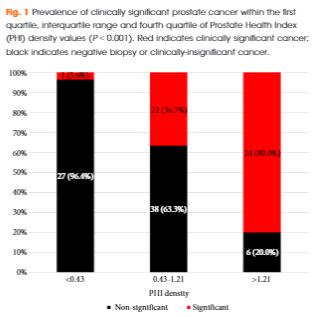
Of 118 men with PHI testing who underwent biopsy, 47 (39.8%) were found to have clinically significant PCa on biopsy. The median (interquartile range [IQR]) PHI density was 0.70 (0.43–1.21), and was 0.53 (0.36–0.75) in men with negative biopsy or clinically insignificant PCa and 1.21 (0.74–1.88) in men with clinically significant PCa (P < 0.001). Clinically significant PCa was detected in 3.6% of men in the first quartile of PHI density (<0.43), 36.7% of men in the IQR of PHI density (0.43–1.21), and 80.0% of men with PHI density >1.21 (P < 0.001). Using a threshold of 0.43, PHI density was 97.9% sensitive and 38.0% specific for clinically significant PCa, and 100% sensitive for Gleason score ≥7 disease. Compared with PSA (area under the curve [AUC] 0.52), PSA density (AUC 0.70), %free PSA (AUC 0.75), the product of %free PSA and prostate volume (AUC 0.79), and PHI (AUC 0.76), PHI density had the highest discriminative ability for clinically significant PCa (AUC 0.84).

Conclusions

Based on the present prospective single-centre experience, PHI density could be used to avoid 38% of unnecessary biopsies, while failing to detect only 2% of clinically significant cancers.

Read more articles of the week

Editorial: Prostate cancer biomarkers: new scenarios in the multi-parametric magnetic resonance imaging era

The management of prostate cancer poses difficult challenges, which is largely because we lack the necessary tools to predict its presence, and discern between indolent disease with a small chance of clinical manifestation and aggressive tumours that are more likely to be lethal.

Despite the fact that novel blood and urine tests are available, which may predict aggressive disease better than PSA; they are not routinely used due to a lack of clinical validity studies.

Tosoian et al. [1] in the present study explored the utility of prostate health index (PHI) density for detection of clinically significant prostate cancer in a contemporary cohort of men presenting for diagnostic evaluation of prostate cancer. Very interestingly the authors hypothesised that, similar to PSA density, PHI density could further improve upon the discriminative ability of PHI to detect prostate cancer. The PHI density calculation was performed using prostate volume, as determined by TRUS. Logistic regression was used to assess the ability of serum markers to predict clinically significant prostate cancer, defined as any Gleason score ≥7 cancer or Gleason score 6 cancer in >2 cores or >50% of any positive core.

They showed, albeit in a small sample size, that PHI density could further improve upon the discriminative ability of PHI and appears to be superior to PSA and other PSA derivatives for the identification of clinically significant disease [1].

However, it is noteworthy that in all studies on urine or serum biomarkers such as this, the ‘gold standard’ for cancer detection is pathological examination of multiple non-targeted systematic TRUS-guided prostate biopsies, not radical prostatectomy specimens. Intrinsically, this approach implies that no cancer predicted by the biomarker may still mean cancer missed by the biopsy.

Introducing mpMRI before prostate biopsy has the potential to improve prostate cancer sampling ink that is the most practical way to make mpMRI before biopsy economically viable for universal NHS adoption.

The aim should be the development of a clinical decision support system based on mpMRI and circulating biomarkers, as in this case PHI density evaluation, to stratify patients according to their risk of prostate cancer progression, using pathological assessment after prostatectomy as the reference standard.

Francesco Porpiglia and Stefano De Luca
Division of Urology, San Luigi Gonzaga Hospital and University of Torino, Orbassano, Italy


Read the full article


References


1 Tosoian JJDruskin SCAndreas D et al. Prostate health index density improves detection of clinically significant prostate cancer. BJU Int2017; 120: 7938.


2 Mottet NBellmunt JBolla M et al. EAU-ESTRO-SIOG guidelines on prostate cancer. Part 1: Screening, diagnosis, and local treatment with curative intent.  Eur Urol 2016; pii: S0302-2838(16)30470-5. [Epub ahead of print]. doi: 10.1016/j.eururo.2016.08.003.


3 Russo FRegge DArmando E et al. Detection of prostate cancer index lesions with multiparametric magnetic resonance imaging (mp-MRI) using whole-mount histological sections as the reference standard. BJU Int 2016; 118: 8494.


5 Porpiglia FManfredi MMele F et al. Diagnostic pathway with multiparametric magnetic resonance imaging versus standard pathway: results from a randomized prospective study in biopsy-naıve patients with suspected prostate cancer. Eur Urol 2016; pii: S0302-2838(16)30509-7. [Epub ahead of print]. doi: 10.1016/j.eururo.2016.08.041


6 Wegelin Ovan Melick HHHooft L et al. Comparing three different techniques for magnetic resonance imaging-targeted prostate biopsies: a systematic review of in-bore versus magnetic resonance imaging- transrectal ultrasound fusion versus cognitive registration. Is there a preferred technique?. Eur Urol 2016; pii: S0302-2838(16)30446-8. [Epub ahead of print]. doi: 10.1016/j.eururo.2016.07.04


Video: Prostate Health Index density improves detection of clinically significant prostate cancer

Prostate Health Index density improves detection of clinically significant prostate cancer

Read the full article

Abstract

Objectives

To explore the utility of Prostate Health Index (PHI) density for the detection of clinically significant prostate cancer (PCa) in a contemporary cohort of men presenting for diagnostic evaluation of PCa.

Patients and Methods

The study cohort included patients with elevated prostate-specific antigen (PSA; >2 ng/mL) and negative digital rectal examination who underwent PHI testing and prostate biopsy at our institution in 2015. Serum markers were prospectively measured per standard clinical pathway. PHI was calculated as ([{−2}proPSA/free PSA] × [PSA]½), and density calculations were performed using prostate volume as determined by transrectal ultrasonography. Logistic regression was used to assess the ability of serum markers to predict clinically significant PCa, defined as any Gleason score ≥7 cancer or Gleason score 6 cancer in >2 cores or >50% of any positive core.

Results

Of 118 men with PHI testing who underwent biopsy, 47 (39.8%) were found to have clinically significant PCa on biopsy. The median (interquartile range [IQR]) PHI density was 0.70 (0.43–1.21), and was 0.53 (0.36–0.75) in men with negative biopsy or clinically insignificant PCa and 1.21 (0.74–1.88) in men with clinically significant PCa (P < 0.001). Clinically significant PCa was detected in 3.6% of men in the first quartile of PHI density (<0.43), 36.7% of men in the IQR of PHI density (0.43–1.21), and 80.0% of men with PHI density >1.21 (P < 0.001). Using a threshold of 0.43, PHI density was 97.9% sensitive and 38.0% specific for clinically significant PCa, and 100% sensitive for Gleason score ≥7 disease. Compared with PSA (area under the curve [AUC] 0.52), PSA density (AUC 0.70), %free PSA (AUC 0.75), the product of %free PSA and prostate volume (AUC 0.79), and PHI (AUC 0.76), PHI density had the highest discriminative ability for clinically significant PCa (AUC 0.84).

Conclusions

Based on the present prospective single-centre experience, PHI density could be used to avoid 38% of unnecessary biopsies, while failing to detect only 2% of clinically significant cancers.

View more videos

Article of the Week: Effect of MetS on serum PSA levels is concealed by enlarged prostate

Every Week the Editor-in-Chief selects an Article of the Week from the current issue of BJUI. The abstract is reproduced below and you can click on the button to read the full article, which is freely available to all readers for at least 30 days from the time of this post.

In addition to the article itself, there is an accompanying editorial written by a prominent member of the urological community. This blog is intended to provoke comment and discussion and we invite you to use the comment tools at the bottom of each post to join the conversation.

Finally, the third post under the Article of the Week heading on the homepage will consist of additional material or media. This week we feature a video discussing the paper.

If you only have time to read one article this week, it should be this one.

Actual lowering effect of metabolic syndrome on serum prostate-specific antigen levels is partly concealed by enlarged prostate: results from a large-scale population-based study

Sicong Zhao*, Ming Xia*, Jianchun Tang† and Yong Yan*


*Department of Urology, and Department of Cardiology, Beijing Shijitan Hospital, Capital Medical University, Beijing, China


Read the full article

Abstract

Objectives

To clarify the lowering effect of metabolic syndrome (MetS) on serum prostate-specific antigen (PSA) levels in a Chinese screened population.

Subjects and Methods

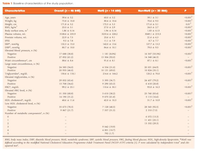
A total of 45 540 ostensibly healthy men aged 55–69 years who underwent routine health check-ups at Beijing Shijitan Hospital between 2008 and 2015 were included in the study. All the men underwent detailed clinical evaluations. PSA mass density was calculated (serum PSA level × plasma volume ÷ prostate volume) for simultaneously adjusting plasma volume and prostate volume. According to the modified National Cholesterol Education Programme–Adult Treatment Panel (NCEP-ATP) III criteria, patients were dichotomized by the presence of MetS, and differences in PSA density and PSA mass density were compared between groups. Linear regression analysis was used to evaluate the effect of MetS on serum PSA levels.

Results

When larger prostate volume in men with MetS was adjusted for, both PSA density and PSA mass density in men with MetS were significantly lower than in men without MetS, and the estimated difference in mean serum PSA level between men with and without MetS was greater than that before adjusting for prostate volume. In the multivariate regression model, the presence of MetS was independently associated with an 11.3% decline in serum PSA levels compared with the absence of MetS. In addition, increasing number of positive MetS components was significantly and linearly associated with decline in serum PSA levels.

Conclusion

The actual lowering effect of MetS on serum PSA levels was partly concealed by the enlarged prostate in men with MetS, and the presence of MetS was independently associated with lower serum PSA levels. Urologists need to be aware of the effect of MetS on serum PSA levels and should discuss this subject with their patients.

Read more articles of the week

Editorial: Anomalous observation with regard to PCa in cancer research

In science, reports showing data deviating from what is expected are called anomalous observations. Metabolic syndrome (MetS) is a promoter of cancer at almost all sites [1]; however, when it comes to prostate cancer (PCa), a series of reports have been published showing an inverse relationship between MetS and its aspects and incident PCa. This lack of coherence in cancer research seriously hampers efforts to fight cancer disorders. It is therefore crucial to find an explanation for this incoherence.

In the search for a reasonable explanation for this anomalous observation, a hypothesis has been formulated, based on the study by Häggström et al. [2], and stating that the PSA-driven diagnostic procedure in PCa, which creates low-stage incident PCa material, is the culprit. The PSA-driven diagnostic procedure introduces several bias mechanisms, which tend to protect men with MetS from being diagnosed with PCa. Thus, men with MetS and its aspects are under-represented in PCa populations generated by PSA-driven diagnostics, thereby creating a distorted incident PCa population. This hypothesis also predicts that high-stage PCa, as well as non-localized and lethal PCa, are not subject to these bias mechanisms, as a minor reduction in the PSA level is of no importance for the PCa diagnosis at these high PSA levels. Finally, the hypothesis predicts that the link between MetS and incident PCa is stage-dependent. A study testing this hypothesis is now in progress.

Several studies have reported that men with MetS had lower PSA levels compared with men without MetS. Zhao et al. [3] address this specific question in this issue of BJUI and confirm that the presence of MetS was independently associated with a lower PSA level and that the enlarged prostate gland, which is an aspect of MetS, partly concealed an even greater PSA level reduction [3]. The findings indicate that a bias mechanism inverses the link between MetS and incident PCa and support the above-mentioned hypothesis.

In short, the following bias mechanisms have been described. MetS is associated with greater body fat with increased aromatase activity, resulting in a reduced testosterone level, which, in turn, is related to a reduced PSA level, as the production of PSA is under androgen control. Another possible bias mechanism, leading to men with MetS being diagnosed less often with PCa, is that these men are more likely to be obese. It is well established that men with a higher BMI also have larger plasma volumes and therefore have greater haemodilution of the PSA production, resulting in a lower PSA level. This means that incident PCa is diagnosed less often in men with MetS, as their PSA level is lower. MetS is also associated with an enlarged prostate gland volume, which means that fewer incident PCas are diagnosed, given the same tumour volume and the same number of biopsies. Another bias mechanism is that a high proportion of men with high socio-economic status undergo PSA testing in the PSA era. It is well established that men with a high socio-economic status have a lower prevalence of MetS and therefore have higher PSA levels, as indicated by the present report in the BJUI [3], and an elevated risk of PCa. Thus, multiple bias mechanisms seem to conceal low-stage PCa in the PSA era.

If it could be confirmed that the negative relationship between MetS and incident PCa is a spurious observation as a result of bias mechanisms, this would open the door for the MetS hypothesis regarding the promotion of multiple cancer disorders. This door has previously been closed by findings in a series of reports of an inverse relationship between MetS and its aspects and incident prostate cancer. Furthermore, this could lead to increased efforts to fight the metabolic aberrations of MetS. It is now well established that MetS and its aspects could be reduced by changes in lifestyle, including physical activity and diet. The most convincing evidence of the effect of diet on MetS comes from studies involving decreased intake of carbohydrates and increased intake of unsaturated fats. Recently, leading authorities in nutrition, endocrinology and metabolism presented a critical review and concluded that carbohydrate restriction is the single most effective intervention to reduce all features of MetS [4]. Another review concluded that carbohydrate restriction is one of the few common interventions that target all features of MetS [5]. This conclusion has recently been confirmed in a meta-analysis by Mansoor et al. [6].

In conclusion, new knowledge challenges the anomalous observation of PCa showing a negative relationship between MetS and PCa. The credibility of the hypothesis that MetS is an important promoting factor for cancer at almost all sites is strengthened. MetS could be treated effectively with a low carbohydrate and high fat diet.

Jan Hammarsten, MD, PhD
Department of Urology, Institute of Clinical SciencesUniversity of Gothenburg, Gothenburg, Sweden


Read the full article


References


1 Esposito K, Chiodini P, Colao AM et al. Metabolic syndrome and risk of cancer. Diabetes Care 2012; 35: 240211 


2Haggstrom C, Stocks T, Ulmert D et al. Prospective study on metabolic factors and risk of prostate cancer. Cancer 2012; 118: 6199206


3 Zhao S, Xia M, Tang J et al. The actual lowering effect of metabolic syndrome on serum prostate-specic antigen levels is partly concealed by enlarged prostate: results from large-scale population-based study. BJU Int 2017; 120: 4829


4 Feinman RD, Pogozelski WK, Astrup A et al. Dietary carbohydrate restriction as the rst approach in diabetes management: critical review and evidence base. Nutrition 2015;31: 113


5 Accurso A, Bernstein RK, Dahlqvist A et al. Dietary carbohydrate restriction in type 2 diabetes mellitus and metabolic syndrome: time for critical appraisal. Nutrition & Metabolism 2008; 5: 9


6 Mansoor N, Vinknes UJ , Veierod MB et al. Effects of low-carbohydrate diets v. low fat diets on body weight and cardiovascular risk factors: meta-analysis of randomized controlled trials. Br J Nutrition 2016; 115: 4667


Video: Effect of MetS on serum PSA levels is concealed by enlarged prostate

Actual lowering effect of metabolic syndrome on serum prostate-specific antigen levels is partly concealed by enlarged prostate: results from a large-scale population-based study

Sicong Zhao*, Ming Xia*, Jianchun Tang† and Yong Yan*


*Department of Urology, and Department of Cardiology, Beijing Shijitan Hospital, Capital Medical University, Beijing, China


Read the full article

Abstract

Objectives

To clarify the lowering effect of metabolic syndrome (MetS) on serum prostate-specific antigen (PSA) levels in a Chinese screened population.

Subjects and Methods

A total of 45 540 ostensibly healthy men aged 55–69 years who underwent routine health check-ups at Beijing Shijitan Hospital between 2008 and 2015 were included in the study. All the men underwent detailed clinical evaluations. PSA mass density was calculated (serum PSA level × plasma volume ÷ prostate volume) for simultaneously adjusting plasma volume and prostate volume. According to the modified National Cholesterol Education Programme–Adult Treatment Panel (NCEP-ATP) III criteria, patients were dichotomized by the presence of MetS, and differences in PSA density and PSA mass density were compared between groups. Linear regression analysis was used to evaluate the effect of MetS on serum PSA levels.

Results

When larger prostate volume in men with MetS was adjusted for, both PSA density and PSA mass density in men with MetS were significantly lower than in men without MetS, and the estimated difference in mean serum PSA level between men with and without MetS was greater than that before adjusting for prostate volume. In the multivariate regression model, the presence of MetS was independently associated with an 11.3% decline in serum PSA levels compared with the absence of MetS. In addition, increasing number of positive MetS components was significantly and linearly associated with decline in serum PSA levels.

Conclusion

The actual lowering effect of MetS on serum PSA levels was partly concealed by the enlarged prostate in men with MetS, and the presence of MetS was independently associated with lower serum PSA levels. Urologists need to be aware of the effect of MetS on serum PSA levels and should discuss this subject with their patients.

Read more articles of the week

Article of the Week: FH as a risk factor for PCa

Every Week the Editor-in-Chief selects an Article of the Week from the current issue of BJUI. The abstract is reproduced below and you can click on the button to read the full article, which is freely available to all readers for at least 30 days from the time of this post.

In addition to the article itself, there is an accompanying editorial written by a prominent member of the urological community. This blog is intended to provoke comment and discussion and we invite you to use the comment tools at the bottom of each post to join the conversation.

If you only have time to read one article this week, it should be this one.

A positive family history as a risk factor for prostate cancer in a population-based study with organised prostate-specific antigen screening: results of the Swiss European Randomised Study of Screening for Prostate Cancer (ERSPC, Aarau)

Marco Randazzo*,, Alexander Muller*, Sigrid Carlsson, Daniel Eberli*, Andreas


Huber, Rainer Grobholz**, Lukas Manka††, Ashkan Mortezavi*, Tullio Sulser*, Franz Recker† and Maciej Kwiatkowski,††


*Department of Urology, University Hospital Zurich, Zurich, Department of Urology, Cantonal Hospital Aarau, Aarau, Switzerland, Department of Surgery (Urology Service), Memorial Sloan Kettering Cancer Center, New York, NY, USA, §Department of Urology, Sahlgrenska Academy at University of Gothenburg, Gothenburg, Sweden, Department of Laboratory Medicine, **Department of Pathology, Cantonal Hospital Aarau, Aarau, Switzerland, and ††Department of Urology, Academic Hospital Braunschweig, Braunschweig, Germany


Read the full article

Objective

To assess the value of a positive family history (FH) as a risk factor for prostate cancer incidence and grade among men undergoing organised prostate-specific antigen (PSA) screening in a population-based study.

Subjects and Methods

The study cohort comprised all attendees of the Swiss arm of the European Randomised Study of Screening for Prostate Cancer (ERSPC) with systematic PSA level tests every 4 years. Men reporting first-degree relative(s) diagnosed with prostate cancer were considered to have a positive FH. Biopsy was exclusively PSA triggered at a PSA level threshold of 3 ng/mL. The primary endpoint was prostate cancer diagnosis. Kaplan–Meier and Cox regression analyses were used.

Results

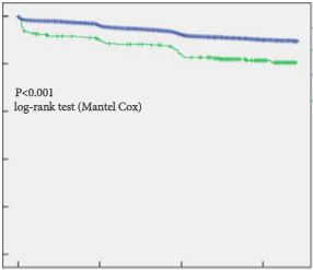
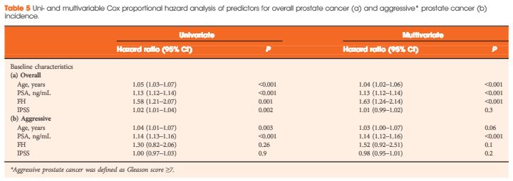
Of 4 932 attendees with a median (interquartile range, IQR) age of 60.9 (57.6–65.1) years, 334 (6.8%) reported a positive FH. The median (IQR) follow-up duration was 11.6 (10.3–13.3) years. Cumulative prostate cancer incidence was 60/334 (18%, positive FH) and 550/4 598 (12%, negative FH) [odds ratio 1.6, 95% confidence interval (CI) 1.2–2.2, P = 0.001). In both groups, most prostate cancer diagnosed was low grade. There were no significant differences in PSA level at diagnosis, biopsy Gleason score or Gleason score on pathological specimen among men who underwent radical prostatectomy between both groups. On multivariable analysis, age (hazard ratio [HR] 1.04, 95% CI 1.02–1.06), baseline PSA level (HR 1.13, 95% CI 1.12–1.14), and FH (HR 1.6, 95% CI 1.24–2.14) were independent predictors for overall prostate cancer incidence (all P < 0.001). Only baseline PSA level (HR 1.14, 95% CI 1.12–1.16, P < 0.001) was an independent predictor of Gleason score ≥7 prostate cancer on prostate biopsy. The proportion of interval prostate cancer diagnosed in-between the screening rounds was not significantly different.

AOTW4Apr

Conclusion

Irrespective of the FH status, the current PSA-based screening setting detects the majority of aggressive prostate cancers and missed only a minority of interval cancers with a 4-year screening algorithm. Our results suggest that men with a positive FH are at increased risk of low-grade but not aggressive prostate cancer.

Read more articles of the week

Editorial: To PSA or not to PSA?

Family history (FH) has long been known to increase a man’s risk of developing prostate cancer (PCa); there is an approximately twofold increased risk with an affected father and a threefold increased risk with an affected brother [1]. Furthermore, FH may increase the risk of more aggressive disease for family members, although results of studies in the PSA screening era have been inconsistent [2, 3]. Using FH as a risk factor, the present analysis of the Swiss arm of the European Randomized Study of Screening for Prostate Cancer (ERSPC) conducted by Randazzo et al. [4] sought to determine whether men with a positive FH of PCa, followed up by PSA screening every 4 years, would have a higher risk of having more aggressive disease.

As expected, the present results show that a significantly higher proportion of men with a FH were diagnosed with PCa compared with those with a negative FH; however, there was no difference in the frequency of more aggressive disease amongst men with a FH, therefore, while FH information can be used to identify men at highest risk of PCa, it does not appear to identify those who are most likely to harbour clinically significant disease.

One reason that a positive FH may not be associated with aggressiveness in the present study is that it has been previously established that PSA screening is associated with a migration towards lower grade and stage disease. It may not be surprising, therefore, that the PSA screened arm may have less aggressive disease. What we ultimately want to understand is whether unscreened men with a FH present with more aggressive disease. This question may have been better addressed by comparing those with a positive FH undergoing screening with those with a positive FH not undergoing screening. Previous studies conducted in this fashion suggested a difference in PCa mortality among men with a positive FH [5]. Unfortunately, these data were not available in the ERSPC. Future studies that continue to evaluate this question may elucidate whether FH predisposes to more aggressive disease.

Another factor that may have impeded the results of the present study is that men aged <55 years were not included in ERSPC. It is possible that FH predisposes to more aggressive cancers earlier in life, in men as young as 40 years. There is evidence that relative risk and risk for early-onset disease increases when a father or brother is diagnosed at a younger age, <60 years [6]. Future studies incorporating this younger cohort of men should therefore be conducted.

Until it has been clarified whether men with a positive FH of PCa are at risk of more aggressive disease, PSA screening strategies that begin at young ages should be used. In this fashion, it will be possible to selectively test populations of men at highest risk of developing PCa. Once these men have been screened, clinicians will be able to distinguish men with aggressive disease from those with more indolent disease. We believe that this shifts the focus to a different question: to treat or not to treat? Future research should continue to focus on methods of identifying those men who will go on to develop aggressive disease. Until then, men with low grade disease should continue to be followed by active surveillance [3].

Read the full article
Christina G. Selkirk* and Brian T. Helfand
*Center for Medical Genetics, Department of Medicine, NorthShore University Health System, and John and Carol Walter Center for Urological Health, Department of Surgery, NorthShor e University HealthSystem, Chicago, IL, USA


References


Article of the Month: ERSPC and PCPT risk calculators in prostate cancer risk prediction

Every Month the Editor-in-Chief selects an Article of the Month from the current issue of BJUI. The abstract is reproduced below and you can click on the button to read the full article, which is freely available to all readers for at least 30 days from the time of this post.

In addition to the article itself, there is an accompanying editorial written by a prominent member of the urological community. This blog is intended to provoke comment and discussion and we invite you to use the comment tools at the bottom of each post to join the conversation.

If you only have time to read one article this week, it should be this one.

Prostate cancer risk prediction using the novel versions of the European Randomised Study for Screening of Prostate Cancer (ERSPC) and Prostate Cancer Prevention Trial (PCPT) risk calculators: independent validation and comparison in a contemporary European cohort

Cedric Poyet, Daan Nieboer*, Bimal Bhindi, Girish S. Kulkarni, Caroline WiederkehrMarian S. Wettstein, Remo Largo, Peter Wild, Tullio Sulser and Thomas Hermanns 


Department of Urology, University Hospital Zurich, University of Zurich, Zurich, Switzerland, *Erasmus MC, University Medical Center Rotterdam, Rotterdam, The Netherlands, Division of Urology, Department of Surgery, University Health Network, University of Toronto, Toronto, ON, Canada, and Institute of Surgical Pathology, University Hospital Zurich, University of Zurich, Zurich, Switzerland


Read the full article

Objectives

To externally validate and compare the two novel versions of the European Randomised Study for Screening of Prostate Cancer (ERSPC)-prostate cancer risk calculator (RC) and Prostate Cancer Prevention Trial (PCPT)-RC.

Patients and Methods

All men who underwent a transrectal prostate biopsy in a European tertiary care centre between 2004 and 2012 were retrospectively identified. The probability of detecting prostate cancer and significant cancer (Gleason score ≥7) was calculated for each man using the novel versions of the ERSPC-RC (DRE-based version 3/4) and the PCPT-RC (version 2.0) and compared with biopsy results. Calibration and discrimination were assessed using the calibration slope method and the area under the receiver operating characteristic curve (AUC), respectively. Additionally, decision curve analyses were performed.

MarchATOM1

Results

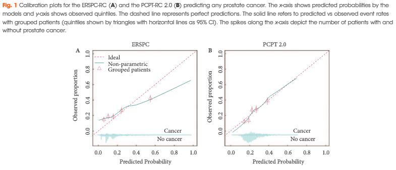
Of 1 996 men, 483 (24%) were diagnosed with prostate cancer and 226 (11%) with significant prostate cancer. Calibration of the two RCs was comparable, although the PCPT-RC was slightly superior in the higher risk prediction range for any and significant prostate cancer. Discrimination of the ERSPC- and PCPT-RC was comparable for any prostate cancer (AUCs 0.65 vs 0.66), while the ERSPC-RC was somewhat better for significant prostate cancer (AUCs 0.73 vs 0.70). Decision curve analyses revealed a comparable net benefit for any prostate cancer and a slightly greater net benefit for significant prostate cancer using the ERSPC-RC.

Conclusions

In our independent external validation, both updated RCs showed less optimistic performance compared with their original reports, particularly for the prediction of any prostate cancer. Risk prediction of significant prostate cancer, which is important to avoid unnecessary biopsies and reduce over-diagnosis and overtreatment, was better for both RCs and slightly superior using the ERSPC-RC.

Read more articles of the week
© 2024 BJU International. All Rights Reserved.