Tag Archive for: Prostate cancer

Posts

Article of the Month: ERSPC and PCPT risk calculators in prostate cancer risk prediction

Every Month the Editor-in-Chief selects an Article of the Month from the current issue of BJUI. The abstract is reproduced below and you can click on the button to read the full article, which is freely available to all readers for at least 30 days from the time of this post.

In addition to the article itself, there is an accompanying editorial written by a prominent member of the urological community. This blog is intended to provoke comment and discussion and we invite you to use the comment tools at the bottom of each post to join the conversation.

If you only have time to read one article this week, it should be this one.

Prostate cancer risk prediction using the novel versions of the European Randomised Study for Screening of Prostate Cancer (ERSPC) and Prostate Cancer Prevention Trial (PCPT) risk calculators: independent validation and comparison in a contemporary European cohort

Cedric Poyet, Daan Nieboer*, Bimal Bhindi, Girish S. Kulkarni, Caroline WiederkehrMarian S. Wettstein, Remo Largo, Peter Wild, Tullio Sulser and Thomas Hermanns 


Department of Urology, University Hospital Zurich, University of Zurich, Zurich, Switzerland, *Erasmus MC, University Medical Center Rotterdam, Rotterdam, The Netherlands, Division of Urology, Department of Surgery, University Health Network, University of Toronto, Toronto, ON, Canada, and Institute of Surgical Pathology, University Hospital Zurich, University of Zurich, Zurich, Switzerland


Objectives

To externally validate and compare the two novel versions of the European Randomised Study for Screening of Prostate Cancer (ERSPC)-prostate cancer risk calculator (RC) and Prostate Cancer Prevention Trial (PCPT)-RC.

Patients and Methods

All men who underwent a transrectal prostate biopsy in a European tertiary care centre between 2004 and 2012 were retrospectively identified. The probability of detecting prostate cancer and significant cancer (Gleason score ≥7) was calculated for each man using the novel versions of the ERSPC-RC (DRE-based version 3/4) and the PCPT-RC (version 2.0) and compared with biopsy results. Calibration and discrimination were assessed using the calibration slope method and the area under the receiver operating characteristic curve (AUC), respectively. Additionally, decision curve analyses were performed.

MarchATOM1

Results

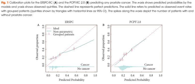
Of 1 996 men, 483 (24%) were diagnosed with prostate cancer and 226 (11%) with significant prostate cancer. Calibration of the two RCs was comparable, although the PCPT-RC was slightly superior in the higher risk prediction range for any and significant prostate cancer. Discrimination of the ERSPC- and PCPT-RC was comparable for any prostate cancer (AUCs 0.65 vs 0.66), while the ERSPC-RC was somewhat better for significant prostate cancer (AUCs 0.73 vs 0.70). Decision curve analyses revealed a comparable net benefit for any prostate cancer and a slightly greater net benefit for significant prostate cancer using the ERSPC-RC.

Conclusions

In our independent external validation, both updated RCs showed less optimistic performance compared with their original reports, particularly for the prediction of any prostate cancer. Risk prediction of significant prostate cancer, which is important to avoid unnecessary biopsies and reduce over-diagnosis and overtreatment, was better for both RCs and slightly superior using the ERSPC-RC.

Editorial: Prostate cancer risk prediction and the persistence of uncertainty

Poyet et al. [1] have performed the largest external validation of the European Randomised Study for Screening of Prostate Cancer (ERSPC) and Prostate Cancer Prevention Trial (PCPT) v2.0 risk calculators (RCs) to date, having retrospectively identified 1996 men undergoing prostate biopsy in a Swiss tertiary care facility.

Asides from the validatory nature of this paper [1], there are several other findings though less novel, which are further important additions to the urological literature.

This study confirms the superior discriminative performance of multi-factorial RCs over PSA alone in the assessment of prostate cancer: where the area under the receiver operating characteristic curve (AUC) for the prediction of significant prostate cancer for PSA alone was 0.65, comparing less favourably than 0.73 and 0.70 for the ERSPC and PCPT v2.0 RCs, respectively.

The authors performed sensitivity analysis showing higher detection rates for prostate cancer (29.4% vs 18.1%) and significant prostate cancer (15.9% vs 5.9%) in patients receiving a 12-core biopsy than in those receiving a 6–8 core biopsy.

Supplementary analysis by the authors evaluated the performance of previous versions of the PCPT-RC, specifically v1.0 and PCPT-RC v1.0 with prostate volume. The inclusion of prostate volume demonstrated an improved predictive ability of this RC. The AUC for the prediction of significant prostate cancer using the PCPT-RC v1.0 with prostate volume was 0.74. This contrasts with the ERSPC risk tool: AUC of 0.73 (which includes a trichotomised estimation of prostate volume), and the novel PCPT-RC v2.0; AUC of 0.70 (which does not include prostate volume as a factor).

The authors conclude that the prediction of significant prostate cancer was superior using the ERSPC-RC compared with the PCPT-RC v2.0, in risk thresholds of 8–35%. Their data also shows that the PCPT-RC v2.0 offers a superior net benefit to the ERPSC-RC to a large number of men outside of this range of threshold probabilities. Their findings suggest that the older PCPT-RC v1.0 with prostate volume may offer benefits superior to both the ERSPC and PCPT v2.0 RCs.

The authors assessment of novel risk tools confirms the rationale for guidelines and consensus statements that PSA testing should not be considered on its own, but rather as part of a multivariate approach [2, 3]. This current work suggests that although calibration of risk tools is still not optimal, they offer superior discriminative ability and superior net benefit in identifying patients with significant prostate cancer. This work affirms the role for variables such as DRE, and the importance of prostate volume in addition to PSA in prostate cancer assessment.

Although further refinement of risk tools is necessary, this work encourages confidence in and should garner further traction for the routine use of such tools in the assessment and counselling of patients before prostate biopsy.

Dara J. Lundon*
*Conway Institute of Biomedical and Biomolecular Science, University College Dublin School of Medicine and Medical Sciences, University College Dublin, Beleld, and Department of Urology, Mater Misericordiae University Hospital, Dublin, Ireland


References


Article of the Week: The impact of PSMs on long-term outcomes after RP

Every Week the Editor-in-Chief selects an Article of the Week from the current issue of BJUI. The abstract is reproduced below and you can click on the button to read the full article, which is freely available to all readers for at least 30 days from the time of this post.

In addition to the article itself, there is an accompanying editorial written by a prominent member of the urological community. This blog is intended to provoke comment and discussion and we invite you to use the comment tools at the bottom of each post to join the conversation.

Finally, the third post under the Article of the Week heading on the homepage will consist of additional material or media. This week we feature a video from Dr. Prabhakar Mithal, discussing his paper.

If you only have time to read one article this week, it should be this one.

Positive Surgical Margins in Radical Prostatectomy Patients Do Not Predict Long-term Oncological Outcomes: Results from SEARCH


Prabhakar Mithal, Lauren E. Howard†‡, William J. Aronson§, Martha K. Terris**††Matthew R. Cooperberg‡‡, Christopher J. Kane§§, Christopher Amling¶¶ and Stephen J. Freedland***


Department of Urology, University of Rochester Medical Center, Rochester, NY, Department of Biostatistics and Bioinformatics, Duke University School of MedicineDivision of Urology, Veterans Affairs Medical Center, Durham, NC, §Urology Section, Department of Surgery, Veterans Affairs Greater Los Angeles Healthcare SystemDepartment of Urology, UCLA School of Medicine, Los Angeles, CA, **Section of Urology, Veterans Affairs Medical Center††Section of Urology, Medical College of Georgia, Augusta, GA, ‡‡Department of Urology, UCSF Helen Diller Family Comprehensive Cancer Center, San Francisco, §§Urology Department, University of California San Diego Health System, San Diego, CA ¶¶Division of Urology, Department of Surgery, Oregon Health and Science University, Portland, OR , and ***Division of Urology, Department of Surgery, Cedars Sinai Medical Center, Los Angeles, CA, USA


Read the full article

Objective

To assess the impact of positive surgical margins (PSMs) on long-term outcomes after radical prostatectomy (RP), including metastasis, castrate-resistant prostate cancer (CRPC), and prostate cancer-specific mortality (PCSM).

Patients and Methods

Retrospective study of 4 051 men in the Shared Equal Access Regional Cancer Hospital (SEARCH) cohort treated by RP from 1988 to 2013. Proportional hazard models were used to estimate hazard ratios (HRs) of PSMs in predicting biochemical recurrence (BCR), CRPC, metastases, and PCSM. To determine if PSMs were more predictive in certain patients, analyses were stratified by pathological Gleason score, stage, and preoperative prostate-specific antigen (PSA) level.

FebAOTW4

Results

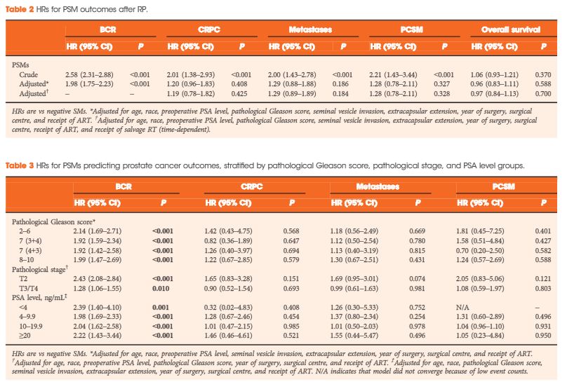
The median (interquartile range) follow-up was 6.6 (3.2–10.6) years and 1 127 patients had >10 years of follow-up. During this time, 302 (32%) men had BCR, 112 (3%) developed CRPC, 144 (4%) developed metastases, and 83 (2%) died from prostate cancer. There were 1 600 (40%) men with PSMs. In unadjusted models, PSMs were significantly associated with all adverse outcomes: BCR, CRPC, metastases and PCSM (all ≤ 0.001). After adjusting for demographic and pathological characteristics, PSMs were associated with increased risk of only BCR (HR 1.98, < 0.001), and not CRPC, metastases, or PCSM (HR ≤1.29, > 0.18). Similar results were seen when stratified by pathological Gleason score, stage, or PSA level, and when patients who underwent adjuvant radiotherapy were excluded.

Conclusions

PSMs after RP are not an independent risk factor for CRPC, metastasis, or PCSM overall or within any subset. In the absence of other high-risk features, PSMs alone may not be an indication for adjuvant radiotherapy.

Editorial: Should we worry about positive surgical margins in prostate cancer?

The debate on the impact of positive surgical margins (PSMs) after radical prostatectomy (RP) continues. The study by Mithal et al. [1] in the present issue contributes further data to an extensive and growing body of literature addressing the clinical significance of a PSM after RP, and ultimately alludes to the question of how to manage these patients.

The authors [1] use the SEARCH database, a large dataset comprised of patients from multiple Veterans Affairs Medical Centers across the USA, to collect and report data on oncological outcomes of PSMs after RP. After adjusting for demographic and pathological confounders, use of adjuvant therapy, and the competing risk of non-prostate-cancer-related death, PSMs were significantly associated with an increased hazard of biochemical recurrence (BCR; hazard ratio 1.99, 95% CI 1.76–2.26), but not castrate resistant prostate cancer (CRPC), metastases, or mortality (prostate-cancer specific or overall). The current study [1], takes such analysis a step further and reports that PSMs were not associated with a negative impact on hard clinical outcomes (CRPC, metastasis, and prostate cancer-specific mortality [PCSM]) in those subgroups at the highest risk of disease progression (high Gleason stage, T-stage, and PSA level). These findings are not necessarily novel, but rather consistent with much of the prior literature. The detrimental impact of PSMs on BCR is well documented [2]. However, the impact of PSMs on hard clinical outcomes such as CRPC, metastasis, and PCSM has not been well demonstrated, despite multiple studies with large cohorts and extensive follow-up.

The negative consequence of a PSM on long-term patient outcome is a very intuitive concept, as it indicates cancer has been inadequately resected or ‘left behind’. Furthermore, the negative impact of a PSM on hard clinical outcomes is well established in many surgically treated malignancies. However, in prostate cancer there remains a disconnect between PSMs and hard clinical outcomes. The authors of the current study [1] provide further evidence to reiterate that the course of this disease after a PSM is not absolute, immediate, or even necessarily concerning (at least in the intermediate follow-up). A PSM after RP may increase the likelihood of BCR but this does not necessarily equal imminent progression and/or death.

PSMs are reported in 10–31% of patients undergoing RP, thus emphasising the clinical importance of this question [3, 4]. Despite the current findings [1], PSMs should not be dismissed. The management of a PSM after RP remains complicated. A proportion of patients will progress and succumb to this disease and, furthermore, therapeutic interventions with shown benefit are available to address such concerns. The decision on how aggressively to manage PSMs may involve an understanding of the patient (comorbidities, lifestyle, and preferences) along with an informed discussion. Furthermore, other pathological details not captured in this study [1] (tumour margin extent and location) [5, 6], along with longer follow-up may be important in identifying drivers for this disease and how to better stratify patients. As with many clinical questions, the approach to PSMs after RP is not necessarily clearly defined and may not apply similarly to all patients across the board. This study [1] may not answer whether or not we should worry about PSMs in prostate cancer, but contributes to our ability to develop clinical algorithms and informed decisions in these patients.

Read the full article


Stanley A. Yap
Department of Urology, University of California Davis Medical Center, 4860 Y Street, Suite 3500, Sacramento, CA, 95817,
USA


References


Video: PSMs in RP patients do not predict long-term oncological outcomes

Positive Surgical Margins in Radical Prostatectomy Patients Do Not Predict Long-term Oncological Outcomes: Results from SEARCH

Prabhakar Mithal, Lauren E. Howard†‡, William J. Aronson§, Martha K. Terris**††Matthew R. Cooperberg‡‡, Christopher J. Kane§§, Christopher Amling¶¶ and Stephen J. Freedland***


Department of Urology, University of Rochester Medical Center, Rochester, NY, Department of Biostatistics and Bioinformatics, Duke University School of MedicineDivision of Urology, Veterans Affairs Medical Center, Durham, NC, §Urology Section, Department of Surgery, Veterans Affairs Greater Los Angeles Healthcare SystemDepartment of Urology, UCLA School of Medicine, Los Angeles, CA, **Section of Urology, Veterans Affairs Medical Center††Section of Urology, Medical College of Georgia, Augusta, GA, ‡‡Department of Urology, UCSF Helen Diller Family Comprehensive Cancer Center, San Francisco, §§Urology Department, University of California San Diego Health System, San Diego, CA ¶¶Division of Urology, Department of Surgery, Oregon Health and Science University, Portland, OR , and ***Division of Urology, Department of Surgery, Cedars Sinai Medical Center, Los Angeles, CA, USA


Read the full article

Objective

To assess the impact of positive surgical margins (PSMs) on long-term outcomes after radical prostatectomy (RP), including metastasis, castrate-resistant prostate cancer (CRPC), and prostate cancer-specific mortality (PCSM).

Patients and Methods

Retrospective study of 4 051 men in the Shared Equal Access Regional Cancer Hospital (SEARCH) cohort treated by RP from 1988 to 2013. Proportional hazard models were used to estimate hazard ratios (HRs) of PSMs in predicting biochemical recurrence (BCR), CRPC, metastases, and PCSM. To determine if PSMs were more predictive in certain patients, analyses were stratified by pathological Gleason score, stage, and preoperative prostate-specific antigen (PSA) level.

FebAOTW4

Results

The median (interquartile range) follow-up was 6.6 (3.2–10.6) years and 1 127 patients had >10 years of follow-up. During this time, 302 (32%) men had BCR, 112 (3%) developed CRPC, 144 (4%) developed metastases, and 83 (2%) died from prostate cancer. There were 1 600 (40%) men with PSMs. In unadjusted models, PSMs were significantly associated with all adverse outcomes: BCR, CRPC, metastases and PCSM (all ≤ 0.001). After adjusting for demographic and pathological characteristics, PSMs were associated with increased risk of only BCR (HR 1.98, < 0.001), and not CRPC, metastases, or PCSM (HR ≤1.29, > 0.18). Similar results were seen when stratified by pathological Gleason score, stage, or PSA level, and when patients who underwent adjuvant radiotherapy were excluded.

Conclusions

PSMs after RP are not an independent risk factor for CRPC, metastasis, or PCSM overall or within any subset. In the absence of other high-risk features, PSMs alone may not be an indication for adjuvant radiotherapy.

Where we are with screening and risk prediction for prostate cancer in 2016

March Editorial ImageThe rate of PSA-based screening over the last 35 years can be compared with driving your car from the Netherlands to Italy. It starts with a rather at drive, perhaps a few hills in the Southern part of the Netherlands, which represents the rate of PSA screening in the late 1980s. Moving with high speed through Germany, one gradually climbs to higher altitudes, i.e. the rate of PSA testing in the 1990s. Then the high (but very difcult to drive) summits and beautiful valleys of Switzerland are there, representing PSA testing practices in the new millennium and the decline in metastatic disease and related mortality [1]. Finally, we descend to Italys Po valley, comparable to PSA testing rates, especially in the USA after the recommendations of the USA Preventive Services Task Force [2,3].
The question is what will we do next? Will we take a left turn and slowly disappear into the sea like Venice? That is, returning to a situation where one out of two or three men died from their prostate cancer? [4] Or will we stop our car, look behind, see the beautiful landscape and return taking the Gotthard road tunnel, avoiding spillage of petrol (i.e. unnecessary PSA testing and potentially harmful prostate biopsies) and go straight to the valleys of Switzerland?
The rst option is obviously not the way to go. Unfortunately, the recommendation to stop the use of the PSA test as a screening tool is direct consequence of the rapid and uncontrolled uptake of the test, often followed by a random biopsy resulting in over-diagnosis and subsequent overtreatment. However, there are ample tools available to turn this situation around and reduce the negative effects of prostate cancer screening [5,6].
An example of such an approach can be found in the publication of Poyet et al. [7] in this issue of BJUI. In this study, the investigators validated updated versions of two multivariate risk-prediction tools, i.e. prostate cancer risk calculators (RCs), in a cohort of 1996 men all biopsied (6-, 8- or 12-core random biopsy) on the basis of an elevated PSA level or abnormal DRE. The data showed that both RCs outperformed the PSA/ DRE-based strategy in reducing unnecessary testing, and in addition avoided over-diagnosis. As said, this approach is one of the many opportunities to reduce the negative aspects of PSA-based screening all summarised in the different guidelines [8]. Reading these guidelines, it soon becomes clear that it is known that repeatedly testing men with low PSA levels is useless. It is known that screening men with a limited life expectancy will only cause harm, and that simply repeating a prostate biopsy after a negative biopsy result (i.e. no prostate cancer detected) is not the way to go. And yet, this is what we see happening in daily clinical practice [9,10].
So, where are we with prostate cancer screening and risk prediction in 2016? We are in a situation that we know that we can reduce suffering and death from (metastatic) prostate cancer, with early detection and treatment, but that we have to selectively identify men that can actually benet. The latter is realistic if we start to implement the knowledge we have acquired over recent decades.


Monique J. Roobol
Department of Urology, Erasmus University Medical Center, Rotterdam, The Netherlands


References


3 Banerji JS, Wolff EM, Massman JD 3rd, Odem-Davis K, Porter CR, Corman JMProstate Needle Biopsy Outcomes in the Era of the U.S. Preventive Services Task Force Recommendation against Prostate Specic Antigen Based Screening. J Urol 2016; 195: 6673


4 Hsing AW, Tsao L, Devesa SS. International trends and patterns of prostate cancer incidence and mortality. Int J Cancer 2000; 85: 607


5 Roobol MJ, Carlsson SV. Risk stratication in prostate cancer screening. Nat Rev Urol 2013; 10: 3848


8 Loeb S. Guideline of guidelines: prostate cancer screening. BJU Int 2014; 114: 3235


Correction: The word “their” was added to this sentence to clarify its meaning: “That is, returning to a situation where one out of two or three men died from their prostate cancer? [4]”


Video: Step-By-Step: Extended PLND – Creating the Spaces

Sequencing robot-assisted extended pelvic lymph node dissection prior to radical prostatectomy: a step-by-step guide to exposure and efficiency

Stephen B. Williams, Yasar Bozkurt , Mary Achim, Grace Achim and John W. Davis


Department of Urology, The University of Texas MD Anderson Cancer Center, Houston, TX, USA


Read the full article
OBJECTIVE

To describe a novel, step-by-step approach to robot-assisted extended pelvic lymph node dissection (ePLND) at the time of robot-assisted radical prostatectomy (RARP) for intermediate–high risk prostate cancer.

PATIENTS AND METHODS

The sequence of ePLND is at the beginning of the operation to take advantage of greater visibility of the deeper hypogastric planes. The urachus is left intact for an exposure/retraction point. The anatomy is described in terms of lymph nodes (LNs) that are easily retrieved vs those that require additional manipulation of the anatomy, and a determined surgeon. A representative cohort of 167 RARPs was queried for representative metrics that distinguish the ePLND: 146 primary cases and 21 with neoadjuvant systemic therapy.

RESULTS

The median (interquartile range, IQR) LN yield was 22 (16–28) for primary surgeries and 21 (16–23) for neoadjuvant cases. The percentage of cases with positive LNs (pN1) was 16.4% for primary and 29% for neoadjuvant. The hypogastric LNs were involved in 75% of pN1 primary cases and uniquely positive in 33%. Each side of ePLND took the attending surgeon a median (IQR) of 16 (13–20) min and trainees 25 (24–38) min.

CONCLUSIONS

Robot-assisted ePLND before RARP provides an anatomical approach to surgical extirpation mimicking the open approach. We think this sequence offers efficiency and efficacy advantages in high-risk and select intermediate-risk patients with prostate cancer undergoing RARP.


Article of the Week: Combination of mpMRI and TTMB of the prostate to identify candidates for hemi-ablative FT

Every Week the Editor-in-Chief selects an Article of the Week from the current issue of BJUI. The abstract is reproduced below and you can click on the button to read the full article, which is freely available to all readers for at least 30 days from the time of this post.

In addition to the article itself, there is an accompanying editorial written by a prominent member of the urological community. This blog is intended to provoke comment and discussion and we invite you to use the comment tools at the bottom of each post to join the conversation.

Finally, the third post under the Article of the Week heading on the homepage will consist of additional material or media. This week we feature a video from Mr. Mark Emberton, discussing his paper. 

If you only have time to read one article this week, it should be this one.

Combination of multi-parametric magnetic resonance imaging (mp-MRI) and transperineal template-guided mapping biopsy (TTMB) of the prostate to identify candidates for hemi-ablative focal therapy

Minh Tran*†‡, James Thompson*§, Maret Bohm†, Marley Pulbrook, Daniel Moses¶, Ron Shnier**, Phillip Brenner*§, Warick Delprado††, Anne-Maree Haynes†, Richard Savdie§ and Phillip D. Stricker*§


*St Vincents Prostate Cancer CentreGarvan Institute of Medical Research & The Kinghorn Cancer Centre, DarlinghurstSchool of Medicine, University of Sydney§School of Medicine, University of New South Wales, SydneySpectrum Medical Imaging , **Southern Radiology, Randwick, and†† Douglass Hanly Moir Pathology, Darlinghurst, NSW, Australia


Read the full article
OBJECTIVE

To evaluate the accuracy of combined multiparametric magnetic resonance imaging (mpMRI) and transperineal template-guided mapping biopsy (TTMB) for identifying lobes with significant prostate cancer (PCa) for the application of hemi-ablative focal therapy (FT).

PATIENTS AND METHODS

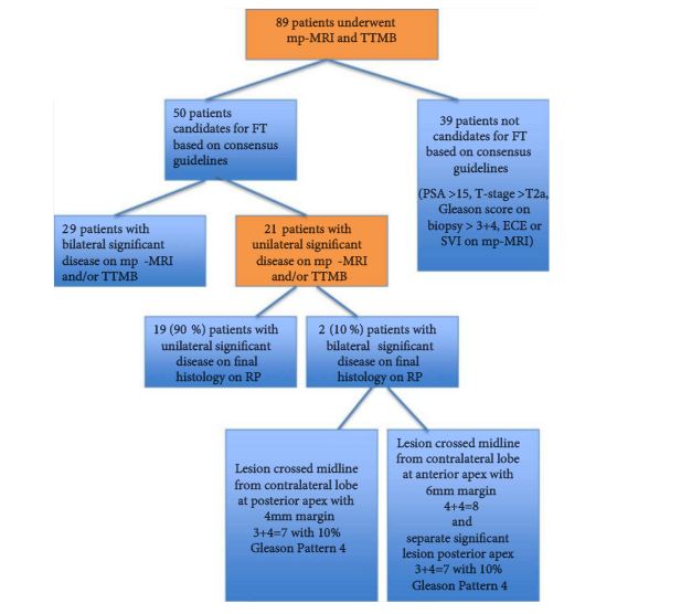
From January 2012 to January 2014, 89 consecutive patients, aged ≥40 years, with a PSA level ≤15 ng/mL, underwent in sequential order: mpMRI, TTMB and radical prostatectomy (RP) at a single centre. Analysis was performed on 50 patients who met consensus guidelines for FT. Lobes were stratified into lobes with significant cancer (LSC), lobes with insignificant cancer and lobes with no cancer. Using histopathology at RP, the predictive performance of combined mpMRI + TTMB in identifying LSC was evaluated.

RESULTS

The sensitivity, specificity and positive predictive value for mpMRI + TTMB for LSC were 97, 61 and 83%, respectively. The negative predictive value (NPV), the primary variable of interest, for mpMRI + TTMB for LSC was 91%. Of the 50 patients, 21 had significant unilateral disease on mpMRI + TTMB. Two of these 21 patients had significant bilateral disease on RP not identified on mpMRI + TTMB.

CONCLUSIONS

In the selection of candidates for FT, a combination of mpMRI and TTMB provides a high NPV in the detection of LSC.

Editorial: Doubling our precision of risk stratification in early prostate cancer? Too good to be true?

It is difficult to over-estimate the enormity of the revolution involved in transitioning away from an organ-based system of care in prostate cancer (prostatectomy or radiotherapy to prostate cancer of any grade, number, location or volume) to one in which the target condition is defined and verified, and then subsequently treated with a margin. Since Hugh Hampton-Young undertook the first radical prostatectomy more than 100 years ago, an organ-based system of care has been the only plausible strategy for men with early-stage prostate cancer. This is because our risk stratification methods could certainly tell us who had prostate cancer when it was indeed identified, but they could not tell us with any degree of precision how much cancer was present (number of foci and volume of cancer), what grade it was and where within the prostate the cancer resided. Surgery or radiotherapy to the whole gland, it was thought, was a reasonable mitigation (at the cost of over-treatment in a significant proportion of men) to the systematic under-estimation of risk to the patient. The degree to which our attempts at risk stratification failed our patients was emphasized in a recent UK report that showed an increase of >50% in histological grade (the most important determinant of risk) at radical prostatectomy compared with the risk the patient was told at the time of diagnosis[1]. ‘Upgrading’ is an inverse measure of the quality of our risk stratification.

With this background it should be of little surprise that there remains considerable skepticism about our ability to localize disease within the prostate, define its volume with some degree of precision and identify the worst histological grade within the tumour most of the time. These conditions, which most patients, quite reasonably, might expect of a modern cancer diagnostic programme, need to be fulfilled if we are to move away from an organ-based strategy for all towards a system of care that risk stratifies with precision, treats only those who are likely to benefit and tries to preserve tissue and function when we are able to do so.

The technologies and developments that have permitted a reduction in risk stratification error from 50 to ~5% are the subject of a paper by Tran et al. [2] in the present issue of BJUI. This group from Australia exploited a cohort of men that underwent a series of tests that culminated in a radical prostatectomy. These comprised: high-quality multiparametric MRI at a range of magnet strengths; formal scoring of the MRI using an ordinal scale of risk; a subsequent 5-mm transperineal template-guided biopsy modified from Winston Barzell’s original account; and additional sampling of prostate sectors that corresponded to a high likelihood of clinically significant cancer based on the MRI reading, a process otherwise known as ‘targeting’. The outputs of these tests were compared with the presence or absence of clinically significant prostate cancer in the 100 prostate lobes evaluated from the 50 eligible men who underwent surgery. The authors’ a priori threshold for declaring clinically significant disease in the tests was the presence or a PIRADS 4–5 MRI lesion (with or without concordant pathology) and/or the presence of exclusive Gleason pattern 3 amounting to ≥4 mm maximum cancer core length or the presence of any Gleason pattern 4 or 5 [3]. At radical prostatectomy slightly different criteria were employed. Clinically significant prostate cancer constituted an exclusive Gleason pattern 3 lesion provided it was ≥1.3 mL. The presence of patterns 4 and 5 within the lesion triggered ‘significance’, as did evidence of capsular invasion or extraprostatic extension.

Although it was a small study, the men included were exposed to the best diagnostic profile that it was possible to have and were subjected to a reference test which most of us would trust. What did they find? Just how well did a modern diagnostic panel rule in or rule out prostate cancer that exceeded a minimum threshold of 4 mm of Gleason pattern 3? Within the hundred lobes that were evaluated, 21 clinically significant cancers were identified in the diagnostic process. At radical prostatectomy two of these were at the midline and therefore attributed, within the rules, to both sides of the gland. In one of these cases there was a 5-mm diameter (0.1 mL) Gleason 3+4 lesion with 10% Gleason pattern 4 that was overlooked by the combined diagnostic process. A lesion of this volume can evade a well-applied sampling strategy based on a 5-mm sampling frame, especially if the lesion is non-spherical. The relatively low component of pattern 4 combined with the relatively low volume means that MRI would also have a hard time detecting it. Under the conditions described in the present paper, the authors concluded that combined MRI and intensive biopsy conferred a sensitivity of 97% and a negative predictive value of 91% for a fairly conservative definition of clinically significant disease. This level of accuracy, which is nearly twice as good as that of which we were previously capable, will result in major benefits for patients in terms of communicating risk with an order of precision that was hitherto not possible. This should translate to more appropriate treatment allocation, both avoiding unnecessary treatment and having treatment when it is likely to be beneficial. It should also result in a significant proportion of patients being offered the option of a tissue-preserving therapy when this is an option [4]. Most importantly, this new precision opens up an opportunity for greater involvement of the patient in the process of informed decision-making [5]. Something that was not really possible in the face of yesterday’s diagnostic uncertainty.

Read the full article
Mark Emberton
Division of Surgery and Interventional Science, University College London, London, UK


References


Video: Combination of mpMRI and TTMB of the prostate to identify candidates for hemi-ablative FT

Combination of multi-parametric magnetic resonance imaging (mp-MRI) and transperineal template-guided mapping biopsy (TTMB) of the prostate to identify candidates for hemi-ablative focal therapy

Minh Tran*†‡, James Thompson*§, Maret Bohm†, Marley Pulbrook, Daniel Moses¶, Ron Shnier**, Phillip Brenner*§, Warick Delprado††, Anne-Maree Haynes†, Richard Savdie§ and Phillip D. Stricker*§


*St Vincents Prostate Cancer CentreGarvan Institute of Medical Research & The Kinghorn Cancer Centre, DarlinghurstSchool of Medicine, University of Sydney§School of Medicine, University of New South Wales, SydneySpectrum Medical Imaging , **Southern Radiology, Randwick, and†† Douglass Hanly Moir Pathology, Darlinghurst, NSW, Australia


Read the full article
OBJECTIVE

To evaluate the accuracy of combined multiparametric magnetic resonance imaging (mpMRI) and transperineal template-guided mapping biopsy (TTMB) for identifying lobes with significant prostate cancer (PCa) for the application of hemi-ablative focal therapy (FT).

PATIENTS AND METHODS

From January 2012 to January 2014, 89 consecutive patients, aged ≥40 years, with a PSA level ≤15 ng/mL, underwent in sequential order: mpMRI, TTMB and radical prostatectomy (RP) at a single centre. Analysis was performed on 50 patients who met consensus guidelines for FT. Lobes were stratified into lobes with significant cancer (LSC), lobes with insignificant cancer and lobes with no cancer. Using histopathology at RP, the predictive performance of combined mpMRI + TTMB in identifying LSC was evaluated.

RESULTS

The sensitivity, specificity and positive predictive value for mpMRI + TTMB for LSC were 97, 61 and 83%, respectively. The negative predictive value (NPV), the primary variable of interest, for mpMRI + TTMB for LSC was 91%. Of the 50 patients, 21 had significant unilateral disease on mpMRI + TTMB. Two of these 21 patients had significant bilateral disease on RP not identified on mpMRI + TTMB.

CONCLUSIONS

In the selection of candidates for FT, a combination of mpMRI and TTMB provides a high NPV in the detection of LSC.

© 2024 BJU International. All Rights Reserved.