Tag Archive for: prognosis

Posts

Video: Prostate cancer in kidney transplant recipients – a nationwide register study

:

Prostate cancer in kidney transplant recipients – a nationwide register study

Abstract

Objective

To investigate whether post‐transplantation immunosuppression negatively affects prostate cancer outcomes in male kidney transplant recipients.

Patients and Methods

We used the Swedish Renal Register and the National Prostate Cancer Register to identify all kidney transplantation recipients diagnosed with prostate cancer in Sweden 1998–2016. After linking these registers with Prostate Cancer Database Sweden (PCBaSe), a case‐control study was designed to compare time period and risk category‐specific probabilities of a prostate cancer diagnosis amongst kidney transplantation recipients versus the male general population. The registers did not include information about the specific immunosuppression agent used in all transplantation recipients. Data from PCBaSe were used to compare prostate cancer characteristics at diagnosis and survival for patients with prostate cancer with versus without a kidney transplant. Propensity score matching, Cox regression analysis and Fisher’s exact test were used and 95% confidence intervals (CIs) calculated.

Results

Almost half of the 133 kidney transplantation recipients were transplanted before the mid‐1990s, when PSA testing became common. The transplant recipients were not more likely than age‐matched control men to be diagnosed with any (odds ratio [OR] 0.84, 95% CI 0.70–0.99) or high‐risk or metastatic prostate cancer (OR 0.84, 95% CI 0.62–1.13). None of the ORs for the different categories of prostate cancer increased with time since transplantation. Cancer characteristics at the time of diagnosis and cancer‐specific survival were similar amongst transplant recipients and the control group of 665 men diagnosed with prostate cancer without a kidney transplant.

Conclusions

This Swedish nationwide, register‐based study gave no indication that immunosuppression after kidney transplantation increases the risk of prostate cancer or adversely affects prostate cancer outcomes. The study suggests that men with untreated low‐grade prostate cancer can be accepted for transplantation.

Article of the week: Targeted deep sequencing of urothelial bladder cancers and associated urinary DNA: a 23‐gene panel with utility for non‐invasive diagnosis and risk stratification

Every week, the Editor-in-Chief selects an Article of the Week from the current issue of BJUI. The abstract is reproduced below and you can click on the button to read the full article, which is freely available to all readers for at least 30 days from the time of this post.

In addition to the article itself, there is an editorial written by a prominent member of the urological community and a video prepared by the authors. These are intended to provoke comment and discussion and we invite you to use the comment tools at the bottom of each post to join the conversation. 

If you only have time to read one article this week, it should be this one.

Targeted deep sequencing of urothelial bladder cancers and associated urinary DNA: a 23‐gene panel with utility for non‐invasive diagnosis and risk stratification

Douglas G. Ward*, Naheema S. Gordon*, Rebecca H. Boucher*, Sarah J. Pirrie*, Laura Baxter, Sascha Ott, Lee Silcock, Celina M. Whalley*, Joanne D. Stockton*, Andrew D. Beggs*, Mike Griffiths§, Ben Abbotts*, Hanieh Ijakipour*, Fathimath N.Latheef*, Robert A. Robinson*, Andrew J. White*, Nicholas D. James*, Maurice P.Zeegers, K. K. Cheng** and Richard T. Bryan*


*Institute of Cancer and Genomic Sciences, University of Birmingham, Birmingham, Department of Computer Science, University of Warwick, Coventry, Nonacus Limited, Birmingham Research Park, §West Midlands Regional Genetics Laboratory, Birmingham Women’s and Children’s NHS Foundation Trust, Birmingham, UK, NUTRIM School for Nutrition and Translational Research in Metabolism and CAPHRI Care and Public Health Research Institute, Maastricht University, Maastricht, The Netherlands and **Institute of Applied Health Research, University of Birmingham, Birmingham, UK

Read the full article

Abstract

Objectives

To develop a focused panel of somatic mutations (SMs) present in the majority of urothelial bladder cancers (UBCs), to investigate the diagnostic and prognostic utility of this panel, and to compare the identification of SMs in urinary cell‐pellet (cp) DNA and cell‐free (cf) DNA as part of the development of a non‐invasive clinical assay.

Patients and Methods

A panel of SMs was validated by targeted deep‐sequencing of tumour DNA from 956 patients with UBC. In addition, amplicon and capture‐based targeted sequencing measured mutant allele frequencies (MAFs) of SMs in 314 urine cpDNAs and 153 urine cfDNAs. The association of SMs with grade, stage and clinical outcomes was investigated by univariate and multivariate Cox models. Concordance between SMs detected in tumour tissue and cpDNA and cfDNA was assessed.


Results

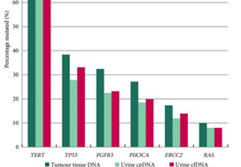
The panel comprised SMs in 23 genes: TERT (promoter), FGFR3, PIK3CA, TP53, ERCC2, RHOB, ERBB2, HRAS, RXRA, ELF3, CDKN1A, KRAS, KDM6A, AKT1, FBXW7, ERBB3, SF3B1, CTNNB1, BRAF, C3orf70, CREBBP, CDKN2A and NRAS; 93.5–98.3% of UBCs of all grades and stages harboured ≥1 SM (mean: 2.5 SMs/tumour). RAS mutations were associated with better overall survival (P = 0.04). Mutations in RXRA, RHOB and TERT (promoter) were associated with shorter time to recurrence (P < 0.05). MAFs in urinary cfDNA and cpDNA were highly correlated; using a capture‐based approach, >94% of tumour SMs were detected in both cpDNA and cfDNA.

Conclusions

SMs are reliably detected in urinary cpDNA and cfDNA. The technical capability to identify very low MAFs is essential to reliably detect UBC, regardless of the use of cpDNA or cfDNA. This 23‐gene panel shows promise for the non‐invasive diagnosis and risk stratification of UBC.

Read more Articles of the week


Editorial: Non‐invasive diagnosis and monitoring of urothelial bladder cancer: are we there yet?

In this issue of BJUI, Ward et al. [1] describe the development of DNA‐based urinary biomarkers for urothelial carcinoma (UC). The genomics of UC have been well characterized through interrogation of tumour issues in institutional series (e.g. the Memorial Sloan Kettering Cancer Center [MSKCC] experience), multi‐institutional collaborations (e.g. The Cancer Genome Atlas [TCGA]) and commercial platforms (e.g. the Foundation Medicine experience) [2]. Until recently, these have been largely academic pursuits, with possible impact on prognostication but limited clinical applicability and utility for therapy selection and monitoring of response; however, with the US Food and Drug Administration approval of erdafitinib several weeks ago, patients with advanced UC will routinely receive genomic assessment for FGFR2/3 mutation or fusion, the targets for this therapy [3]. In due time, it is anticipated that multiple other putative targets with associated therapies (e.g. ERBB2, CDKN2A), as well as potential predictive biomarkers, may also warrant testing.

The evolving landscape in advanced UC makes a non‐invasive biomarker particularly attractive. The authors of the present commentary have previously reported results from a series of 369 patients with advanced UC, demonstrating that genomic alterations in ctDNA could be identified in 91% of patients using a commercially available 73-gene panel [4]. More recently, Christensen et al. [5] assessed a cohort of 68 patients receiving neoadjuvant chemotherapy for muscle‐invasive disease, demonstrating 100% sensitivity and 98% specificity for the detection of relapsed disease with a patient‐specific ctDNA assessment (sequenced to a median target coverage of 105 000×) after cystectomy. Impressively, the data also showed that the dynamics of ctDNA appeared to be more useful than pathological downstaging in predicting relapse.

In contrast to these studies, Ward et al. have developed a 23‐gene panel based on frequently expressed genes in a cohort of 916 UC tissue specimens, largely derived from patients with non‐muscle‐invasive disease. Ultimately, with a cohort of 314 patients with DNA derived from a urinary cell pellet, sequencing identified 645 (71.4%) of 903 mutations detected in tumour. Using urinary supernatant, 353 (80.7%) of 437 mutations were detected. These relatively high sensitivities, if they can be interpreted as such, are promising but do not rise to the level of replacing existing strategies for UC detection, staging and monitoring. Notably, another study demonstrated that urinary ctDNA can be detected with high sensitivity and specificity in patients with localized early‐stage bladder cancer and for after‐treatment surveillance, providing the foundation for further studies evaluating the role of ctDNA in non‐invasive detection, genotyping and monitoring [6].

Beyond its use as a diagnostic tool, it is hoped that urinary ctDNA may also find applications in the selection of therapeutics. To this end, Ward et al. identified FGFR3, PIK3CA, ERCC2 and ERBB2 mutations in 45%, 32%, 14% and 7% of patients, respectively. The frequency of FGFR3 alteration decreased with increasing stage and grade, ranging from 72% in pTaG1 disease to just 13% in ≥pT2 disease, consistent with other reports [7]. These results may guide forthcoming studies evaluating FGFR inhibitors in non‐muscle‐invasive, muscle‐invasive and metastatic disease, where studies are ongoing. In reviewing the potential link between genomic alterations and clinical outcomes, perhaps the most curious finding is that between RAS mutations and improved overall survival (P = 0.04), the only such association found in multivariate analysis. These results stand in sharp contrast to reports in lung cancer, colorectal cancer and multiple other tumour types [8]. A closer look at the deleterious nature and functional impact of NRAS and KRAS mutations seen in this series is certainly warranted, along with further external validation in a more homogenous and larger patient population. There is also the potential application of monitoring treatment response by assessing eradication of urinary ctDNA, a hypothesis that is being evaluated in ongoing studies [9].

How will the results of this and other emerging urinary biomarker studies eventually make their way to the clinic? The answer is simple: incorporation of these biomarkers in prospective therapeutic trials. As the bladder cancer investigative community formulates novel trials for non‐muscle‐invasive and muscle‐invasive disease using targeted therapies, an excellent opportunity exists to correlate urinary, blood and tissue‐based biomarkers and to assess their relative predictive capabilities and clinical utility. Furthermore, with clinical surrogate endpoints likely to drive regulatory approval (e.g. landmark complete response rates for non‐muscle‐invasive disease, or pT0N0 rate for muscle‐invasive disease), a validated urinary biomarker could ultimately offer an alternative biological surrogate endpoint [10]. In an era of genomic revolution, prospective validation can help establish the potential clinical utility of promising biomarkers and help realize the dream of ‘precision oncology’.

by Rohit K. Jain, Petros Grivas and Sumanta K. Pal

References

  1. Ward DGGordon NSBoucher RH et al. Targeted deep sequencing of urothelial bladder cancers and associated urinary DNA: a 23‐gene panel with utility for non‐invasive diagnosis and risk stratification. BJU Int 2019
  2. Schiff JPBarata PCYu EYGrivas PPrecision therapy in advanced urothelial cancer. Expert Rev Precis Med Drug Dev 2019481– 93
  3. FDA grants accelerated approval to erdafitinib for metastatic urothelial carcinoma [press release] 2019.
  4. Agarwal NPal SKHahn AW et al. Characterization of metastatic urothelial carcinoma via comprehensive genomic profiling of circulating tumor DNA. Cancer 20181242115– 24
  5. Christensen EBirkenkamp‐Demtroder KSethi H et al. Early detection of metastatic relapse and monitoring of therapeutic efficacy by ultra‐deep sequencing of plasma cell‐free DNA in patients with urothelial bladder carcinoma. J Clin Oncol 2019371547– 57
  6. Dudley JCSchroers‐Martin JLazzareschi DV et al. Detection and surveillance of bladder cancer using urine tumor DNA. Cancer Discov 20199500– 9
  7. Tomlinson DCBaldo OHarnden PKnowles MAFGFR3 protein expression and its relationship to mutation status and prognostic variables in bladder cancer. J Pathol 200721391– 8
  8. Zhuang RLi SLi Q et al. The prognostic value of KRAS mutation by cell‐free DNA in cancer patients: a systematic review and meta‐analysis. PLoS One 201712e0182562
  9. Abbosh PHPlimack ERMolecular and clinical insights into the role and significance of mutated DNA repair genes in bladder cancer. Bladder Cancer 201849– 18
  10. Jarow JPLerner SPKluetz PG et al. Clinical trial design for the development of new therapies for nonmuscle‐invasive bladder cancer: report of a Food and Drug Administration and American Urological Association public workshop. Urology 201483262– 4


Video: Targeted deep sequencing of urothelial bladder cancers and associated urinary DNA

Targeted deep sequencing of urothelial bladder cancers and associated urinary DNA: a 23‐gene panel with utility for non‐invasive diagnosis and risk stratification

Read the full article

Abstract

Objectives

To develop a focused panel of somatic mutations (SMs) present in the majority of urothelial bladder cancers (UBCs), to investigate the diagnostic and prognostic utility of this panel, and to compare the identification of SMs in urinary cell‐pellet (cp) DNA and cell‐free (cf) DNA as part of the development of a non‐invasive clinical assay.

Patients and Methods

A panel of SMs was validated by targeted deep‐sequencing of tumour DNA from 956 patients with UBC. In addition, amplicon and capture‐based targeted sequencing measured mutant allele frequencies (MAFs) of SMs in 314 urine cpDNAs and 153 urine cfDNAs. The association of SMs with grade, stage and clinical outcomes was investigated by univariate and multivariate Cox models. Concordance between SMs detected in tumour tissue and cpDNA and cfDNA was assessed.

Results

The panel comprised SMs in 23 genes: TERT (promoter), FGFR3, PIK3CA, TP53, ERCC2, RHOB, ERBB2, HRAS, RXRA, ELF3, CDKN1A, KRAS, KDM6A, AKT1, FBXW7, ERBB3, SF3B1, CTNNB1, BRAF, C3orf70, CREBBP, CDKN2A and NRAS; 93.5–98.3% of UBCs of all grades and stages harboured ≥1 SM (mean: 2.5 SMs/tumour). RAS mutations were associated with better overall survival (P = 0.04). Mutations in RXRA, RHOB and TERT (promoter) were associated with shorter time to recurrence (P < 0.05). MAFs in urinary cfDNA and cpDNA were highly correlated; using a capture‐based approach, >94% of tumour SMs were detected in both cpDNA and cfDNA.

Conclusions

SMs are reliably detected in urinary cpDNA and cfDNA. The technical capability to identify very low MAFs is essential to reliably detect UBC, regardless of the use of cpDNA or cfDNA. This 23‐gene panel shows promise for the non‐invasive diagnosis and risk stratification of UBC.


View more videos

Article of the week: The World Health Organization 1973 classification system for grade is an important prognosticator in T1 non‐muscle‐invasive bladder cancer

Every week, the Editor-in-Chief selects an Article of the Week from the current issue of BJUI. The abstract is reproduced below and you can click on the button to read the full article, which is freely available to all readers for at least 30 days from the time of this post.


In addition to the article itself, there is an accompanying editorial written by a prominent member of the urological community. These are intended to provoke comment and discussion and we invite you to use the comment tools at the bottom of each post to join the conversation.

If you only have time to read one article this week, it should be this one.

The World Health Organization 1973 classification system for grade is an important prognosticator in T1 non‐muscle‐invasive bladder cancer

Elisabeth E. Fransen van de Putte*, Judith Bosschieter*, Theo H. van der Kwast§, Simone Bertz, Stefan Denzinger**, Quentin Manach††, Eva M. Compérat‡‡,
Joost L. Boormans§§, Michael A.S. Jewett¶¶, Robert Stoehr, Geert J.L.H. van Leenders§, Jakko A. Nieuwenhuijzen, Alexandre R. Zlotta¶¶***, Kees Hendricksen*,
Morgan Rouprêt††, Wolfgang Otto**, Maximilian Burger**, Arndt Hartmannand Bas W.G. van Rhijn***§§¶¶

*Department of Surgical Oncology (Urology), Netherlands Cancer Institute, Antoni van Leeuwenhoek Hospital, Department of Urology, VU University Medical Centre, Amsterdam, §Department of Pathology, §§Department of Urology, Erasmus MC Cancer Institute, Erasmus MC, Rotterdam, The Netherlands, Department of Pathology, ¶¶Department of Surgical Oncology (Urology), Princess Margaret Cancer Center, University Health Network, ***Department of Urology, Mount Sinai Hospital, University of Toronto, Toronto, ON, Canada, Department of Pathology, University of Erlangen, Erlangen, **Department of Urology, Caritas St. Josef Medical Centre, University of Regensburg, Regensburg, Germany, ††Academic Department of Urology and ‡‡Department of Pathology, Pitie-Salpétrière Hospital, Assistance-Publique pitaux de Paris, Pierre et Marie Curie Medical School, University Paris, Paris, France

Read the full article

Abstract

Objectives

To compare the prognostic value of the World Health Organization (WHO) 1973 and 2004 classification systems for grade in T1 bladder cancer (T1‐BC), as both are currently recommended in international guidelines.

Patients and Methods

Three uro‐pathologists re‐revised slides of 601 primary (first diagnosis) T1‐BCs, initially managed conservatively (bacille Calmette–Guérin) in four hospitals. Grade was defined according to WHO1973 (Grade 1–3) and WHO2004 (low‐grade [LG] and high‐grade [HG]). This resulted in a lack of Grade 1 tumours, 188 (31%) Grade 2, and 413 (69%) Grade 3 tumours. There were 47 LG (8%) vs 554 (92%) HG tumours. We determined the prognostic value for progression‐free survival (PFS) and cancer‐specific survival (CSS) in Cox‐regression models and corrected for age, sex, multiplicity, size and concomitant carcinoma in situ.

Results

At a median follow‐up of 5.9 years, 148 patients showed progression and 94 died from BC. The WHO1973 Grade 3 was negatively associated with PFS (hazard ratio [HR] 2.1) and CSS (HR 3.4), whilst WHO2004 grade was not prognostic. On multivariable analysis, WHO1973 grade was the only prognostic factor for progression (HR 2.0). Grade 3 tumours (HR 3.0), older age (HR 1.03) and tumour size >3 cm (HR 1.8) were all independently associated with worse CSS.

Conclusion

The WHO1973 classification system for grade has strong prognostic value in T1‐BC, compared to the WHO2004 system. Our present results suggest that WHO1973 grade cannot be replaced by the WHO2004 classification in non‐muscle‐invasive BC guidelines.


Read more Articles of the week


Editorial: Predicting progression in T1 non‐muscle‐invasive bladder cancer: back to histology

Stage pT1 bladder carcinomas (BCs) represent a difficult clinical scenario as they have different outcomes and are associated with a high risk of progression to muscle‐invasive tumours. The optimal therapeutic approach for individual patients in this setting is still unclear: conservative treatment with BCG instillation and intravesical chemotherapy may lead to disease progression and death, while radical cystectomy may represent a mutilating overtreatment for patients with tumours that may have low potential for progression.

The ability to discriminate those patients who will probably progress to carcinoma invading bladder muscle is therefore crucial. Among prognostic factors associated with progression to muscle invasion, tumour grade is one of the most important. In their important paper, van de Putte et al. [1] aimed to compare the prognostic value of the WHO 1973 and 2004 grading systems, the latter being recommended by the AUA guidelines as the most widely accepted in the USA [2], although it has not been proven superior to the other [3].

The authors collected transurethral resections from 601 primary T1 BCs, initially managed conservatively (BCG), from four institutions, and three pathologists reviewed the slides. Importantly, a second transurethral resection was performed if the muscularis propria was absent and/or the initial resection was incomplete. Grade was assigned according to the WHO 1973 (G1–3) and WHO 2004 (low grade [LG] and high grade [HG]) systems. None of the cases was classified as G1. The prognostic value of both grading systems for progression‐free and cancer‐specific survival was then assessed. Notably, the author found WHO1973 G3 to be significantly negatively associated with progression‐free survival and cancer‐specific survival on multivariable analysis, while the WHO 2004 grading system was not. Importantly, intra‐observer variability was assessed in 66 cases and was found to be almost perfect for the WHO 1973 and moderate to substantial for the WHO 2004 system, while inter‐observer variability ranged from moderate to substantial for both systems. One of the reasons for the lack of prognostic potential of the WHO 2004 system, as underscored by the authors, is the fact that the morphological criteria defined in the WHO 2004 system cause an important shift of many cases from the G2 to HG category, rendering it an almost one‐tier system with consequently very few LG tumours. Other studies have assessed the prognostic value of the WHO 1973 and WHO 2004 systems [3] but so far no clear superiority emerged for one system over the other, probably because of relatively low sample sizes.

Other clinical prognostic factors associated with progression to muscle‐invasive tumours include tumour dimension, the presence of multiple lesions, the presence of carcinoma in situ, lymphovascular invasion and level of lamina propria invasion. Regarding the latter prognostic factor, different studies have defined T1 sub‐staging according to invasion above (T1a), within (T1b) or beyond (T1c) the muscularis mucosae and vascular plexus; however, this approach has been found not to be applicable in >40% of cases because of difficulties in identifying the vascular plexus or lack of orientation of the specimens. A more friendly and reproducible method has been proposed by some of the authors of the study, consisting of a categorization of T1 BCs into microinvasive (T1m) and extensively invasive (T1e) tumours, which has been demonstrated to be applicable in 100% of cases and more reproducible [4]. Further study incorporating T1 sub‐staging together with grade may prove very useful.

Different studies have been performed to identify prognostic markers at the molecular level; however, despite huge efforts, no molecular biomarker with prognostic potential is currently suitable for clinical application [5]. Moreover, in six studies that investigated T1 sub‐stage and molecular markers in the same series, T1 sub‐stage showed the highest prognostic value [4]. More recently, subtyping BC into basal‐like and genomically unstable or squamous cell carcinoma‐like tumours has emerged as a promising tool for dividing T1 BCs into low‐ and high‐risk categories [6]; however, such an approach must be combined with the prognostic value of the classic histological variables discussed so far before eventually being integrated into prognostic tools.

In this regard, van de Putte et al. [1] have shown that tumour grade still represents a powerful marker in T1 BC and that the WHO 2004 grading system cannot replace the WHO 1973 system as a prognosticator of T1 BC; therefore, as recommended by the European Association of Urology guidelines, the WHO 1973 grading system categories should always be present in the pathology reports.


References

  1. van de Putte EEF, Bosschieter J, van der Kwast TH et al. The World Health Organization 1973 classification system for grade is an important prognosticator in T1 non‐muscle‐invasive bladder cancer. BJU Int 2018; 122: 978–85
  2. Chang SS, Boorjian SA, Chou R et al. Diagnosis and treatment of non‐muscle invasive bladder cancer: AUA/SUO guideline. J Urol 2016; 196: 1021–93
  3. Babjuk M, Bohle A, Burger M et al. EAU guidelines on non‐muscle‐invasive urothelial carcinoma of the bladder: update 2016. Eur Urol 2017; 71: 447–614
  4. van Rhijn BW, Liu L, Vis AN et al. Prognostic value of molecular markers, sub‐stage and European Organisation for the Research and Treatment of Cancer risk scores in primary T1 bladder cancer. BJU Int 2012; 110: 1169–76
  5. Munari E, Chaux A, Maldonado L et al. Cyclin A1 expression predicts progression in pT1 urothelial carcinoma of bladder: a tissue microarray study of 149 patients treated by transurethral resection. Histopathology 2015; 66: 262–9
  6. Patschan O, Sjodahl G, Chebil G et al. A molecular pathologic framework for risk stratification of stage T1 urothelial carcinoma. Eur Urol 2015; 68: 824–32


What’s the diagnosis?

scsd

Test yourself against our experts with our weekly quiz. You can type your answers here if you want to compare with our answers.

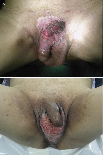
These images are taken from a recent BJUI paper: Primary invasive carcinoma associated with penoscrotal extramammary Paget’s disease: a clinicopathological analysis of 56 cases (Dai et al. BJUI 2015).

No such quiz/survey/poll

Article of the Month: Neutrophil–lymphocyte ratio helps predict survival in patients with upper tract urothelial carcinoma

Every week the Editor-in-Chief selects the Article of the Week from the current issue of BJUI. The abstract is reproduced below and you can click on the button to read the full article, which is freely available to all readers for at least 30 days from the time of this post.

In addition to the article itself, there is an accompanying editorial written by a prominent member of the urological community. This blog is intended to provoke comment and discussion and we invite you to use the comment tools at the bottom of each post to join the conversation.

Finally, the third post under the Article of the Week heading on the homepage will consist of additional material or media. This week we feature a video from Georg Hutterer discussing his paper.

If you only have time to read one article this week, it should be this one.

Validation of pretreatment neutrophil–lymphocyte ratio as a prognostic factor in a European cohort of patients with upper tract urothelial carcinoma

Orietta Dalpiaz, Georg C. Ehrlich, Sebastian Mannweiler*, Jessica M. Martín Hernández, Armin Gerger, Tatjana Stojakovic, Karl Pummer, Richard Zigeuner, Martin Pichler and Georg C. Hutterer

Department of Urology, *Institute of Pathology, Division of Oncology, Department of Internal Medicine, and Clinical Institute of Medical and Chemical Laboratory Diagnostics, Medical University of Graz, Graz, Austria

Read the full article

OBJECTIVE

• To investigate the potential prognostic significance of the neutrophil–lymphocyte ratio (NLR) in a large European cohort of patients with upper urinary tract urothelial cell carcinoma (UUT-UCC).

PATIENTS AND METHODS

• We retrospectively evaluated data from 202 consecutive patients with non-metastatic upper urinary tract urothelial cell carcinoma (UUT-UCC), who underwent surgery between 1990 and 2012 at a single tertiary academic centre.

• Patients’ cancer-specific survival (CSS) and overall survival (OS) were assessed using the Kaplan–Meier method.

• To evaluate the independent prognostic significance of the NLR, multivariate proportional Cox regression models were applied for both endpoints.

RESULTS

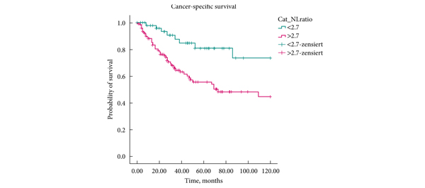
• A higher NLR was significantly associated with shorter CSS (P = 0.002, log-rank test), as well as with shorter OS (P < 0.001, log-rank test).

• Multivariate analysis identified a high NLR as an independent prognostic factor for patients’ CSS (hazard ratio 2.72, 95% CI 1.25–5.93, P = 0.012), and OS (hazard ratio 2.48, 95% CI 1.31–4.70, P = 0.005).

CONCLUSIONS

• In the present cohort, patients with a high preoperative NLR had higher cancer-specific and overall mortality after radical surgery for UUT-UCC, compared with those with a low preoperative NLR.

• This easily identifiable laboratory measure should be considered as an additional prognostic factor in UUT-UCC in future.

Read more articles of the week

Editorial: Neutrophil-to-lymphocyte ratio as a prognostic factor in upper tract urothelial cancer

The immune system response is critical to cancer development, treatment and progression. Dalpiaz et al. [1]. show that patients with a higher neutrophil-to-lymphocyte ratio (NLR) have a higher cancer-specific and overall mortality when undergoing radical nephroureterectomy for upper tract urothelial cell cancer (UTUC). The study is the first and largest one to evaluate the impact of preoperative NLR on UTUC and proposes its incorporation into our risk assessment tools as an independent predictor of survival.

Pathological prognostic factors such as tumour stage and grade have established importance in UTUC [2]. Additionally, lymphovascular invasion and tumour necrosis have been shown to be independent predictors of survival [3]. Preoperative markers have the advantage of prospective planning and counselling for treatment. The NLR has been studied in various cancers, including renal and gastric, and was recently incorporated into a risk stratification scheme for radical cystectomy patients as an independent prognostic factor for survival [4].

Dalpiaz et al. retrospectively reviewed 202 patients with UTUC who underwent radical nephroureterectomy. A threshold NLR value of 2.7 was used to discriminate between patients. NLR was significantly associated with lymphovascular invasion, but not with age, gender, tumour site, vascular invasion, tumour grade, pathological T-stage, tumour site, tumour location or presence of tumour necrosis. The mean follow-up was 45 months. The median survival was 44.5 months in the low-NLR group and 27 months in the high-NLR group. Multivariate analysis showed that T-stage and NLR were predictors of cancer-specific survival. High NLR and muscle invasion were shown to be independent predictors of overall survival.

Although interesting, these results should be interpreted cautiously as it is very difficult to control all confounders in a retrospective study. The authors did try to address aspects of the inflammatory response by incorporating Eastern Cooperative Oncology Group Performance Status and Charlson Comorbidity Index into their analysis. They found no statistically significant association between NLR and Eastern Cooperative Oncology Group Performance Status or Charlson Comorbidity Index. When adjusting for these variables, the relationships between NLR and cancer-specific survival and between NLR and overall survival were maintained. Although helpful in supporting the conclusions, using the Eastern Cooperative Oncology Group Performance Status and Charlson Comorbidity Index as markers of the inflammatory response should be approached carefully, as many other factors, such as hydronephrosis, tumour invasion, and pre-procedure treatments, which were not evaluated could have a more significant effect on the NLR than general measures of chronic conditions.

The threshold value of the NLR (2.7) was obtained by testing all possible thresholds and choosing a value based on its ability to predict survival and mathematical convenience. Thus the threshold value is self-serving to the conclusion. The statistical analysis suffers due to the dichotomous discrimination as opposed to further divisions like quartiles, but nonetheless shows the value of NLR as an important predictor, the threshold value of which might differ from cohort to cohort.

The present study shows that NLR as an important predictor of survival in UTUC. NLR is easy to perform, relatively inexpensive and is probably already available as part of the standard evaluation of patients with UTUC. It is therefore easy to assess. How should it change our practices? For example, should we be considering neoadjuvant chemotherapy, lymph node dissections or earlier radical surgery in patients with high NLR? The present study develops the hypothesis that can serve as the basis of future validation in a larger cohort or in a prospective fashion.

Read the full article

Moben Mirza
Department of Urology, University of Kansas, Kansas City, KS, USA

References
  1. Rouprêt M, Hupertan V, Seisen T et al.; French National Database on Upper Tract Tumors; Upper Tract Urothelial Carcinoma Collaboration. Prediction of cancer specific survival after radical nephroureterectomy for upper tract urothelial carcinoma: development of an optimized postoperative nomogram using decision curve analysis. J Urol 2013; 189: 1662–1669
  2. Zigeuner R, Shariat SF, Margulis V et al. Tumour necrosis is an indicator of aggressive biology in patients with urothelial carcinoma of the upper urinary tract. Eur Urol 2010; 57: 575
  3. Gondo T, Nakashima J, Ohno Y et al. Prognostic value of neutrophil-to-lymphocyte ratio and establishment of novel preoperative risk stratification model in bladder cancer patients treated with radical cystectomy. Urology 2012; 79: 1085


Video: Prognostic value of neutrophil–lymphocyte ratio in patients with UTUC

Validation of pretreatment neutrophil–lymphocyte ratio as a prognostic factor in a European cohort of patients with upper tract urothelial carcinoma

Orietta Dalpiaz, Georg C. Ehrlich, Sebastian Mannweiler*, Jessica M. Martín Hernández, Armin Gerger, Tatjana Stojakovic, Karl Pummer, Richard Zigeuner, Martin Pichler and Georg C. Hutterer

Department of Urology, *Institute of Pathology, Division of Oncology, Department of Internal Medicine, and Clinical Institute of Medical and Chemical Laboratory Diagnostics, Medical University of Graz, Graz, Austria

Read the full article

OBJECTIVE

• To investigate the potential prognostic significance of the neutrophil–lymphocyte ratio (NLR) in a large European cohort of patients with upper urinary tract urothelial cell carcinoma (UUT-UCC).

PATIENTS AND METHODS

• We retrospectively evaluated data from 202 consecutive patients with non-metastatic upper urinary tract urothelial cell carcinoma (UUT-UCC), who underwent surgery between 1990 and 2012 at a single tertiary academic centre.

• Patients’ cancer-specific survival (CSS) and overall survival (OS) were assessed using the Kaplan–Meier method.

• To evaluate the independent prognostic significance of the NLR, multivariate proportional Cox regression models were applied for both endpoints.

RESULTS

• A higher NLR was significantly associated with shorter CSS (P = 0.002, log-rank test), as well as with shorter OS (P < 0.001, log-rank test).

• Multivariate analysis identified a high NLR as an independent prognostic factor for patients’ CSS (hazard ratio 2.72, 95% CI 1.25–5.93, P = 0.012), and OS (hazard ratio 2.48, 95% CI 1.31–4.70, P = 0.005).

CONCLUSIONS

• In the present cohort, patients with a high preoperative NLR had higher cancer-specific and overall mortality after radical surgery for UUT-UCC, compared with those with a low preoperative NLR.

• This easily identifiable laboratory measure should be considered as an additional prognostic factor in UUT-UCC in future.

Read more articles of the week

© 2025 BJU International. All Rights Reserved.