Tag Archive for: #PCSM

Posts

Article of the Month: ProCare Trial

Every Month the Editor-in-Chief selects an Article of the Month from the current issue of BJUI. The abstract is reproduced below and you can click on the button to read the full article, which is freely available to all readers for at least 30 days from the time of this post.

In addition to the article itself, there is an accompanying editorial written by a prominent member of the urological community. This blog is intended to provoke comment and discussion and we invite you to use the comment tools at the bottom of each post to join the conversation.

If you only have time to read one article this week, it should be this one.

ProCare Trial: a phase II randomized controlled trial of shared care for follow-up of men with prostate cancer

Jon D. Emery*,,, Michael Jefford§,¶, Madeleine King**,††, Dickon Hayne‡‡,§§, Andrew Martin¶¶, Juanita Doorey, Amelia Hyatt, Emily Habgood*, Tee Lim***Cynthia Hawks‡‡,§§, Marie Pirotta*, Lyndal Trevena††† and Penelope Schoeld§,¶,‡‡‡


*Department of General Practice, University of Melbourne, Carlton, Western Health and the Victorian Comprehensive Cancer Centre, Melbourne, Vic., School of Primary Aboriginal and Rural Health Care, University of Western Australia, Crawley, WA, §Sir Peter MacCallum Department of Oncology, Faculty of Medicine, Dentistry and Health Sciences, The University of Melbourne, Parkville, Department of Cancer Experiences Research, Peter MacCallum Cancer Centre, East Melbourne, Vic., **Quality of Life Ofce, Psycho-oncology Co-operative Research Group, School of Psychology, University of Sydney, ††Sydney Medical School, University of Sydney, Sydney, NSW, ‡‡School of Surgery, University of Western Australia, Crawley,WA, §§Department of Urology, Fiona Stanley Hospital, Perth, WA, ¶¶NHMRC Clinical Trials Centre, University of Sydney, Sydney, NSW, ***Genesis Cancer Care, Department of Radiation Oncology, Fiona Stanley Hospital, Perth, WA, †††Primary Health Care, Sydney School of Public Health, University of Sydney, Sydney, NSW, and ‡‡‡Department of Psychology, Swinburne University of Technology, Melbourne, Vic., Australia


Abstract

Objectives

To test the feasibility and efficacy of a multifaceted model of shared care for men after completion of treatment for prostate cancer.

Patients and Methods

Men who had completed treatment for low- to moderate-risk prostate cancer within the previous 8 weeks were eligible. Participants were randomized to usual care or shared care. Shared care entailed substituting two hospital visits with three visits in primary care, a survivorship care plan, recall and reminders, and screening for distress and unmet needs. Outcome measures included psychological distress, prostate cancer-specific quality of life, satisfaction and preferences for care and healthcare resource use.

march-2017-aotm

Results

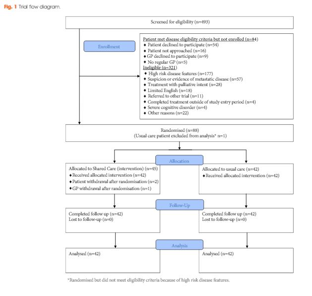
A total of 88 men were randomized (shared care n = 45; usual care n = 43). There were no clinically important or statistically significant differences between groups with regard to distress, prostate cancer-specific quality of life or satisfaction with care. At the end of the trial, men in the intervention group were significantly more likely to prefer a shared care model to hospital follow-up than those in the control group (intervention 63% vs control 24%; P<0.001). There was high compliance with prostate-specific antigen monitoring in both groups. The shared care model was cheaper than usual care (shared care AUS$1411; usual care AUS$1728; difference AUS$323 [plausible range AUS$91–554]).

Conclusion

Well-structured shared care for men with low- to moderate-risk prostate cancer is feasible and appears to produce clinically similar outcomes to those of standard care, at a lower cost.

Editorial: Rethinking cancer surveillance with shared-care models and survivorship plans: the time is now!

Urologists are increasingly facing significant practice concerns related to timely access, surgeon availability, clinical throughput and rising cost of care, yet little has changed over the years regarding the routine postoperative surveillance of urological cancers. While urologists have appropriately focused evaluations on oncological outcomes and procedure-specific quality-of-life concerns, the ability to maintain this practice model in the setting of more new patients (and subsequently more cancer survivors) seems unrealistic. In addition, gaps exist with the current model related to timely and effective communication to the local care team and assurances that specialists comprehensively address all concerns raised by patients. Furthermore, the role of the local care team in cancer survivorship remains poorly defined. Recognising these and other unmet needs in cancer care survivorship, the American Cancer Society (ACS) and the American Society of Clinical Oncology (ASCO) recently published guidelines on cancer survivorship [1-3]. The guidelines recommend a standardised approach to follow-up with emphasis on quality, comprehensive patient assessments, value, and shared use of a multidisciplinary team. With prostate cancer survivorship, for instance, ASCO recommends PSA checks every 6–12 months for the first 5 years and then annually (higher-risk patients can have more frequent checks), adherence to ACS guidelines for early detection of prostate cancer, assessment of physical and psychological effects of prostate cancer and it’s treatments, and annual assessments for long-term or late side-effects [3]. To help with the coordination of care between the patient, the oncological specialist, and the local primary care provider, survivorship care plans have been developed. [4]. While use of survivorship care plans has been sparse in urology to date, new mandates will spur their use in the coming years and development will likely involve innovative healthcare delivery solutions.

Leading the way in this nascent field, Emery et al. [4] report, in this issue of BJUI, an innovative phase II prospective randomised study on the feasibility of a novel shared-care model for follow-up of patients with prostate cancer. Men who had completed treatment for low- and moderate-risk prostate cancer were randomised to undergo usual care or shared care with the assistance of the patient’s primary care team. The novel shared-care model substituted two postoperative urology visits with three postoperative visits in primary care, provided patients and primary care providers a survivorship care plan, included appointment reminders, and provided a novel mechanism to screen for distress and other unmet needs. Among the 88 men randomised in the prospective study, no significant differences were noted between delivery models for satisfaction of care, overall quality of life, incidence of distress, or compliance with serum PSA testing. Patients in the shared-care model were significantly more likely to prefer the new model compared to normal care (cases, 63% vs controls, 24%, P < 0.001). Importantly, the shared-care model was also more economical, saving 323 Australian dollars compared to usual care [4].

The authors should be congratulated for their well-designed study and early contribution to the field. Rethinking all aspects of care delivery will become increasingly important as the practice of urology responds to access limitations, the shortage of urologists, and financial pressures of value-based reimbursement. The report also engenders many questions about the ideal care model of the future, composition of the collaborative care team, and the importance of making evidence-based clinical recommendations. For instance, are already overburdened primary care providers ideal or realistic in shared-care models? Should care remain primarily under the control of urologist with assistance provided by other current (e.g. advance practice providers, urology nurses) or future team member roles (e.g. survivorship care coordinators)? What role can the patient alone play in a self-guided survivorship care plan under the watchful eye of the collaborative care team acting asynchronously? How can enabling technologies such as smartphones, mobile applications, wearables, and video-conferencing contribute to high-value cancer surveillance building upon the principles highlighted in the current article and further engaging patients in their cancer survivorship care? [5]. Lastly, what actually are the evidence-based imperatives of survivorship care (what risk groups, what testing intervals and duration of testing) that provide measurable value to the patient experience? In the current study [4], for instance, high risk patients were excluded but ultimately these patients may be best suited for comprehensive survivorship care. Future work on survivorship and care models will hopefully continue to advance ‘win-win’ situations where patients and providers alike experience increasingly high-value systems of healthcare delivery.

Read the full article
Matthew T. Gettman


Mayo Clinic Department of Urology, 200 First Street, SW, Rochester, MN 55905, USA


References


1 Mayer DK, Nekhlyudov L, Snyder CF, Merrill JK , Wollins DS, Shulman LN. American Society of Clinical Oncology clinical expert statement on cancer survivorship care planning. J Oncol Pract 2014; 10: 34551


2 Skolarus TA, Wolf AM, Erb NL et al. American Cancer Society prostate cancer survivorship care guidelines. CA Cancer J Clin 2014; 64: 22549


Article of the Month: 68Ga-PSMA PET/CT for LN staging in PCa

Every Month the Editor-in-Chief selects an Article of the Month from the current issue of BJUI. The abstract is reproduced below and you can click on the button to read the full article, which is freely available to all readers for at least 30 days from the time of this post.

In addition to the article itself, there is an accompanying editorial written by a prominent member of the urological community. This blog is intended to provoke comment and discussion and we invite you to use the comment tools at the bottom of each post to join the conversation.

If you only have time to read one article this week, it should be this one.

Prospective evaluation of 68Gallium-prostate-specific membrane antigen positron emission tomography/computed tomography for preoperative lymph node staging in prostate cancer

Pim J. van Leeuwen*, Louise Emmett,§, Bao Ho, Warick Delprado, Francis Ting*Quoc Nguyen† and Phillip D. Stricker*


*St Vincents Prostate Cancer Centre, St Vincents Clinic, Australian Prostate Cancer Research Centre, New South Wales, The Garvan Institute of Medical Research/The Kinghorn Cancer Centre, Department of Diagnostic Imaging, St Vincents Public Hospital, §University of New South Wales, Sydney, and University of Notre Dame, Darlinghurst, NSW, Australia


Read the full article

Abstract

Objectives

To assess the accuracy of 68Gallium-prostate-specific membrane antigen (68Ga-PSMA) positron emission tomography/computed tomography (PET/CT) for lymph node (LN) staging in intermediate- and high-risk prostate cancer (PCa).

Materials and Methods

From April to October 2015, 30 patients with intermediate- (n = 3) or high-risk (n = 27) PCa were prospectively enrolled. Patients underwent preoperative 68Ga-PSMA PET/CT. Both visual and semi-quantitative analyses were undertaken. Subsequently, all patients underwent radical prostatectomy (RP) with an extended pelvic lymph node dissection. The sensitivity, specificity, and positive (PPV) and negative predictive value (NPV) for LN status of 68Ga-PSMA were calculated using histopathology as reference.

aotmfeb2017-results

Results

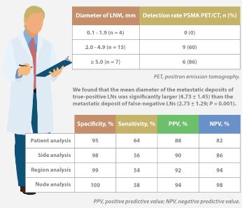
Eleven patients (37%) had lymph node metastases (LNMs); 26 LNMs were identified in the 11 patients. Patient analysis showed that 68Ga-PSMA PET/CT had a sensitivity of 64% for the detection of LNMs, its specificity was 95%, the PPV was 88%, and the NPV was 82%. In total, 180 LN fields were analysed. In the LN-region-based analysis, the sensitivity of 68Ga-PSMA PET/CT for detection of LNMs was 56%, the specificity was 98%, the PPV was 90% and the NPV was 94%. The mean size of missed LNMs was 2.7 mm. Receiver-operating characteristic curve analysis showed a high accuracy of maximum standardized uptake value (SUVmax) for the detection of LNMs, with an area under the curve of 0.915 (95% confidence interval 0.847–0.983); the optimum SUVmax was 2.0.

Conclusions

In patients with intermediate- to high-risk PCa, 68Ga-PSMA PET/CT had a high specificity and a moderate sensitivity for LNM detection. 68Ga-PSMA PET/CT had the potential to replace current imaging for LN staging of patients with PCa scheduled for RP.

info-feb-2017

Click on image for full infographic


Editorial: Bringing clarity or confusion? The role of prostate-specific membrane antigen positron-emission/computed tomography for primary staging in prostate cancer

The use of 68Ga-labelled prostate-specific membrane antigen (PSMA) positron-emission tomography (PET)/CT for staging prostate cancer in Australia has reached almost plague-like proportions. Despite what must be admitted is little high-level evidence to guide us in the accuracy or appropriateness of this imaging technique for either primary staging or prostate cancer recurrence, hundreds of these scans are being performed every week around Australia, and in many cases we simply do not know what to do with the results. We performed the first such scan at our centre in Melbourne in August 2014, and were soon receiving 10 requests per day, with patients waiting up to 3 months to be scanned. Fast-forward 2 years, and there are now eight centres offering PSMA PET/CT in Melbourne, a city of 4.5 million people. Scans can be obtained within 24 h of referral and costs have dropped to €500. A similar situation exists in Germany where this imaging method was pioneered [1], and interest is also growing in Belgium, Italy, India and a number of other countries (the USA being a notable exception). But do we really understand the impact of the decision to perform PSMA/PET scanning, and do we have enough evidence to guide us on the most appropriate setting for its use?

The current interest in PSMA PET/CT has been triggered by the development of small molecule ligands which bind to the extracellular domain of the PSMA molecule, leading to increased sensitivity and specificity when compared with conventional imaging [2]. Previously, the use of PET imaging for prostate cancer detection was greatly limited by the relatively poor performance characteristics of choline-based PET/CT, and limited availability and high costs associated with this type of imaging. The introduction of 68Ga-labelled PSMA PET/CT has addressed many of these concerns, although high-quality evidence is still lacking to help guide its most appropriate utility. The best data exist for identification of prostate recurrence in patients with biochemical recurrence (BCR) after previous definitive therapy. In our recent systematic review and meta-analysis of this topic, we reported pooled data on 1309 men with BCR undergoing PSMA PET/CT [3]. When stratified by PSA level post-radical prostatectomy, positive scans are reported in 42, 58, 76 and 95% of patients with PSA levels of 0–0.2, 0.2–1, 1–2, and >2 ng/mL, respectively. Fewer data exist for the role of PSMA PET/CT in the primary staging setting.

In this interesting paper from some of our Australian colleagues, van Leeuwen et al. [4] report their experience of PSMA PET/CT in the primary staging setting, in particular to evaluate the performance of PSMA PET/CT to evaluate lymph node positivity in patients with intermediate- and high-risk disease, scheduled for radical prostatectomy. A total of 30 patients underwent preoperative PSMA PET/CT, of which 27 were stratified as high risk, and all subsequently underwent radical prostatectomy and pelvic lymph node dissection. In total, 11 patients (37%) had histologically proven lymph node metastases. On a per-patient basis, PSMA PET had a sensitivity of 64%, specificity of 95%, positive predictive value of 88%, and negative predictive value of 82%. The average size of positive lymph nodes not detected by PSMA PET/CT was 2.7 mm; therefore, in this population of patients with predominately high-risk prostate cancer, PSMA PET/CT had very high specificity and moderate sensitivity for lymph node metastasis detection.

In a larger experience from Munich, Maurer et al. [5] compared pathology findings of 130 patients with intermediate- and high-risk disease who underwent radical prostatectomy and pelvic lymph node dissection, with preoperative PSMA PET/CT or PET/MRI findings. They reported similar sensitivity, specificity and accuracy of 65.9, 98.9 and 88.5%, respectively. On receiver-operating characteristic analysis, PSMA-PET performed significantly better than conventional imaging alone on patient and template-based analyses (P = 0.002 and <0.001, respectively).

Just as there appears to be some clarity, however, in the role of PSMA PET/CT in patients with BCR, and in improving the detection of lymph node metastases preoperatively, there are many instances in which the high specificity of this scanning method leaves us in a decision-making quandary. As van Leeuwen et al. identified in their paper, and as we have frequently observed ourselves, PSMA PET/CT may identify prostate cancer in hitherto unidentified and unusual locations such as the mesorectum (Fig. 1). Disease may also be identified in quite distant locations despite relatively low PSA levels, thereby disrupting traditional management algorithms including the use of postoperative radiotherapy [6]. Should we alter patients’ management based on novel imaging, or should we assess the decision impact more formally in prospective studies? The answer should obviously be the latter, but the current plague of PSMA PET imaging means such decisions are already being taken in the absence of high-quality evidence.

image

Figure 1. 68Ga-labelled prostate-specific membrane antigen (PSMA) positron-emission tomography (PET)/CT in a 72-year-old man with biochemical recurrence after previous radical prostatectomy. His PSA level was 0.21 ng/mL and conventional staging including CT and bone scan showed no evidence of disease. PSMA PET/CT demonstrates intense avidity in an 11-mm mesorectal node near the recto-sigmoid junction on the left side. (a) CT demonstrates non-specific findings in area of subsequent avidity; (b) PSMA PET raw data demonstrating avidity in mesorectal node; (c) fused PSMA PET/CT image provides anatomical correlation; (d) coronal fused PET/CT image.

Nonetheless, PSMA PET imaging is here to stay, and will doubtless have a positive impact in improving decision-making in prostate cancer management as a result of the more accurate staging which it heralds. We must await more formal evaluation of the decision impact before defining the patient population who will benefit the most from this exciting imaging method.

Read the full article
Declan G. Murphy, Urologist*,, Michael Hofman, Nuclear Medicine Physician, Nathan Lawrentschuk, Urologist*,§ and Tobias Maurer, Urologist


*Division of Cance r Surgery, Peter MacCallum Cancer Centre, University of Melbourne, Epworth Prostate Centre, Epworth Hospital, Department of Cancer Imaging, Peter MacCallum Cancer Centre, Melbourne, §Department of Surgery, The Austin Hospital, University of Melbourne, Heidelberg, Vic.Australia and Department of Urology, Technische Universitat Munchen, Klinikum rechts der Isar, Munich, Germany


References


Infographic: 68Ga-PSMA PET/CT for LN staging in PCa

Infographic to accompany the February 2017 Article of the Month

68ga-psma-pet-ct-infographic

Read the full article

See more infographics

Article of the Week: TRT and rates of PCa or LUTS

Every Week the Editor-in-Chief selects an Article of the Week from the current issue of BJUI. The abstract is reproduced below and you can click on the button to read the full article, which is freely available to all readers for at least 30 days from the time of this post.

In addition to the article itself, there is an accompanying editorial written by a prominent member of the urological community. This blog is intended to provoke comment and discussion and we invite you to use the comment tools at the bottom of each post to join the conversation.

If you only have time to read one article this week, it should be this one.

Testosterone treatment is not associated with increased risk of prostate cancer or worsening of lower urinary tract symptoms: prostate health outcomes in the Registry of Hypogonadism in Men

Frans M.J. Debruyne*, Hermann M. Behre, Claus G. Roehrborn, Mario Maggi§Frederick C.W. Wu, Fritz H. Schroder**, Thomas Hugh Jones††, Hartmut Porst‡‡Geoffrey Hackett§§, Olivia A. Wheaton¶¶, Antonio Martin-Morales***, Eric J. Meuleman†††, Glenn R. Cunningham‡‡‡, Hozefa A. Divan¶¶ and Raymond C. Rosen ¶¶ for the RHYME Investigators


*Andros Mens Health Institutes, Arnhem, The Netherlands, Center for Reproductive Medicine and Andrology, Martin Luther University Halle-Wittenberg, Halle, Germany, Southwestern Medical Center, University of Texas, Dallas, TX, USA, §Sexual Medicine and Andrology Unit, Department of Experimental and Clinical Biomedical Sciences, University of Florence, Florence, Italy, ¶ University of Manchester, Manchester, UK, **Erasmus Medical Center, Rotterdam, The Netherlands, ††Barnsley Hospital NHS Foundation Trust, Barnsley, UK, ‡‡Private Practice of Urology/Andrology, Hamburg, Germany, §§Holly Cottage Clinic, Licheld, Staffordshire, UK, ¶¶New England Research Institutes, Inc., Watertown, MA, USA, ***Carlos Haya University Hospital, Malaga, Spain, †††VU Medical Center, Amsterdam, The Netherlands, and ‡‡‡Baylor College of Medicine, St. Lukes Episcopal Hospital, Houston, TX, USA


Read the full article

Abstract

Objectives

To evaluate the effects of testosterone-replacement therapy (TRT) on prostate health indicators in hypogonadal men, including rates of prostate cancer diagnoses, changes in prostate-specific antigen (PSA) levels and lower urinary tract symptoms (LUTS) over time.

Patients and Methods

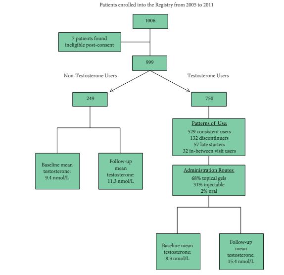
The Registry of Hypogonadism in Men (RHYME) is a multi-national patient registry of treated and untreated, newly-diagnosed hypogonadal men (n = 999). Follow-up assessments were performed at 3–6, 12, 24, and 36 months. Baseline and follow-up data collection included medical history, physical examination, blood sampling, and patient questionnaires. Prostate biopsies underwent blinded independent adjudication for the presence and severity of prostate cancer; PSA and testosterone levels were measured via local and central laboratory assays; and LUTS severity was assessed via the International Prostate Symptom Score (IPSS). Incidence rates per 100 000 person-years were calculated. Longitudinal mixed models were used to assess effects of testosterone on PSA levels and IPSS.

feb-aotw-2-results

Results

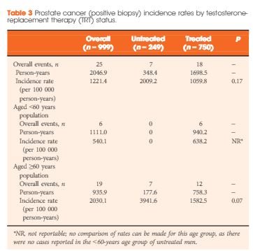
Of the 999 men with clinically diagnosed hypogonadism (HG), 750 (75%) initiated TRT, contributing 23 900 person-months of exposure. The mean testosterone levels increased from 8.3 to 15.4 nmol/L in treated men, compared to only a slight increase from 9.4 to 11.3 nmol/L in untreated men. In all, 55 biopsies were performed for suspected prostate cancer, and 12 non-cancer related biopsies were performed for other reasons. Overall, the proportion of positive biopsies was nearly identical in men on TRT (37.5%) compared to those not on TRT (37.0%) over the course of the study. There were no differences in PSA levels, total IPSS, or the IPSS obstructive sub-scale score by TRT status. Lower IPSS irritative sub-scale scores were reported in treated compared to untreated men.

Conclusions

Results support prostate safety of TRT in newly diagnosed men with HG.


Editorial: Mythology and Reality Should Never Be Confused (But Often Are)

The debating halls of learned urological societies often attempt to define theories based on little substance. Unfortunately, although this type of scientific discourse should be encouraged, it can create both content for unrepeatable ‘late breaking’ abstracts and headlines for mass circulation newspapers. Once started, the momentum can make a perception become a ‘reality’ that is invariably difficult to dislodge. An area of particular current interest to both the scientific and lay press, and indeed the regulatory authorities in the USA and the European Union, is testosterone replacement. Within this context, an important potential myth-buster is described in the article by DeBruyne et al. [1]. The hypothesis (aka myth) that was being examined was that application of exogenous testosterone, irrespective of formulation, could exacerbate both benign and malignant prostatic disease. To understand the importance of this type of definitive study we must start at the beginning.

The foundation of the mythology, or in reality a Canadian-US urological tragedy, was ironically in the Nobel Prize winning work of Charles B Huggins. The ‘good news’ was that prostatic tumour regression could be affected by androgen deprivation, a finding certainly worthy of global recognition and acceptance; however, this became embellished to a certain extent, incorporating other aliquots of somewhat circumstantial evidence, leading to a suggestion that testosterone replacement could increase the probability and/or rate of prostate cancer progression. As a result, various societies perhaps understandably adopted their normal conservative approach of inserting a warning (with varying degrees of emphasis) in their guidelines [2]. Over the last decade, with the increasing use of testosterone replacement in the treatment of men with hypogonadism, the issue of benefit–risk has become more relevant, indeed is often transposed to risk–benefit, and is increasingly likely to feature in the popular press. Within the last couple of years, we have seen the spectre of testosterone and cardiovascular risk played out in full public view, but the issue of testosterone and exacerbation of prostatic disease has continued to smoulder in the background.

The RHYME study [1], although it is largely confirmatory as other studies have described similar conclusions, is of considerable ‘real-life’ value. The power of the study is that it should be considered to be representative of the potential hypogonadal population likely to present to the physician. Although not used for regulatory approval purposes, a registry study is considered to be the ‘gold standard’ for this type of analysis. So what are the conclusions of this and the majority of other studies? In essence, there is no evidence that restoration of testosterone to the normal range does increase the incidence or progression of either benign (BPH) or malignant prostatic disease. Any slight changes in PSA level were considered not to be clinically relevant and likely to be as a consequence of the direct pharmacology of increased testosterone levels [3]. This does not imply that exacerbations will never be seen or that appropriate monitoring should not be carried out; more that the conventional wisdom on testosterone and prostatic disease has gone (or should go) the way of antimuscarinic agents, invariably causing urinary retention in patients with BPH. It is to be hoped that studies such as the RHYME study will continue to be undertaken and help provide perspective for specialist and primary care physicians alike in areas of emergent clinical interest.

Read the full article
Michael Wyllie
Global Pharma Consulting Ltd, Stratton House, Shenington, Banbury, Oxfordshire


References


Article of the Week: Accuracy of prostate biopsies for predicting Gleason score in radical prostatectomy specimens: nationwide trends 2000–2012

Every Week the Editor-in-Chief selects an Article of the Week from the current issue of BJUI. The abstract is reproduced below and you can click on the button to read the full article, which is freely available to all readers for at least 30 days from the time of this post.

In addition to the article itself, there is an accompanying editorial written by a prominent member of the urological community. This blog is intended to provoke comment and discussion and we invite you to use the comment tools at the bottom of each post to join the conversation.

If you only have time to read one article this week, it should be this one.

Accuracy of prostate biopsies for predicting Gleason score in radical prostatectomy specimens: nationwide trends 2000–2012

Daniela Danneman*, Linda Drevin, Brett Delahunt, Hemamali Samaratunga§¶David Robinson**, Ola Bratt††‡‡, Stacy Loeb§§ Par Stattin¶¶*** and Lars Egevad*†††


*Department of Oncology-Pathology, Karolinska Institute, Stockholm, † Regional Cancer Centre, Uppsala University Hospital, Uppsala, Sweden, Wellington School of Medicine and Health Sciences, University of Otago, Wellington, New Zealand, §Aquesta Pathology, The University of Queensland School of Medicine, Brisbane, Qld, Australia, **Department of Urology, Ryhov County Hospital, Jonkoping, Sweden, ††Department of Urology, Cambridge University Hospitals, Cambridge, UK, ‡‡Department of Translational Medicine, Lund University, Lund, Sweden, §§Department of Urology and Population Health, New York University and Manhattan Veterans Affairs Medical Centre, New York, NY, USA, ¶¶Department of Surgical and Perioperative Sciences, Urology and Andrology, Umea University, Umea, ***Department of Surgical Sciences, Uppsala University, Uppsala, Umea, and †††Department of Pathology, Karolinska University Hospital, Stockholm, Sweden


Read the full article

Abstract

Objectives

To investigate how well the Gleason score in diagnostic needle biopsies predicted the Gleason score in a subsequent radical prostatectomy (RP) specimen before and after the 2005 International Society of Urological Pathology (ISUP) revision of Gleason grading, and if the recently proposed ISUP grades 1–5 (corresponding to Gleason scores 6, 3 + 4, 4 + 3, 8 and 9–10) better predict the RP grade.

Patients and Methods

All prostate cancers diagnosed in Sweden are reported to the National Prostate Cancer Register (NPCR). We analysed the Gleason scores and ISUP grades from the diagnostic biopsies and the RP specimens in 15 598 men in the NPCR who: were diagnosed between 2000 and 2012 with clinical stage T1–2 M0/X prostate cancer on needle biopsy; were aged ≤70 years; had serum PSA concentration of <20 ng/mL; and underwent a RP <6 months after diagnosis as their primary treatment.

aotw-jan-4-2017-results

Results

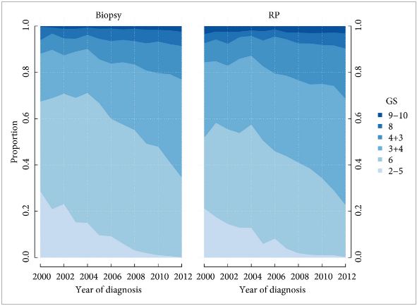
Prediction of RP Gleason score increased from 55 to 68% between 2000 and 2012. Most of the increase occurred before 2005 (nine percentage points; P < 0.001); however, when adjusting for Gleason score and year of diagnosis in a multivariable analysis, the prediction of RP Gleason score decreased over time (odds ratio [OR] 0.98; P < 0.002). A change in the ISUP grades would have led to a decreasing agreement between biopsy and RP grades over time, from 68% in 2000 to 57% in 2012, with an OR of 0.95 in multivariable analysis (P < 0.001).

Conclusion

Agreement between biopsy and RP Gleason score improved from 2000 to 2012, with most of the improvement occurring before the 2005 ISUP grading revision. Had ISUP grades been used instead of Gleason score, the agreement between biopsy and RP grade would have decreased, probably because of its separation of Gleason score 7 into ISUP grades 2 and 3 (Gleason score 3 + 4 vs 4 + 3).

Editorial: The prognostic value of prostate biopsy grade – Forever a product of sampling

The ability to project clinical outcomes based on limited data is crucial to the practice of medicine. This principle is particularly germane to the management of prostate cancer, where clinical outcomes vary widely. In the current issue of BJUI, Danneman et al. [1] assess pathological grade concordance between diagnostic needle biopsy and subsequent radical prostatectomy specimens from 2000 to 2012. The authors observed increased concordance of biopsy and prostatectomy Gleason scores over the time period (from 55% in 2000 to 68% in 2012) with the majority of improvement occurring before 2005. Interestingly, concordance decreased over time (from 68 to 57%) with use of the newly revised grading system. These and other findings led to the proposal that increased concordance was attributable more to the elimination of Gleason scores 2–5 than the systematic change in grading itself.

We commend the authors for exploring this important topic. Our ability to derive meaningful information on disease biology and behaviour from biopsy specimens is essential to counselling patients on the many available management options. At the same time, biopsy grading is inherently limited in its ability to predict overall prostate pathology because it is not only dependent on architecture and morphology, but also on the, admittedly minimal, sample of tissue obtained. As such, we should be cautious in using terms such as ‘undergrading’ in describing biopsy specimens, which may have been properly graded, but simply lacked the higher grade tumour observed at prostatectomy. In reality, such a phenomenon represents undersampling rather than undergrading, and there is hope that such undersampling will decrease with improved methods of detection, such as multiparametric MRI/TRUS fusion-guided biopsy.

Notably, the authors refer to the updated grading system, which was first described by Dr Epstein and validated in a multi-institutional study [2] before the 2014 International Society of Urological Pathology (ISUP) consensus conference, as ISUP grades 1–5. For clarity, it should be noted that the initial report and validation of the new system [2], the 2014 ISUP consensus conference proceedings [3] and the WHO 2016 edition of Pathology and Genetics: Tumours of the Urinary System and Male Genital Organs [4], have all described the new system based on grade groups 1–5. Consistent use of the adopted terminology will be helpful moving forward.

Nonetheless, there are several potential explanations for the patterns observed in the present study. As the authors note, lower concordance based on the grade group system can be largely explained by the more precise classification of Gleason score 7 cancers. Based on evidence of disparate outcomes in Gleason score 3+4 = 7 and 4+3 = 7 disease [5], the ISUP system distinctly classifies these cancers as prognostic grades 2 and 3, respectively. Certainly, when compared with a system in which Gleason score 7 represents a single classification, one would expect poorer concordance in the more widely distributed group. We believe the clinical utility of separating these classifications far outweighs a modest decrease in concordance, which may be explained by other factors in any case. Previous studies have shown the importance of subdividing the Gleason score 7 population when comparing grading systems [6]. Furthermore, details are not provided as to whether a global grade was assigned to biopsy, a common practice in Sweden, which is not the currently recommended practice. That 5–7% of specimens received a Gleason score < 6 calls into question whether contemporary recommendations were fully adopted during the study period.

Regardless, Danneman et al. elegantly highlight the frequency with which biopsy and prostatectomy grades are discordant, and the fact that, to date, pathological grading remains a subjective practice. As noted, there are widespread efforts to address both of these issues, including the use of targeted biopsies and tissue-based genomic markers. Until these practices are well-validated and widely implemented, there are several reasons to believe the most recent grade group system will improve contemporary practice, despite limited concordance. For one, use of a more intuitive scale ranging from 1 to 5 should prove easier for patients to understand, a significant consideration in light of the information overload patients absorb with a new diagnosis of cancer. Furthermore, available data to this point demonstrate excellent prognostic value. In one study from Johns Hopkins, the revised Grade Group system showed improved accuracy for predicting 5-year metastasis (C-index 0.80 vs 0.70) and 10-year prostate cancer-specific mortality (C-index 0.77 vs 0.64) as compared with the original Gleason score [7].

Until truly objective methods of pathological assessment emerge, additional validation of the new grade group system is likely to further support its use moving forward. As Danneman et al. point out, however, we must keep in mind that biopsy, although perhaps our most useful tool, captures only a small fraction of the overall picture.

Read the full article
Jeffrey J. Tosoian* and Jonathan I. Epstein*,,‡ *James Buchanan


Brady Urological Institute and Department of Urology, Department of Pathology, Johns Hopkins University School of Medicine, and Department of Oncology, Johns Hopkins University School of Medicine, Baltimore, MD, USA


References


2 Pierorazio PM, Walsh PC, Partin AW, Epstein JI. Prognostic gleason grade grouping: data based on the modied gleason scoring system. BJU Int 2013; 111: 75360


4 Moch H, Humphrey P, Ulbright T, Reuter V. WHO classication of tumours: pathology and genetics.Tumours of the Urinary and Male Reproductive System. Lyon, France:IARC Press; 2016.


5 Eggener SE, Scardino PT, Walsh PC et al. Predicting 15-year prostate cancer specic mortality after radical prostatectomy. J Urol 2011; 185: 86975


6 Lee MC, Dong F, Stephenson AJ, Jones JS, Magi-Galluzzi C, Klein EAThe Epstein criteria predict for organ-conned but not insignicant disease and a high likelihood of cure at radical prostatectomy. Eur. Urol 2010; 58: 905


Video: International Consultation on Urological Diseases and European Association of Urology International Consultation on Minimally Invasive Surgery in Urology: laparoscopic and robotic adrenalectomy

International Consultation on Urological Diseases and European Association of Urology International Consultation on Minimally Invasive Surgery in Urology: laparoscopic and robotic adrenalectomy

Mark W. Ball*, Ashok K. Hemal† and Mohamad E. Allaf*


*James Buchanan Brady Urological Institute and Department of Urology, Johns Hopkins University School of Medicine, Baltimore, MD, and Department of Urology, Wake Forest School of Medicine, Winston-Salem, NC, USA


Read the full article

Abstract

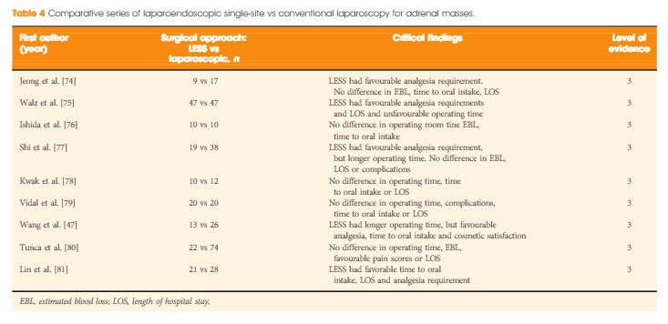
The aim of this study was to provide an evidence-based systematic review of the use of laparoscopic and robotic adrenalectomy in the treatment of adrenal disease as part of the International Consultation on Urological Diseases and European Association of Urology consultation on Minimally Invasive Surgery in Urology. A systematic literature search (January 2004 to January 2014) was conducted to identify comparative studies assessing the safety and efficacy of minimally invasive adrenal surgery. Subtopics including the role of minimally invasive surgery for pheochromocytoma, adrenocortical carcinoma (ACC) and large adrenal tumours were examined. Additionally, the role of transperitoneal and retroperitoneal approaches, as well as laparoendoscopic single-site (LESS) and robotic adrenalectomy were reviewed. The major findings are presented in an evidence-based fashion. Large retrospective and prospective data were analysed and a set of recommendations provided by the committee was produced. Laparoscopic surgery should be considered the first-line therapy for benign adrenal masses requiring surgical resection and for patients with pheochromocytoma. While a laparoscopic approach may be feasible for selected cases of ACC without adjacent organ involvement, an open surgical approach remains the ‘gold standard’. Large adrenal tumours without preoperative or intra-operative suspicion of ACC may be safely resected via a laparoscopic approach. Both transperitoneal and retroperitoneal approaches to laparoscopic adrenalectomy are safe. The approach should be chosen based on surgeon training and experience. LESS and robotic adrenalectomy should be considered as alternatives to laparoscopic adrenalectomy but require further study.


aotwjan3-reults


© 2024 BJU International. All Rights Reserved.