Tag Archive for: nephrolithiasis

Posts

Article of the Week: Accuracy of ultrasonography for renal stone detection and size determination: is it good enough for management decisions?

Every week the Editor-in-Chief selects an Article of the Week from the current issue of BJUI. The abstract is reproduced below and you can click on the button to read the full article, which is freely available to all readers for at least 30 days from the time of this post.

In addition to the article itself, there is an accompanying editorial written by a prominent member of the urological community. This blog is intended to provoke comment and discussion and we invite you to use the comment tools at the bottom of each post to join the conversation.

Finally, the third post under the Article of the Week heading on the homepage will consist of additional material or media. This week we feature a video discussing the paper.

If you only have time to read one article this week, it should be this one.

Accuracy of ultrasonography for renal stone detection and size determination: is it good enough for management decisions?

Vishnu Ganesan*,, Shubha De*, Daniel Greene*, Fabio Cesar Miranda Torricelli* and Manoj Monga*


*Glickman Urological Kidney Institute, and Lerner College of Medicine, Cleveland Clinic, Cleveland, OH, USA


Abstract

Objectives

To determine the sensitivity and specificity of ultrasonography (US) for detecting renal calculi and to assess the accuracy of US for determining the size of calculi and how this can affect counselling decisions.

Materials and Methods

We retrospectively identified all patients at our institution with a diagnosis of nephrolithiasis who underwent US followed by non-contrast computed tomography (CT) within 60 days. Data on patient characteristics, stone size (maximum axial diameter) and stone location were collected. The sensitivity, specificity and size accuracy of US was determined using CT as the standard.

aotw-mar

Results

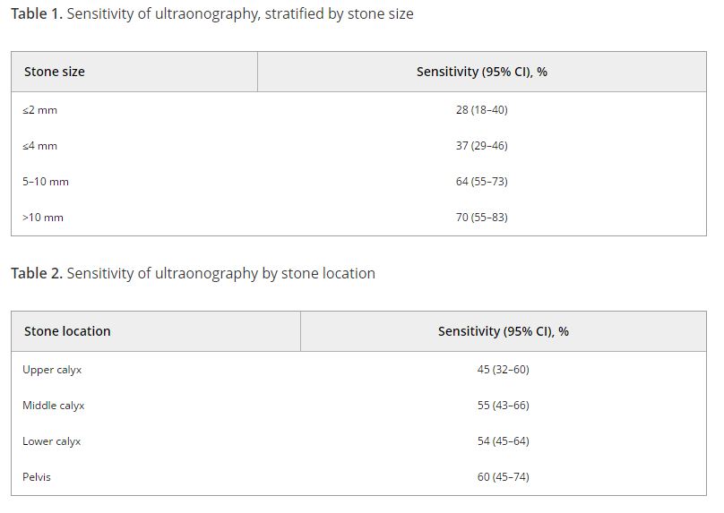
A total of 552 US and CT examinations met the inclusion criteria. Overall, the sensitivity and specificity of US was 54 and 91%, respectively. There was a significant association between sensitivity of US and stone size (P < 0.001), but not with stone location (P = 0.58). US significantly overestimated the size of stones in the 0–10 mm range (P < 0.001). Assuming patients with stones 0–4 mm in size will be selected for observation and those with stones ≥5 mm could be counselled on the alternative of intervention, we found that in 14% (54/384) of cases where CT would suggest observation, US would lead to a recommendation for intervention. By contrast, when CT results would suggest intervention as management, US would suggest observation in 39% (65/168) of cases. An average of 22% (119/552) of patients could be inappropriately counselled. Stones classified as 5–10 mm according to US had the highest probability (43% [41/96]) of having their management recommendation changed when CT was performed. The use of plain abdominal film of kidney, ureter and bladder and US increases sensitivity (78%), but 37% (13/35) of patients may still be counselled inappropriately to undergo observation.

Conclusions

Using US to guide clinical decision-making for residual or asymptomatic calculi is limited by low sensitivity and inability to size the stone accurately. As a result, one in five patients may be inappropriately counselled when using US alone.

Editorial: Ultrasonography vs computed tomography for stone size

In this edition of the BJUI Ganesan et al. [1] report a retrospective analysis of 552 ultrasonography (US) examinations that were followed by a non-contrast CT within 60 days in 486 patients collected over an 18-year period (1995–2012). The sensitivity of US for stone detection was 54% and its specificity was 91% when compared to CT, and sensitivity was positively associated with stone size (increasing from 73% for stones of 0–4 mm to 77% for 5–10 mm, and 89% for >10 mm; P < 0.001), but not with intra-renal location of stones (P = 0.58). US overestimated the size of stones that were <10 mm (P < 0.001), and had a tendency to underestimate size for those >10 mm (P = 0.05).

Stones were grouped into three size categories, based on clinical relevance to stone management: ≤4 mm (where observation would likely be recommended), 5–10 mm (where shockwave lithotripsy [SWL] would be chosen) or >10 mm where an endoscopic approach would be undertaken). Using these thresholds, 39% of cases would have been misassigned to observation and 14% of patients would have been inappropriately advised to undergo active treatment.

One may question the use of CT as the ‘gold standard’, as CT is also prone to sizing inaccuracy. Nevertheless, the headline findings that the inaccuracies inherent in US diagnosis and sizing may compromise clinical management are important. Other authors have made similar observations: in a literature review, Ray et al. [2] reported that US sensitivity was 45% for the detection of renal and ureteric calculi, with specificity up to 94% for ureteric stones and 88% for renal stones and that US overestimated stone size by a mean of 1.9 mm over CT, especially with stones of <5 mm. Similarly, Sternberg et al. [3] showed that the largest stone diameter was over-estimated by an average of 2.2 mm with US, and that errors increased with reducing stone size, rising from a 3% difference in stones >10 mm to 27% for those of 5–10 mm, and an 85% difference in stones ≤5 mm.

It is well established that, whilst having the advantage of no radiation dose, that US is a ‘user dependent’ study but there are also inherent limitations of US compared to CT for stone imaging. CT is capable of much finer spatial resolution, whilst US is prey to more diagnostic confounders. Reflectivity arising from sinus fat or the edges of the papillae may be mistaken for small calculi. For size, it can be difficult to delineate stone edges with the same precision as with CT. The sensitivity of US for stone detection can be improved by adjusting the imaging modalities between ray line (the conventional form of US), spatial compound and harmonic imaging (the most accurate stone size modality). Techniques such as increasing the gain and the transducer-to-stone depth and identifying ‘twinkle artefact’ using colour Doppler have also been used to improve stone detection [4].

However, manoeuvres to improve sensitivity of US may also compromise size measurement. An in vitro study has shown that each 2 cm increase in depth setting increases the size overestimation of stones by ~22% [5]. Using calcium oxalate monohydrate stones, the same group have shown that measuring the posterior acoustic shadow provided a more precise assessment of stone size than measurement of the stone itself [4]. Interestingly, the accuracy of stone width measurement was worse with greater transducer-to-stone depth, but measurement of the shadow width was independent of depth, and all US modalities (ray line, spatial compound, and harmonic imaging) performed similarly for shadow size. Shadow measurement was accurate to within 1 mm of the stone size [4], and similar findings have been shown in vivo, where 73% of the stone measurements and 85% of the shadow measurements were within 2 mm of the size on CT [6].

Unfortunately, not all stones cast an acoustic shadow, particularly the smaller ones, which are most likely to be over-sized. May et al. [6] showed that 89% of stones >5 mm, but only 53% of stones <5 mm demonstrated a posterior acoustic shadow. However, this may provide a further value for US-based clinical decision making, as stones that do not shadow are most likely <5 mm and are small enough to pass spontaneously, and therefore to be managed conservatively.

It is also important to be aware that CT stone measurements are also prone to error and inter-observer variability. Comparing in vitro CT measurements of stones in a ‘kidney sized potato model’, Eisner et al. [7] have shown that the most accurate measurements were obtained using magnified ‘bone window’ settings, which showed a mean 0.13 mm difference compared to a ‘gold standard’ measurement using callipers. This study also included a comparison of size estimate for spontaneously passed ureteric stones (thus a true reference standard) demonstrating that magnified ‘bone window’ measurements were equivalent to digital calliper measurements (the mean underestimation vs digital callipers was only 0.3 mm, P = 0.4), while measurements using magnified soft tissue windows were statistically different (mean underestimation 1.4 mm, P = 0.001) [7].

With its safety and accessibility, US should be the ideal modality for postoperative follow-up, both for assessment of stone recurrence, monitoring for enlargement of residual fragments, and for identifying the rare but important finding of ‘silent obstruction’, with the potential to lose renal function. However, given the ‘real-life’ data reported in this edition of the BJUI [1], and particularly the findings that 22% of patients might have been managed inappropriately when using US for decision making alone, increasing to 43% of patients who had stones between 5 and 10 mm on US, the authors have concluded that patients monitored by US might benefit from an additional CT if intervention is being considered, particularly for stones in the 5–10 mm range by US measurement.

Given the key importance of stone size to the outcome of interventions for stone disease, accurate imaging should translate into improved decision making and patient counselling and allow fairer inter-surgeon and departmental comparisons. Until the best US protocol and settings have been established, we recommend that, when US is used for diagnosis or follow-up, careful optimisation of the settings is crucial. Colour Doppler for ‘twinkle artefact’, and a high gain setting can be used to reduce the risk of missing stones, combined with removing all filtering and compressing the grey scale range to enhance the posterior shadowing. Harmonic imaging (which is now available on most commercial machines) is more accurate than cross beam or compound beams (that are used for standard renal US settings). When decisions need to be made, particularly those based on stone size, CT of the kidneys, ureters and bladder remains invaluable, from which the longest stone diameter should be measured, using magnified images and the ‘bone window’ setting. Current methods for accurate estimation of stone volume are impractical or imprecise. Manual segmentation can be accurate but is laborious, whilst standard semi-ellipsoid formulae cannot account for the wide variety of stone shapes seen in practice. Further studies devoted to simplifying stone volume estimation are necessary. There is also the wider challenge of how best to report stone imaging data. The key variables are stone size, density and location; and the morphology of the collecting system. Agreement between the various stakeholders – sonographers, radiologists and endourologists – over imaging standards and a minimal data set for stone imaging would improve management.

Daron Smith* and Uday Patel


*Institute of Urology, University College Hospital, and Department of Radiology, St Georges Hospital, London, UK


References


2 Ray AA, Ghiculete D, Pace KT, Honey RJ. Limitations to ultrasound in the detection and measurement of urinary tract calculi. Urology 2010; 76: 295300


3 Sternberg KM, Eisner B, Larson T, Hernandez N, Han J, Pais VMUltrasonography signicantly overestimates stone size when compared to low-dose, noncontrast computed tomography. Urology 2016; 95: 6771


4 Dunmire B, Harper JD, Cunitz BW et al. Use of the acoustic shadow width to determine kidney stone size with ultrasound. J Urol 2016; 195: 1717


5 Dunmire B, Lee FC, Hsi RS et al. Tools to improve the accuracy of kidney stone sizing with ultrasound. J Endourol 2015; 29: 14752


6 May PC, Haider Y, Dunmire B et al. Stone-mode ultrasound for determining renal stone size. J Endourol 2016; 30: 95862


Video: Accuracy of ultrasonography for renal stone detection and size determination: is it good enough for management decisions?

Accuracy of ultrasonography for renal stone detection and size determination: is it good enough for management decisions?


Abstract

Objectives

To determine the sensitivity and specificity of ultrasonography (US) for detecting renal calculi and to assess the accuracy of US for determining the size of calculi and how this can affect counselling decisions.

Materials and Methods

We retrospectively identified all patients at our institution with a diagnosis of nephrolithiasis who underwent US followed by non-contrast computed tomography (CT) within 60 days. Data on patient characteristics, stone size (maximum axial diameter) and stone location were collected. The sensitivity, specificity and size accuracy of US was determined using CT as the standard.

Results

A total of 552 US and CT examinations met the inclusion criteria. Overall, the sensitivity and specificity of US was 54 and 91%, respectively. There was a significant association between sensitivity of US and stone size (P < 0.001), but not with stone location (P = 0.58). US significantly overestimated the size of stones in the 0–10 mm range (P < 0.001). Assuming patients with stones 0–4 mm in size will be selected for observation and those with stones ≥5 mm could be counselled on the alternative of intervention, we found that in 14% (54/384) of cases where CT would suggest observation, US would lead to a recommendation for intervention. By contrast, when CT results would suggest intervention as management, US would suggest observation in 39% (65/168) of cases. An average of 22% (119/552) of patients could be inappropriately counselled. Stones classified as 5–10 mm according to US had the highest probability (43% [41/96]) of having their management recommendation changed when CT was performed. The use of plain abdominal film of kidney, ureter and bladder and US increases sensitivity (78%), but 37% (13/35) of patients may still be counselled inappropriately to undergo observation.

Conclusions

Using US to guide clinical decision-making for residual or asymptomatic calculi is limited by low sensitivity and inability to size the stone accurately. As a result, one in five patients may be inappropriately counselled when using US alone.

View more videos

Rocktober – Keep on rocking #urojc

We celebrated the two-year anniversary of the international urology journal club this month (@iurojc) with record participation. There were over 500 tweets in the 48 hour discussion of this month’s article, published in New England Journal of Medicine on September 18, 2014, Ultrasound versus Computed Tomography for Suspected Nephrolithiasis. It was a true multidisciplinary discussion with nephrologists, EM docs, and study author, radiologist Rebecca Smith-Bindman tweeting.

This was a multi-institutional prospective randomized control study evaluating bedside and radiology ultrasonography versus CT as the first test performed for patients presenting to the ED with flank or abdominal pain. Patients who initially underwent ultrasound could also receive a CT if the provider felt necessary based on clinical presentation and ultrasound findings. In terms of the primary endpoints, authors found no significant difference across the three groups in high-risk diagnoses with complications related to missed or delayed diagnoses. There was significantly less 6-month cumulative radiation exposure in patients assigned to the ultrasonography groups compared to those assigned to CT. Conclusions of this study in the form of a tweet: Get US first #noharmdone #lessradiation.

Conversation first focused on clarifying the main conclusions of this article. Notably, ED physicians were the focus of this study, who care whether the patient will be admitted or sent home. Information about size, location, etc of stones was omitted from the study since the goal was not definitive stone treatment.

Some of the limitations of the study were brought up early on. First of all, obese patients were excluded (men >129 kg and women >113 kg).

Additionally, the definition of being diagnosed with a stone only applied to individuals who reported passing a stone or having a stone surgically removed.

Much of the conversation focused on how this approach may be beneficial in recurrent stone-formers, although at least a KUB likely needed before taking a patient to the OR.

One of the main issues seemed to be the practicality of universally applying the “ultrasound first” approach. Many institutions do not have ultrasound readily available during night or weekend hours.

ER folks disagreed, and thought that point-of-care ultrasound could be easily adopted.

@soph_cash suggested urologists be the ones to perform ultrasound. Although an important skill to learn, the idea was quickly put to rest.

Author Rebecca Smith-Bindman made a brief appearance in support of the evidence in the study.

Coincidentally, the twitter-based nephrology journal club, #nephjc, discussed the same article this month. @hswapnil tweeted a useful chart comparing radiation doses (think about this next time you eat a banana) https://www.xkcd.com/radiation/

Although the conclusions among nephrologists were similar, @uretericbud said it best:

Overall, the consensus seemed to be that the paper presents good evidence for starting with ultrasound in the ED but applying this in all institutions may be difficult. Ultrasound also has limited use for urologists who are focused on stone treatment rather than catastrophic misses. Finally, some concluding thoughts from participants:

Thank you to all the tweeps over the last two years who have provided knowledge, insight, and a healthy dose of comedy to make #urojc such a huge success. Plugging an idea floated by @CanesDavid

This month’s best tweet prize was sponsored by one beautiful thing vintage furniture.

Lastly, here are the symplur analytics for the month.

Ariel Fredrick is a PGY-2 urology resident at Lahey Hospital in Burlington, Massachusetts.


Article of the week: Stent QOL comes with strings attached

Every week the Editor-in-Chief selects the Article of the Week from the current issue of BJUI. The abstract is reproduced below and you can click on the button to read the full article, which is freely available to all readers for at least 30 days from the time of this post.

In addition to the article itself, there is an accompanying editorial written by a prominent member of the urological community. This blog is intended to provoke comment and discussion and we invite you to use the comment tools at the bottom of each post to join the conversation.

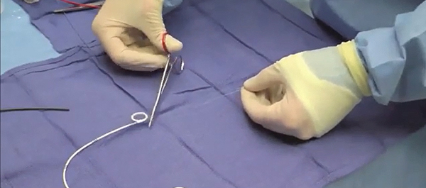
Finally, the third post under the Article of the Week heading on the homepage will consist of additional material or media. This week we feature a video from Dr. Barnes and colleagues summarising their paper on stent extraction strings.

If you only have time to read one article this week, it should be this one

Do ureteric stent extraction strings affect stent-related quality of life or complications after ureteroscopy for urolithiasis: a prospective randomised control trial

Kerri T. Barnes, Megan T. Bing and Chad R. Tracy

Department of Urology, University of Iowa, Iowa City, IA, USA

Read the full article
OBJECTIVE

• To determine whether ureteric stent extraction strings affect stent-related quality of life (QoL) or increase complications after ureteroscopy (URS) for stone disease.

PATIENTS AND METHODS

• In all, 68 patients undergoing URS (October 2011 to May 2013) for stone disease were randomised to receive a ureteric stent with or without an extraction string.

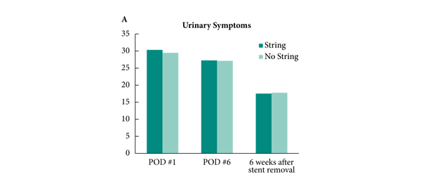
• Patients completed the Ureteric Stent Symptom Questionnaire (USSQ) on postoperative days 1 and 6, and 6 weeks after stent removal.

• Pain was assessed at stent removal.

• Adverse events, including early stent removal, stent migration, retained stent, urinary tract infection (UTI), emergency room (ER) visits and postoperative phone calls were monitored.

RESULTS

• There was no difference in stent-related QoL as measured by the USSQ between those with and without a stent extraction string, pain at stent removal between those who pulled their stent independently vs those who underwent cystoscopy for stent removal, or in the rate of UTIs, ER visits or phone calls between groups.

• Five patients (four female, one male) removed their stent early by inadvertently pulling the string; none required replacement.

• Patients without a string had a significantly longer period with the postoperative ureteric stent (10.6 vs 6.3 days, P < 0.001).

• One patient without a stent string retained her ureteric stent for 6 months, which was removed by cystoscopy without incident.

CONCLUSION

• Ureteric stent extraction strings may offer several advantages without increasing stent-related urinary symptoms, complications, or postoperative morbidity.


Editorial: Tether your stents!

Ureteric stents are commonly placed after ureteroscopy to protect the ureter and to facilitate subsequent stone fragment passage. They are known to be a cause of significant morbidity as judged by standardised validated questionnaires [1]. Whether placement of a stent is required at all is debatable, with randomised studies suggesting they are unnecessary after routine ureteroscopy [2]. The European Association of Urology (EAU) guidelines recommend stent insertion only ‘in patients who are at increased risk of complications’ and ‘in all doubtful cases to avoid stressful emergency situations’. Despite this, available evidence would suggest that we continue to commonly place stents [3].

If a stent is placed, the principal means of reducing morbidity is by minimising the stent dwell-time. One of the ways of doing this is to leave a stent with extraction strings/tether. This obviates the delay associated with scheduling cystoscopic extraction, the morbidity of cystoscopy and potentially reduces additional hospital visits if the patient is able to remove the stent at home.

Tethered stents are not widely used due to preconceptions about their tolerability, increased risk of complications (e.g. infection, migration) and accidental removal. Perhaps for this reason there have been few studies into the effectiveness of tethered stents in minimising stent-related morbidity to date, with only a handful in the past 30 years that have specifically addressed this issue.

In this issue of BJUI, Barnes et al. [4] report on the results of a prospective randomised trial analysing stented patients with or without the extraction strings attached, for both quality of life and postoperative complications after ureteroscopy for stone disease. This follows on from a retrospective series previously reported by the same group [5]. It is pleasing to see the authors, who originally concluded that randomised trials are needed in this area, actually get on and do the trial!

Two aspects of the trial methodology are worth highlighting: (i) the surgeons were not told that the patient was part of the study until they had made the decision to stent to minimise selection bias; (ii) patients completed the Ureteric Stent Symptom Questionnaire (USSQ) 6 weeks after stent removal as a control for their USSQ scores at postoperative days 1 and 6.

The headline results showed that there was no difference in quality of life and stent-related symptoms between patients with and without the extraction strings. There was also no difference in postoperative complications, emergency room visits or phone calls between the groups. What is surprising is that they found no difference in pain scores between self-removal and cystoscopic removal. This has not been our experience with tethered stents and may be due to the few men in the study. However, stent dwell-time was significantly less for patients with tethers compared with those without (10.6 vs 6.3 days, P < 0.001).

For urologists planning on using this technique it should be noted that the authors removed the original knot and shortened the string considerably to reduce the risk of accidental removal. For this reason the string was not attached to the patient’s skin.

This trial addresses many of the reservations urologists have about the use of tethered stents. Furthermore, reducing accidental removal and encouraging self-removal should be possible with improved patient education and selection. This was addressed by a study in New Zealand [6], which showed the feasibility of self-removal of stents.

The authors also acknowledged weaknesses in their study, which included failure to reach target enrolment, a 68% completion of trial surveys and a larger proportion of women in the study group due to male anxiety about self-removal of stents. In all, 15% of stents were inadvertently removed early and thus this technique should be used with caution in patients where early removal may be detrimental, e.g. in single kidneys. This does of course prompt the question: ‘If you are going to place a stent, how long does the stent need to stay for?’ and hopefully future trials may address this unanswered question.

Archana Fernando and Matthew Bultitude
Urology Department, Guy’s and St Thomas’ NHS Trust, London, UK

References

  1. Joshi HB, Newns N, Stainthorpe A et al. Ureteral stent symptom questionnaire: development and validation of a multidimensional quality of life measure. J Urol 2003; 169: 1060–1064
  2. Song T, Liao B, Zheng S, Wei Q. Meta-analysis of postoperatively stenting or not in patients underwent ureteroscopic lithotripsy. Urol Res 2012; 40: 67–77
  3. Mangera A, Parys B. BAUS Section of Endourology national ureteroscopy audit: setting the standards for revalidation. J Clin Urol 2012; 6: 45–49
  4. Barnes KT, Bing MT, Tracy CR. Do ureteric stent extraction strings affect stent-related quality of life or complications after ureteroscopy for urolithiasis: a prospective randomised control trial. BJU Int 2014; 113: 605–609
  5. Bockholt N, Wild T, Gupta A et al. Ureteric stent placement with extraction strings: no strings attached? BJU Int 2012; 110: 1069–1073
  6. York N, English S. Self-removal of ureteric JJ stents: analysis of patient experience. Presented at AUA 2013, May 7; San Diego, CA, USA. Abstract no. 1979. J Urol 2013; 189 (Suppl. 4): e812


Video: Stent extraction strings after ureteroscopy

Do ureteric stent extraction strings affect stent-related quality of life or complications after ureteroscopy for urolithiasis: a prospective randomised control trial

Kerri T. Barnes, Megan T. Bing and Chad R. Tracy

Department of Urology, University of Iowa, Iowa City, IA, USA

Read the full article
OBJECTIVE

• To determine whether ureteric stent extraction strings affect stent-related quality of life (QoL) or increase complications after ureteroscopy (URS) for stone disease.

PATIENTS AND METHODS

• In all, 68 patients undergoing URS (October 2011 to May 2013) for stone disease were randomised to receive a ureteric stent with or without an extraction string.

• Patients completed the Ureteric Stent Symptom Questionnaire (USSQ) on postoperative days 1 and 6, and 6 weeks after stent removal.

• Pain was assessed at stent removal.

• Adverse events, including early stent removal, stent migration, retained stent, urinary tract infection (UTI), emergency room (ER) visits and postoperative phone calls were monitored.

RESULTS

• There was no difference in stent-related QoL as measured by the USSQ between those with and without a stent extraction string, pain at stent removal between those who pulled their stent independently vs those who underwent cystoscopy for stent removal, or in the rate of UTIs, ER visits or phone calls between groups.

• Five patients (four female, one male) removed their stent early by inadvertently pulling the string; none required replacement.

• Patients without a string had a significantly longer period with the postoperative ureteric stent (10.6 vs 6.3 days, P < 0.001).

• One patient without a stent string retained her ureteric stent for 6 months, which was removed by cystoscopy without incident.

CONCLUSION

• Ureteric stent extraction strings may offer several advantages without increasing stent-related urinary symptoms, complications, or postoperative morbidity.


Second-degree burn after shock wave lithotripsy: an unusual complication

We present an unreported complication of a second-degree burn sustained by a 62-year-old male who underwent shockwave lithotripsy for two renal calculi. 


Authors: Sriram Rangarajan1, Hossein Mirheydar1, Roger L. Sur1,2

1. UC San Diego Health Sciences, Department of Surgery, Division of Urology; San Diego, CA
2. VA San Diego Medical Center, San Diego, CA

Corresponding Author: Roger L. Sur, MD, UC San Diego Health Sciences, Division of Urology, 200 W Arbor Dr. #8897, San Diego, CA 92103-8897. T: 619-543-2630   E-mail: [email protected]

Abstract
We present an unreported complication of a second-degree burn sustained by a 62-year-old male who underwent shockwave lithotripsy for two renal calculi.  The patient was treated conservatively for his burn injury but did require several ancillary procedures to render him stone free.  We speculate that the number of shocks and bubbles associated with an unapproved coupling medium for this particular lithotripter may have led to this highly unusual adverse event.


Introduction
 
Shockwave lithotripsy is considered a minimally invasive procedure for upper tract urinary stone disease with the obvious benefit of minimal side effects and rare adverse events.  These advantages have helped evolve it into a common surgical treatment modality especially due to its safety profile.  We report an unusual adverse event following shockwave lithotripsy involving a second degree skin burn at the coupling site.


Case Report
 
A 62-year-old Caucasian male presented with gross hematuria and left-flank pain. He had been seen in the emergency department 18 months prior for left-flank pain associated with two calcifications (a 9 mm midcalyceal calculus and an 8 mm lower pole calculus).  The patient’s past medical history was unremarkable. His medications included allopurinol, 300 mg, and aspirin, 81 mg, both of which he had discontinued one week prior to procedure. Physical examination revealed mild left costovertebral angle tenderness.  Computerized tomography (CT) urogram of the abdomen and pelvis demonstrated the two previously noted renal calculi  (10 mm,1500 HU in the left calyx and 9mm,1000 HU in the left calyx ).  Cystoscopy and cytological examination of two urine specimens did not reveal any abnormality.
The patient opted for shockwave lithotripsy (SWL). An electromagnetic lithotripter (Medispec EM1000, Germantown, MD), which our institution leases from Medispec, was used, and 4000 shocks (2000 to each calculus) were delivered at 90 shocks/minute after gradually increasing the power to 20kV. The Medispec technician did not have the standard coupling gel recommended by Medispec and instead used ultrasound gel available from the hospital.  In the recovery unit, the entire skin overlying the area where the gel had been placed was noted to be moderately erythematous with 3-4 cm central area of subcutaneous hemorrhage (figure 1).


Figure 1. demonstrates the immediate post-operative changes noted in the recovery unit: erythema and central region of petechiae
The patient returned on postoperative day 1 with significantly increased erythema and the development of large blisters (figure 2).  He also noted some abdominal pain, gross hematuria and passage of small fragments. The burn surgery service was consulted. A second-degree burn was confirmed, and local debridement was performed along with application of Collegnase, polymyxin B, xeroform, and gauze dressings.


Figure 2 demonstrates development of marked erythema and bullae on post-operative day 1


The patient returned again on postoperative day 5 complaining of left-flank pain and vomiting. The  wound remained erythematous with denuded skin. A noncontrast CT of his abdomen and pelvis CT  demonstrated an interval decrease in stone burden but multiple fragmented calculi (900HU) in the lower calyx as well as at least two adjacent 6 mm calculi in the proximal ureter (HU 1000). The patient consented to ureteroscopy with Ho:YAG laser lithotripsy.  During the left ureteroscopy with Ho:YAG laser lithotripsy and ureteric stenting, the ureteric calculi migrated into the kidney and one large calculus was fragmented.  A significant amount of mucous material in the urine precluded good visualization. The indwelling stent was removed 6 days later at outpatient cystoscopy.
Renal ultrasound performed 5 weeks later demonstrated a moderate left hydronephrosis and CT of the abdomen and pelvis revealed a 2 cm column of distal ureteral calculi. Subsequent ureteroscopy was aborted because of an iatrogenic extraperitoneal bladder perforation during dilation of an unexpected bulbar urethral stricture encountered during initial cystoscopy.  After documenting healing of the bladder, left ureteroscopy with Ho:YAG laser lithotripsy and ureteraic stenting was performed 10 days later. This rendered the patient stone free, as confirmed by direct visualization. Outpatient cystoscopic stent removal was performed 5 days later. He was asymptomatic two months afterwards with normal voiding and a  normal appearance on renal ultrasound.


Discussion
 
Complications following SWL are rare but include renal hematoma 1-13 % [1-3], steinstrasse 2-10% [4], acute renal injury, and theoretically, chronic renal injury such as a risk of hypertension [5].  To our knowledge this is the first case documenting a second-degree skin burn following SWL treatment.  There is no obvious explanation for this unusual adverse event, but some facts specific to this individual case deserve mention.  Firstly, we acknowledge that 4000 shocks might be considered marginally high. However, this has been routinely delivered with this particular lithotriptor in appropriate cases where calculi were in different renal locations, and this adverse event had never been experienced previously.  The two stones was located in different regions of the kidney and required 2000 shocks to adequately fragment under fluoroscopy.  It is tempting to surmise that a fewer shocks would have prevented this outcome.
We also note that conventional ultrasound gel was used in lieu of standard coupling gel approved for use with the Medispec EM1000 lithotripter.  The Medispec-recommended coupling medium is Lithoclear gel (Sonotech, Bellingham, WA). It has a density of 1.013 g/cc and is made of water-based couplants with a general composition of >80% water, 5 to 15% propylene glycol, 1 to 5 % glycerine, an acrylic polymer and a cosmetic-grade preservative.  The Lithoclear gel is filled and reportedly packaged under tighter controls than ultrasound gel to ensure that the containers are absolutely free of micro- and macrobubbles that would cause reflection or scattering of shock wave acoustics during SWL [6].  Anecdotally, it has higher viscosity than ultrasound gel. Higher viscosity may result in lower tendency to form bubbles [7].  The ideal medium would mimic the viscosity of water to minimize loss of energy but would be viscous enough that it would not run off the patient during the procedure.  The medium should also be devoid of air bubbles, as they are thought to further attenuate the energy transmission to the patient. Just as cavitation functions to fragment calculi during lithotripsy, some have proposed that cavitation of bubbles within the coupling medium is responsible for skin pain [7].  Could the skin burn be attributed to cavitation of excessive bubbles within the nonapproved ultrasound gel? Ultrasound gel is routinely used as a coupling agent in some centers, though ensuring the presence of bubbles is minimized is commonly advocated [8].  We can only speculate that excessive cavitation from the coupling medium associated with an elevated number of delivered shocks may have been sufficient to generate a thermal injury.  Another explanation is heat generated from the water-filled cushion, though there is no plausible reason to think this is possible.  The specific lithotripter used for this case was evaluated by the maker, Medispec, and no mechanical abnormalities were discovered.


References


1. Evan AP, Willis LR (2007). Extracorporeal shock wave lithotripsy: complications. In: Smith AD, Badlani G, Bagley D, eds. Smith’s Textbook of Endourology. 2nd ed. Hamilton, ON, BC Decker, 2007; chap 41: 353–65.
2. Orozco Farinas R, Iglesias Prieto JI, Massarrah Halabi J, Mancebo Gómez JM, Perez-Castro Ellendt E. Renal hematoma after extracorporeal shockwave lithotripsy in a series of 324 consecutive sessions with the DOLI-S lithotripter: incidents, characteristics, multifactorial analysis and review. Arch Espan Urol. 2008; 61: 889–914.
3. Mobley TB. Low energy lithotripsy with the lithostar: treatment results with 19,962 renal and ureteral stones. J Urol. 1993; 149: 1419–24.
4. Madbouly K, Sheir KZ, Elsobky E, Eraky I, Kenawy M. Risk factors for the formation of a steinstrasse after extracorporeal shock wave lithotripsy: a statistical model. J Urol. 2002; 167: 1239–42.
5. Lingeman JE, McAteer JA, Assimos DG, Baxley J, Kahn RI, Krambeck A, Matlaga BR, Penson D, Preminger GM, Zhong P. White Paper: Current Perspective on Adverse Effects in Shock Wave Lithotripsy. American Urological Association Education and Research, 2009.
6. Email communication with Sonotech.
7. Heidenreich A, Bonfig R, Wilbert DM, Engelmann UH. Painless ESWL by cutaneous administration of Vaseline. Urologe A. 1995 Jul;34(4):343-7. German
8. Cartledge JJ, CrossWR, Lloyd SN, Joyce AD. The efficacy of a range of contact media as coupling agents in extracorporeal shockwave lithotripsy. BJU Int. 2001; 88: 321–4.


Date added to bjui.org: 25/04/2012 


DOI: 10.1002/BJUIw-2012-006-web


© 2024 BJU International. All Rights Reserved.