Tag Archive for: multiparametric MRI

Posts

Editorial: Re‐thinking active surveillance for the multiparametric magnetic resonance imaging era

The last decade has seen a dramatic change in the management of low‐risk prostate cancer. Active surveillance (AS) has moved from a controversial management strategy to the preferred option for men with low‐risk disease. Despite widespread acceptance, there remain aspects of the pathway that men find difficult to accept, including the need for numerous repeat surveillance biopsies. In this issue of the BJUI, Gallagher et al. [1] report the outcomes of an AS programme using selective repeat biopsy based on multiparametric MRI (mpMRI) and PSA dynamics. The authors address the important issue of whether mpMRI can be used to safely avoid repeat biopsies in AS protocols.

The evidence for repeat biopsies in AS is based on studies from the pre‐MRI era, where up to 30% of men were upgraded on repeat systematic TRUS biopsy [2]. It has been established that TRUS biopsy is a highly unreliable test and misses a substantial proportion of clinically significant disease. The current approach requiring the repeated application of an unreliable test will not improve the systematic error inherent to the test. It is clear that the pathway needs to be updated for the mpMRI era, and the cohort of men in Gallagher et al. [1] provides valuable real‐life clinical data of an mpMRI‐based AS programme with a unique 4‐year follow‐up period.

The results are encouraging, with upgrading occurring in only 1.8% of men with a prior negative MRI. With follow‐up, progression to radical treatment was 12.8%, which is consistent with the established diagnostic performance of mpMRI. The authors seek further improvements by investigating if PSA dynamics can identify men with a negative MRI at risk of progression. They find that PSA velocity is strongly associated with subsequent progression (AUC 0.95, P < 0.001) and conclude that men on AS with low‐risk disease can safely avoid biopsy in favour of MRI, PSA monitoring and selective re‐biopsy. This study [1] supports a growing body of evidence that mpMRI may be adopted as the primary surveillance tool for men on AS. The finding regarding PSA velocity should be interpreted carefully as it contrasts with previous studies, which found that PSA dynamics have a limited role as independent predictors of disease progressions in AS [3]. A non‐invasive alternative to biopsy would be a valuable addition to AS and improve its acceptability as a management option. The burden of repeat surveillance biopsies for men on AS should not be underestimated. Indeed, in the present study ~30% of men declined biopsy in favour of continued mpMRI surveillance. The question is can we adapt our current standard AS approach for the mpMRI era? There are still many challenges and many unanswered questions. The cost‐effectiveness of mpMRI surveillance programmes needs to be established and the lack of MRI capacity remains a significant obstacle in introducing mpMRI pathways. The optimal imaging interval and the natural history of mpMRI lesions are just a few of the questions that need further research. These are exciting times to be a researcher in this field and there is much work to do as we start to build the new evidence‐base covering all the questions required for the mpMRI era.

References

  1. Gallagher KM, Christopher E, Cameron AJ et al. Four‐year outcomes from a multiparametric magnetic resonance imaging (MRI)‐based active surveillance programme: PSA dynamics and serial MRI scans allow omission of protocol biopsies. BJU Int 2019; 123: 429–38.
  2. Dall’Era MA, Albertsen PC, Bangma C et al. Active surveillance for prostate cancer: a systematic review of the literature. Eur Urol 2012; 62:976–83
  3. Loblaw A, Zhang L, Lam A et al. Comparing prostate specific antigen triggers for intervention in men with stable prostate cancer on active surveillance. J Urol 2010; 184: 1942–6


Video: Four-year outcomes from a multiparametric MRI based active surveillance programme

Four‐year outcomes from a multiparametric magnetic resonance imaging (MRI)‐based active surveillance programme: PSA dynamics and serial MRI scans allow omission of protocol biopsies


Abstract

Objectives

To report outcomes from a multiparametric (mp) magnetic resonance imaging (MRI)‐based active surveillance programme that did not include performing protocol biopsies after the first confirmatory biopsy.

Patients and Methods

All patients diagnosed with Gleason 3 + 3 prostate cancer because of a raised PSA level who underwent mpMRI after diagnosis were included. Patients were recorded in a prospective clinical database and followed up with PSA monitoring and repeat MRI. In patients who remained on active surveillance after the first MRI (with or without confirmatory biopsy), we investigated PSA dynamics for association with subsequent progression. Comparison between first and second MRI scans was undertaken. Outcomes assessed were: progression to radical therapy at first MRI/confirmatory biopsy and progression to radical therapy in those who remained on active surveillance after first MRI.

Results

A total of 211 patients were included, with a median of 4.2 years of follow‐up. The rate of progression to radical therapy was significantly greater at all stages among patients with visible lesions than in those with initially negative MRI (47/125 (37.6%) vs 11/86 (12.8%); odds ratio 4.1 (95% CI 2.0–8.5), P < 0.001). Only 1/56 patients (1.8%) with negative initial MRI scans who underwent a confirmatory systematic biopsy had upgrading to Gleason 3 + 4 disease. PSA velocity was significantly associated with subsequent progression in patients with negative initial MRI (area under the curve 0.85 [95% CI 0.75–0.94]; P <0.001). Patients with high‐risk visible lesions on first MRI who remained on active surveillance had a high risk of subsequent progression 19/76 (25.0%) vs 9/84 (10.7%) for patients with no visible lesions, despite reassuring targeted and systematic confirmatory biopsies and regardless of PSA dynamics.

Conclusion

Men with low‐risk Gleason 3 + 3 prostate cancer on active surveillance can forgo protocol biopsies in favour of MRI and PSA monitoring with selective re‐biopsy.


View more videos


Residents’ podcast: Implementation of mpMRI technology for evaluation of PCa in the clinic

Giulia Lane M.D. is a Fellow in Neuro-urology and Pelvic Reconstruction in the Department of Urology at the University of Michigan; Kyle Johnson is a Urology Resident in the same department.

In this podcast they discuss the following BJUI Article of the Month:

Implementation of multiparametric magnetic resonance imaging technology for evaluation of patients with suspicion for prostate cancer in the clinical practice setting

Abstract

Objectives

To investigate the impact of implementing magnetic resonance imaging (MRI) and ultrasonography fusion technology on biopsy and prostate cancer (PCa) detection rates in men presenting with clinical suspicion for PCa in the clinical practice setting.

Patients and Methods

We performed a review of 1 808 consecutive men referred for elevated prostate‐specific antigen (PSA) level between 2011 and 2014. The study population was divided into two groups based on whether MRI was used as a risk stratification tool. Univariable and multivariable analyses of biopsy rates and overall and clinically significant PCa detection rates between groups were performed.

Results

The MRI and PSA‐only groups consisted of 1 020 and 788 patients, respectively. A total of 465 patients (45.6%) in the MRI group and 442 (56.1%) in the PSA‐only group underwent biopsy, corresponding to an 18.7% decrease in the proportion of patients receiving biopsy in the MRI group (P < 0.001). Overall PCa (56.8% vs 40.7%; P < 0.001) and clinically significant PCa detection (47.3% vs 31.0%; P < 0.001) was significantly higher in the MRI vs the PSA‐only group. In logistic regression analyses, the odds of overall PCa detection (odds ratio [OR] 1.74, 95% confidence interval [CI] 1.29–2.35; P < 0.001) and clinically significant PCa detection (OR 2.04, 95% CI 1.48–2.80; P < 0.001) were higher in the MRI than in the PSA‐only group after adjusting for clinically relevant PCa variables.

Conclusion

Among men presenting with clinical suspicion for PCa, addition of MRI increases detection of clinically significant cancers while reducing prostate biopsy rates when implemented in a clinical practice setting.

Read the full article

BJUI Podcasts now available on iTunes, subscribe here https://itunes.apple.com/gb/podcast/bju-international/id1309570262


Article of the month: Implementation of multiparametric MRI technology for evaluation of PCa in the clinic

Every month, the Editor-in-Chief selects an Article of the Month from the current issue of BJUI. The abstract is reproduced below and you can click on the button to read the full article, which is freely available to all readers for at least 30 days from the time of this post.

In addition to the article itself, there is an editorial written by a prominent member of the urological community, and a podcast produced by our current Resident Podcasters. These are intended to provoke comment and discussion and we invite you to use the comment tools at the bottom of each post to join the conversation. 

If you only have time to read one article this week, it should be this one.

Implementation of multiparametric magnetic resonance imaging technology for evaluation of patients with suspicion for prostate cancer in the clinical practice setting

Paras H. Shah*, Vinay R. Patel, Daniel M. Moreira, Arvin K. George§, Manaf Alom*, Zachary Kozel, Vidhu Joshi*, Eran Ben-Levi**, Robert Villani**, Oksana Yaskiv††Louis R. Kavoussi, Manish Vira, Carl O. Olsson‡‡ and Ardeshir R. Rastinehad


*Department of Urology, Mayo Clinic, Rochester, MN, Department of Urology, Icahn Smith Institute for Urology, Northwell Health, New York, NY, Department of Urology, University of Illinois at Chicago, Chicago, IL, §Department of Urology, University of Michigan, Ann Arbor, MI, Department of Urology, Smith Institute for Urology, Northwell Health, **Department of Radiology, Hofstra Northwell School of Medicine, ††Department of Pathology, Hofstra Northwell School of Medicine, New Hyde Park, and ‡‡Integrated Medical Professionals, Melville, NY, USA


Read the full article

Abstract

Objectives

To investigate the impact of implementing magnetic resonance imaging (MRI) and ultrasonography fusion technology on biopsy and prostate cancer (PCa) detection rates in men presenting with clinical suspicion for PCa in the clinical practice setting.

Patients and Methods

We performed a review of 1 808 consecutive men referred for elevated prostate‐specific antigen (PSA) level between 2011 and 2014. The study population was divided into two groups based on whether MRI was used as a risk stratification tool. Univariable and multivariable analyses of biopsy rates and overall and clinically significant PCa detection rates between groups were performed.

Results

The MRI and PSA‐only groups consisted of 1 020 and 788 patients, respectively. A total of 465 patients (45.6%) in the MRI group and 442 (56.1%) in the PSA‐only group underwent biopsy, corresponding to an 18.7% decrease in the proportion of patients receiving biopsy in the MRI group (P < 0.001). Overall PCa (56.8% vs 40.7%; P < 0.001) and clinically significant PCa detection (47.3% vs 31.0%; P < 0.001) was significantly higher in the MRI vs the PSA‐only group. In logistic regression analyses, the odds of overall PCa detection (odds ratio [OR] 1.74, 95% confidence interval [CI] 1.29–2.35; P < 0.001) and clinically significant PCa detection (OR 2.04, 95% CI 1.48–2.80; P < 0.001) were higher in the MRI than in the PSA‐only group after adjusting for clinically relevant PCa variables.

Conclusion

Among men presenting with clinical suspicion for PCa, addition of MRI increases detection of clinically significant cancers while reducing prostate biopsy rates when implemented in a clinical practice setting.

Read more Articles of the week


Editorial: Multiparametric MRI for prostate cancer detection: do clinical trial findings reflect real‐world practice?

‘First, do no harm’; with this in mind, researchers in urology strive to minimize the burden of overdiagnosis and overtreatment of prostate cancer. A promising tool in this arena is multiparametric (mp)MRI, which has been shown in a large‐scale randomized clinical trial to enhance the ability of prostate biopsy to detect clinically significant prostate cancer [1]. The extent to which findings from an idealized trial protocol extend to ‘real‐world’ clinical practice, however, remains largely unknown.

In this issue of BJUI, Shah et al. [2] aimed to fill this knowledge gap by investigating the impact of mpMRI‐guided biopsy on the detection rates of clinically significant prostate cancer in two large academic centres. The authors studied men with an elevated PSA presenting over a 3‐year span (2011–2014); 1020 men underwent mpMRI and 788 did not. Those in the MRI group had higher detection rates of both overall and clinically significant prostate cancer, defined as any Gleason score ≥7 on fusion or standard 12‐core TRUS biopsies, Gleason 6 with a lesion volume >0.5 cm3 volume on MRI, or Gleason 6 with >2 cores positive and/or >50% of any core involved with cancer on biopsy according to Epstein’s criteria, as well as a lower detection rate of clinically insignificant cancer.

The study provides timely implications for both patients and physicians, providing further insight into how findings from clinical trials [1,3] compare with real‐life practice. In fairness, the bulk of patients and clinicians do not follow strict study protocols for both decision‐making and interpretation of results, but rather assess very individual situations. A recent study by Bukavina et al. [4] showed that urologists and radiation oncologists largely perceive mpMRI guidance for targeted biopsies as valuable tools to improve prostate cancer stratification, but only a quarter of respondents reported implementation into their own clinical practice. This underlines some of the challenges of widespread implementation of mpMRI despite strong belief in its value.

Another strength of the study by Shah et al. is the exclusion of men who underwent mpMRI after negative biopsy in the PSA‐only group. This allows the isolation of the impact of mpMRI on downstream biopsy outcomes. A previous study that investigated targeted vs non‐targeted biopsies enrolled a cohort of men who all underwent mpMRI [5], which precludes any assessment of how mpMRI may impact the detection of clinically significant prostate cancer. Shah et al. [2] also astutely tracked detection rates of clinically significant and insignificant prostate cancer. Since the process of diagnosing prostate cancer is not without morbidity, it is crucial to understand the extent to which mpMRI can prevent the diagnosis of clinically indolent cancers.

Important questions regarding the challenges of widespread implementation of mpMRI for prostate cancer detection remain unanswered by the study of Shah et al. The study participants were gathered from large academic centres with readily available equipment, infrastructure and physician expertise to maximize favourable detection outcomes; however, these results may not be representative of the community setting. Additionally, >20% of men who did not undergo mpMRI did not do so because of a lack of insurance approval. This may reflect socio‐economic differences between the groups and also relates to the high costs of mpMRI that make routine implementation difficult [6]. Lastly, the presented findings mostly apply to positive mpMRI scans; the number of underdiagnosed men with negative scans may only be speculated upon, given the lack of follow‐up data in this population. It remains fundamentally important to improve the management of men with elevated PSA levels and negative findings on MRI.

Nonetheless, the present study demonstrates that research findings find their way into clinical practice. In essence, we are doing well, but we can do better.

by Marieke J. Krimphove, Sean A. Fletcher and Quoc‐Dien Trinh


References

  1. Kasivisvanathan V, Rannikko AS, Borghi M et al. MRI‐targeted or standard biopsy for prostate‐cancer diagnosis. N Engl J Med 2018378: 1767–77
  2. Shah PH, Patel VR, Moreira DM et al. Implementation of multiparametric magnetic resonance imaging technology for evaluation of patients with suspicion for prostate cancer in the clinical practice setting. BJU Int 2019123: 239–45
  3. Ahmed HU, El‐Shater Bosaily A, Brown LC et al. Diagnostic accuracy of multi‐parametric MRI and TRUS biopsy in prostate cancer (PROMIS): a paired validating confirmatory study. Lancet 2017389: 815–22
  4. Bukavina L, Tilburt JC, Konety B et al. Perceptions of prostate MRI and fusion biopsy of radiation oncologists and urologists for patients diagnosed with prostate cancer: results from a national survey. Eur Urol Focus 2018; [Epub ahead of print]
  5. Pokorny MR, de Rooij M, Duncan E et al. Prospective study of diagnostic accuracy comparing prostate cancer detection by transrectal ultrasound–guided biopsy versus magnetic resonance (MR) imaging with subsequent MR‐guided biopsy in men without previous prostate biopsies. Eur Urol 201466: 22–9
  6. Kim SJ, Vickers AJ, Hu JC. Challenges in adopting level 1 evidence for multiparametric magnetic resonance imaging as a biomarker for prostate cancer screening. JAMA Oncol 2018; [Epub ahead of print]


Re: Does the introduction of prostate multiparametric MRI into the active surveillance protocol for localized PCa improve patient re-classification?

Letter to the Editor

Does the introduction of prostate multiparametric magnetic resonance imaging into the active surveillance protocol for localized prostate cancer improve patient re-classification?

Dear Sir,

Recently Bryant and colleagues published an important series on multiparametric magnetic resonance imaging (mpMRI) in active surveillance (AS) for prostate cancer [1]. The work was deservedly highlighted as Paper of the Week for its potential impact on clinical practice. Meanwhile, it caught our attention that the authors – as many before them – use the term “significant prostate cancer” throughout their paper. We feel that this issue needs to be addressed and discussed further.

As most prostate cancers have an indolent course, the term “significant prostate cancer” must be reserved for cancers, which are potentially lethal [2]. From earlier studies, we know that this group of cancers are characterized by high tumor stages, high Gleason scores, and a high percentage of tumor involvement on biopsies. However, this knowledge is based on systematic prostate biopsies. Thus, findings on mpMRI targeted biopsies, including increase in Gleason grade group, number of positive cores, and maximum cancer core length, cannot readily be translated to the clinical setting of earlier times. Tumor grade and volume of the disease may simply be upgraded even with no change in the underlying cancers. Nevertheless, this is usually how such findings are used and the focus is mainly on moving patients from AS to active treatment.

The potential problems of this approach are illustrated in the results of Bryant et al., as 30% of the patient cohort is taken out of the AS program after a median time of only 1.55 years with mpMRI based findings as the most common cause. In previous publications, men have been able to stay on AS for considerably longer with limited consequences on metastasis free survival [3]. The issue is further highlighted by results from the ProtecT trial in which there was a median of about 4 years before 30% of the active monitoring group had gone on to active treatment, without this causing a significant increase in either overall survival or disease specific survival [4]. In addition, data from prostate cancer cohorts not treated with curative intent show us that even men with localized intermediate risk prostate cancer have a low risk of dying from prostate cancer [5]. Seen in this context, it is plausible that some cancers are falsely classified as “significant” cancers as a result of mpMRI. Indeed, Bryant and colleagues illustrate that men undergoing mpMRI have an increased risk of discontinuing AS which may result in increased over-treatment. The issue is further exacerbated by the tendency to treat increasingly older patients even though a survival benefit from active treatment is questionable in this group.

There is no doubt that mpMRI may provide important and valuable information and it is a massive leap forward that we might be able to identify occult high-grade cancers early. However, this new tool must be used with consideration and respect for the gaps in our knowledge. In that context, it is likely that current indications for AS can be expanded to include more favorable intermediate risk cancers when the diagnosis is made based on mpMRI targeted biopsies combined with systematic biopsies. In addition, mpMRI should be utilized to reduce the proportion of patients who opt for active treatment due to anxiety or the burden of multiple sets of systematic biopsies. Exploring these issues are crucial because staying on AS is not just about safely avoiding surgery or radiotherapy. Rather it is about avoiding or postponing highly prevalent and clinically significant functional side effects with considerable implications for quality of life. Thus, we want to underline that the use of mpMRI should not only be used to exclude patients from AS protocols but also in future protocols to include men in such programs.


Conflicts of interest: Mikkel Fode is Advisory Board member for Astellas Pharma A/S and has received honoraria as speaker from Astellas Pharma A/S and Ferring Pharmaceuticals. Peter B. Østergren has received honoraria as speaker from Astellas Pharma A/S, Ferring Pharmaceuticals and IPSEN. Dr. Østergren has received honoraria for consultancies from Astellas Pharma A/S and as Advisory Board member for IPSEN. Kasper D. Berg has no conflicts of interest to declare.


Mikkel Fode MD, PhD, FECSM*, Kasper Drimer Berg MD, PhD†**, Peter Busch Østergren* MD, PhD

*Department of Urology, Herlev and Gentofte Hospital, Herlev, Denmark, Department of Urology, Regional Hospital West Jutland, Holstebro, Denmark, **Copenhagen Prostate Cancer Center, Department of Urology, Rigshospitalet, University of Copenhagen, Copenhagen, Denmark.


References

  1. Bryant RJ, Yang B, Philippou Y, Lam K, Obiakor M, Ayers J, et al. Does the introduction of prostate multiparametric magnetic resonance imaging into the active surveillance protocol for localized prostate cancer improve patient re-classification? BJU Int.; 2018;122(5):794–800.
  2. Popiolek M, Rider JR, Andren O, Andersson S-O, Holmberg L, Adami H-O, et al. Natural history of early, localized prostate cancer: a final report from three decades of follow-up. Eur Urol.; 2013;63(3):428–35.
  3. Bokhorst LP, Valdagni R, Rannikko A, Kakehi Y, Pickles T, Bangma CH, et al. A Decade of Active Surveillance in the PRIAS Study: An Update and Evaluation of the Criteria Used to Recommend a Switch to Active Treatment. Eur Urol.; 2016;70(6):954–60.
  4. Hamdy FC, Donovan JL, Lane JA, Mason M, Metcalfe C, Holding P, et al. 10-Year Outcomes after Monitoring, Surgery, or Radiotherapy for Localized Prostate Cancer. N Engl J Med.; 2016;375(15):1415–24.
  5. Rider JR, Sandin F, Andren O, Wiklund P, Hugosson J, Stattin P. Long-term outcomes among noncuratively treated men according to prostate cancer risk category in a nationwide, population-based study. Eur Urol.; 2013;63(1):88–96.


Read the article


Reply by the authors

We thank Dr Fode and colleagues for their correspondence regarding our recently published manuscript [1]. We wish to clarify some of the issues and comments they highlight. Regarding the term “significant prostate cancer”, we agree that this definition should be reserved for cancers that are potentially lethal. Historically, and currently, in the context of Active Surveillance (AS), these cancers are characterised by a higher tumour stage, increased number of positive biopsy cores and/or longer cancer length, and higher Gleason grade group compared to baseline findings, in the absence of routine clinical use of any potential molecular determinants of a lethal phenotype. The clinical question addressed in our recent manuscript was whether introducing multi-parametric magnetic resonance imaging (mpMRI) into our AS protocol for localised prostate cancer improved patient re-classification. We did not define “significant prostate cancer”, but rather focused on the drivers for men to abandon AS and receive active treatment. We agree that the use of this imaging adjunct may therefore lead to disease upgrading, even without a biological change in the underlying cancer per se, and hence our use of the term “re-classification”, rather than disease “progression”.

The results of the targeted (plus systematic) biopsy following the initial undertaking of mpMRI during AS likely accounts for much of the 30% of our cohort receiving radical treatment after a median time of 1.55 years, as our protocol had been to undertake that first mpMRI around one year after starting AS. The reported cohort of AS patients had not received a baseline pre-biopsy mpMRI, as this practice was introduced at our institution in 2016 [2], after these men had started AS. Hence, it is likely that the timing of men leaving AS to receive treatment was triggered by the first mpMRI as a follow-up measure and subsequent repeat targeted biopsies, rather than true biological disease progression. The use of indiscriminate repeat biopsies at pre-specified and arbitrary intervals during any AS protocol (as practiced historically before the availability of mpMRI) is likely to have driven men towards active treatment unnecessarily, and added to potential adverse effects of repeat biopsies on functional outcomes after radical prostatectomy [3].

With regards to the ProtecT study [4], men in the non-intervention arm received Active Monitoring (AM), rather than intensive AS. In ProtecT, men on AM did not receive regular imaging by mpMRI, nor did they receive repeat biopsies unless triggered by a suspicion of disease progression. This is very different from contemporary AS protocols. It is therefore inappropriate to draw parallels between AM in ProtecT and contemporary AS.

We agree that benefits of mpMRI in AS programs may be to reduce the proportion of patients who opt for active treatment due to anxiety, as well as decreasing the burden of multiple sets of unnecessary systematic biopsies.

The true definition of “clinically significant” prostate cancer is subject to much controversy, and many prostate cancers are classified inappropriately as “significant”. This depends on complex biological and molecular features of aggressive and lethal disease – yet to be defined by ongoing research – as well as competing co-morbidity. We agree entirely with Fode and colleagues that many men with localised intermediate-risk prostate cancer have a relatively low risk of dying from prostate cancer. This underpins the recommendation from the UK National Institute for Health and Care Excellence (NICE) that patients with low-volume low- and intermediate-risk localised prostate cancer may consider AS as a viable management option, re-iterated in its recently updated guidelines under consultation [5,6,7].


Richard J. Bryant*† , Prasanna Sooriakumaran†**, Freddie C. Hamdy*† and Simon F. Brewster*

*Department of Urology, Oxford University Hospitals NHS Foundation Trust, Oxford, UK, †Nuffield Department of Surgical Sciences, University of Oxford, Oxford, UK, and **Department of Uro-Oncology, University College London Hospital NHS Foundation Trust, London, UK


References

  1. Bryant RJ, Yang B, Philippou Y, Lam K, Obiakor M, Ayers J, et al. Does the introduction of prostate multiparametric magnetic resonance imaging into the active surveillance protocol for localized prostate cancer improve patient re-classification? BJU Int.; 2018;122(5):794–800.
  2. Bryant RJ, Hobbs CP, Eyre KS, Davies LC, Sullivan ME, Shields W, et al. Comparison of prostate biopsy with or without pre-biopsy multi-parametric MRI in prostate cancer detection: an observational cohort study. J Urol. 2018. [Epub ahead of print]
  3. Sooriakumaran P, Calaway A, Sagalovich D, Roy S, Srivastava A, Joneja J, et al. The impact of multiple biopsies on outcomes of nerve-sparing robotic-assisted radical prostatectomy. Int J Impot Res. 2012;24(4):161-4.
  4. Hamdy FC, Donovan JL, Lane JA, Mason M, Metcalfe C, Holding P, et al. 10-Year Outcomes after Monitoring, Surgery, or Radiotherapy for Localized Prostate Cancer. N Engl J Med.; 2016;375(15):1415–24.
  5. National Institute for Health and Care Excellence. Prostate Cancer: Diagnosis and Management (CG175). National Institute for Clinical Excellence; 2014.
  6. Graham J, Kirkbride P, Cann K, Hasler E, Prettyjohns M. Prostate cancer: summary of updated NICE guidance. BMJ 2014; 348: f7524
  7. https://www.nice.org.uk/guidance/GID-NG10057/documents/evidence-review-7.


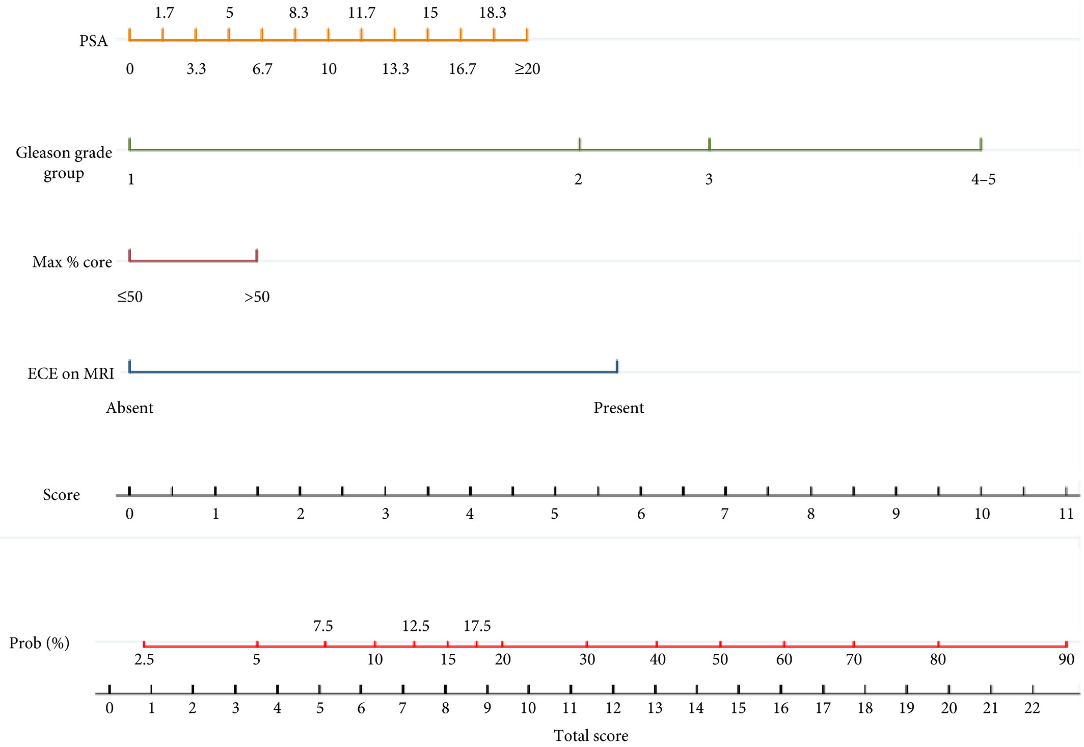
Infographic: Development of a side‐specific, mpMRI‐based nomogram for the prediction of extracapsular extension of PCa

Infographic: Development of a side‐specific, mpMRI‐based nomogram for the prediction of extracapsular extension of PCa

Read the full article

See more infographics

Article of the week: Development of a side‐specific, mpMRI‐based nomogram for the prediction of extracapsular extension of PCa

Every week, the Editor-in-Chief selects an Article of the Week from the current issue of BJUI. The abstract is reproduced below and you can click on the button to read the full article, which is freely available to all readers for at least 30 days from the time of this post.

In addition to the article itself, there are two accompanying editorials written by prominent members of the urological community. These are intended to provoke comment and discussion and we invite you to use the comment tools at the bottom of each post to join the conversation. There is also a video produced by the authors. 

If you only have time to read one article this week, it should be this one.

Development and internal validation of a side‐specific, multiparametric magnetic resonance imaging‐based nomogram for the prediction of extracapsular extension of prostate cancer

Alberto Martini*, Akriti Gupta*, Sara C. Lewis, Shivaram Cumarasamy*, Kenneth G. Haines III§, Alberto Briganti, Francesco Montorsiand Ashutosh K. Tewari*


Departments of *Urology, Radiology, §Pathology, Icahn School of Medicine at Mount Sinai, New York, NY, USA and Department of Urology, Vita-Salute San Raffaele University, Milan, Italy

Read the full article

Abstract

Objectives

To develop a nomogram for predicting side‐specific extracapsular extension (ECE) for planning nerve‐sparing radical prostatectomy.

Materials and Methods

We retrospectively analysed data from 561 patients who underwent robot‐assisted radical prostatectomy between February 2014 and October 2015. To develop a side‐specific predictive model, we considered the prostatic lobes separately. Four variables were included: prostate‐specific antigen; highest ipsilateral biopsy Gleason grade; highest ipsilateral percentage core involvement; and ECE on multiparametric magnetic resonance imaging (mpMRI). A multivariable logistic regression analysis was fitted to predict side‐specific ECE. A nomogram was built based on the coefficients of the logit function. Internal validation was performed using ‘leave‐one‐out’ cross‐validation. Calibration was graphically investigated. The decision curve analysis was used to evaluate the net clinical benefit.

Results

The study population consisted of 829 side‐specific cases, after excluding negative biopsy observations (n = 293). ECE was reported on mpMRI and final pathology in 115 (14%) and 142 (17.1%) cases, respectively. Among these, mpMRI was able to predict ECE correctly in 57 (40.1%) cases. All variables in the model except highest percentage core involvement were predictors of ECE (all P ≤ 0.006). All variables were considered for inclusion in the nomogram. After internal validation, the area under the curve was 82.11%. The model demonstrated excellent calibration and improved clinical risk prediction, especially when compared with relying on mpMRI prediction of ECE alone. When retrospectively applying the nomogram‐derived probability, using a 20% threshold for performing nerve‐sparing, nine out of 14 positive surgical margins (PSMs) at the site of ECE resulted above the threshold.

Conclusion

We developed an easy‐to‐use model for the prediction of side‐specific ECE, and hope it serves as a tool for planning nerve‐sparing radical prostatectomy and in the reduction of PSM in future series.

Read more Articles of the week


Editorial: A novel nomogram for predicting ECE of prostate cancer

We read with great interest the publication on the side‐specific multiparametric magnetic resonance imaging (mpMRI)‐based nomogram from Martini et al. [1].

The prediction of extracapsular extension (ECE) of prostate cancer is of utmost importance to inform accurate surgical planning before radical prostatectomy (RP).

Today, surgical strategy is tailored to the patient’s characteristics, and the need for a correct prediction of ECE is of paramount importance to guarantee oncological safety, as well as optimal functional outcome. The most up‐to‐date guidelines suggest referring to nomograms to decide whether or not to perform nerve‐sparing (NS) surgery. Since the first version of the Partin Tables in 1993, several models have been developed based on PSA, Gleason score at prostate biopsy, and clinical staging, as the most used covariates.

Furthermore, mpMRI is increasingly used in the diagnostic pathway of prostate cancer to aid prostate biopsy targeting and to attain a more accurate diagnosis of clinically significant prostate cancer. Despite its recognised role in the detection of cancer, the accuracy for local staging is poor, providing a low and heterogeneous sensitivity for the detection of ECE [2].

Given this limitation, the addition of MRI to clinically derived nomograms might result in an improved assessment of preoperative local staging. In a retrospective analysis of 501 patients who underwent RP, MRI + clinical models outperformed clinical‐based models alone for all staging outcomes, with better discrimination in predicting ECE with MRI + Partin Tables and MRI + Cancer of the Prostate Risk Assessment (CAPRA) score than nomograms alone [3].

In the current article, Martini et al. [1] suggest a novel nomogram for predicting ECE that includes the presence of a ‘documented definite ECE at mpMRI’ as an additional variable beyond PSA, Gleason score, and maximum percentage of tumour in the biopsy core with the highest Gleason score. Readers should recognise that this is the first model integrating side‐specific MRI findings together with side‐specific biopsy data to provide a ‘MRI‐based side‐specific prediction of ECE’, in an effort to support the surgical decision for a uni‐ or bilateral NS approach.

However, given the frail generalisability of nomograms in different datasets even after external validation [4], a predictive tool has to be built on a rigorous methodology with clear reproducibility of all steps the covariates derive from.

In this respect, the current model raises some concerns.

The schedule of preoperative MRI assessment is arbitrary, with imaging being performed either before (23.9%) or after systematic biopsy (76.1%), and amongst patients with a MRI prior to biopsy, only 94 of 134 patients underwent additional targeted sampling. As a result, MRI is applied by chance in three different ways: before prostate biopsy without targeted sampling, before prostate biopsy with targeted sampling, and after prostate biopsy.

Based upon this heterogeneous MRI timing, the performance of such a model in a novel population may be biased depending on the diagnostic pathway applied at each institution.

The choice of the variables included represents another point of concern. The output of two out of four covariates, ECE depiction at mpMRI and the percentage of tumour in the biopsy core, have been deliberately dichotomised, without taking into account the continuous trend intrinsic to both variables.

Actually, local staging in the European Society of Urogenital Radiology (ESUR) guidelines has been scored on a 1–5 point scale to grade the likelihood of an ECE event. The authors deliberately dichotomised mpMRI findings, considering ‘the loss of prostate capsule and its irregularity’ as suggestive of ECE and ‘broad capsular contact, abutment or bulge without gross ECE’ evocative of organ‐confined disease. As a result, the included MRI covariate may account for a gross prediction of ECE, maintaining the inaccurate and inter‐reader subjective interpretation of local staging intrinsic to MRI.

Beyond those methodological concerns and the moderate sample size that may limit the reproducibility of the model, we wonder if such a prediction really assists the surgeon’s capability to perform a tailored surgery.

The ‘all or none’ era of NS surgery is over, and we are currently able to grade NS according to different approaches reported in the literature. Particularly, Tewari et al. [5] proposed a NS approach based on four grades of dissection, with the veins on the lateral aspect as vascular landmarks to gain the correct dissection planes. Patel et al. [6] described a five‐grade scale of dissection, using the arterial periprostatic vasculature as a landmark to the same purpose.

If we are able to grade a NS surgery, the prediction of ECE should be graded as well and should answer the prerequisite of knowing the amount of prostate cancer extent outside the capsule. How does a surgeon make the decision to follow a more or less conservative dissection otherwise?

We tried to address this issue by using a tool aimed at predicting the amount of ECE [the Predicting ExtraCapsular Extension in Prostate cancer tool] [6] and supporting the choice of the correct plane of dissection with a suggested decision rule. In our study, developed on a large sample of nearly 12 000 prostatic lobes and several combined clinicopathological variables, the absence of imaging characterization was the major point of weakness.

To date, the ideal predictive tool has yet to be described. However, in the modern era of precision surgery, we think that a model should encompass the surgical knowledge and techniques currently available.

Future developments will probably include three‐dimensional surgical navigation models displayed on the TilePro™ function of the robotic console (Intuitive Surgical Inc., Sunnyvale, CA, USA), based on the integration of MRI (for the number, size and location of disease) and predictive tools (to define the amount of ECE).


References

  1. Martini A, Gupta A, Lewis SC et al. Development and internal validation of a side‐specific, multiparametric magnetic resonance imaging‐based nomogram for the prediction of extracapsular extension of prostate cancer. BJU Int 2018; 122: 1025–33
  2. de Rooij M, Hamoen EH, Witjes JA, Barentsz JO, Rovers MM. Accuracy of magnetic resonance imaging for local staging of prostate cancer: a diagnostic meta‐analysis. Eur Urol 2016; 70: 233–45
  3. Morlacco A, Sharma V, Viers BR et al. The incremental role of magnetic resonance imaging for prostate cancer staging before radical prostatectomy. Eur Urol 2017; 71: 701–4
  4. Bleeker SE, Moll HA, Steyerberg EW et al. External validation is necessary in prediction research: a clinical example. J Clin Epidemiol 2003; 56: 826–32
  5. Tewari AK, Srivastava A, Huang MW et al. Anatomical grades of nerve sparing: a risk‐stratified approach to neural‐hammock sparing during robot‐assisted radical prostatectomy (RARP). BJU Int 2011; 108: 984–92
  6. Patel VR, Sandri M, Grasso AA et al. A novel tool for predicting extracapsular extension during graded partial nerve sparing in radical prostatectomy. BJU Int 2018; 121: 373–82


Editorial: A picture is worth a thousand words… but does it add utility to a nomogram to predict extraprostatic extension?

Martini et al. [1] ask whether adding in prostate MRI data to a preoperative nomogram can usefully aid in the decision to nerve‐spare on one or both sides in men undergoing radical prostatectomy, using a dataset of 829 positive prostate lobes in 561 men. The nomogram includes PSA, maximum ipsilateral Gleason grade, percentage core involvement, and presence of extracapsular extension (ECE) on MRI, although the percentage core involvement (< or >50%) was not found to be significant. Pathological ECE was noted in 142 (17.1%) of the lobes, and radiological suspicion of ECE was noted in 115 (14%) lobes.

The incorporation of MRI in the decision‐making process is to be welcomed. However, MRI only correctly predicted ECE in 57/142 (40.1%) cases, showing significant over‐ and under‐detection on MRI criteria alone. Nerve‐sparing was done in 78% of men, and 30 men had a positive surgical margin. The authors found the nomogram to have greater accuracy in predicting ECE than MRI alone, with an area under the curve for MRI alone of 68.83%, compared to 82.92% for the nomogram. The use of the nomogram to inform a decision to nerve‐spare, made independently for each side, is proposed.

We need to be clear about the different definitions that are being applied here. The MRI features used for assessing ECE, namely bulging/irregular margin, obliteration of the rectoprostatic angle, >1 cm capsular abutment, and neurovascular bundle invasion, set a somewhat high threshold, which we would expect to correlate with significant histological burden and ECE. The exact pathological definition of ECE is not described by the authors and so presumably includes presence of any cancer outside the surgical capsule, whilst the presence of a positive surgical margin is defined as any tumour touching an inked margin. This difference in the threshold for radiological and pathological significance of ECE has been noted by others [2]. In addition, there is some discussion of the long‐term clinical significance of a positive surgical margin of <3 mm [3], although both ECE and PSM are recognised as predictors of recurrence.

Even given this discrepancy in definitions, there are other possible reasons why MRI was less predictive than might be expected [4]. The majority (76%) of the MRI scans were done after biopsy, which is known to reduce the accuracy of MRI, resulting in both under‐ and over‐staging. These post‐biopsy effects can persist for some considerable time, often past the 4 week post‐biopsy recovery period used as the minimum in this series, and in many institutions [5]. Differences in prevalence of pathological ECE (17% in this series [1] vs 32.4% in the series reported by Gaunay et al. [4]) could also affect the performance characteristics of MRI for staging.

An alternative to the preoperative nomogram approach is the use of techniques such as neurovascular structure‐adjacent frozen‐section examination (NeuroSAFE) [6]. This allows an intraoperative decision on the extent of excision, based on frozen‐section examination, and it has been shown to increase the ability to nerve‐spare, with associated improved functional outcomes, whilst reducing positive surgical margins. However, it does have significant cost implications and is not widely available.

It makes sense to use preoperative MRI, currently widely recommended for staging, in combination with clinical parameters, to maximise the use of nerve‐sparing to favour functional outcomes, whilst minimising positive surgical margins. Martini et al. [1] present a nomogram based on readily available parameters, which could be readily adopted in the routine setting. The move towards MRI before first biopsy is likely to give us more accurate imaging data, which should help us to further refine the decision to nerve‐spare for men undergoing radical prostatectomy.

References

  1. Martini A, Gupta A, Lewis S et al. Development and internal validation of a side‐specific, multiparametric magnetic resonance imaging‐based nomogram for the prediction of extracapsular extension of prostate cancer. BJU Int 2018; 122: 1025–33
  2. Dev HS, Wiklund P, Patel V et al. Surgical margin length and location affect recurrence rates after robotic prostatectomy. Urol Oncol 2015; 33: 109.e7‐13
  3. Gaunay GS, Patel V, Shah P et al. Multi‐parametric MRI of the prostate: factors predicting extracapsular extension at the time of radical prostatectomy. Asian J Urol 2017; 4: 31–6
  4. Latifoltojar A, Dikaios N, Ridout A et al. Evolution of multi‐parametric MRI quantitative parameters following transrectal ultrasound‐guided biopsy of the prostate. Prostate Cancer Prostatic Dis 2015; 18: 343–51
  5. Mirmilstein G, Rai BP, Gbolahan O et al. The neurovascular structure‐adjacent frozen‐section examination (NeuroSAFE) approach to nerve sparing in robot‐assisted laparoscopic radical prostatectomy in a British setting ‐ a prospective observational comparative study. BJU Int 2018; 121: 854–62


© 2024 BJU International. All Rights Reserved.