Tag Archive for: metastasis

Posts

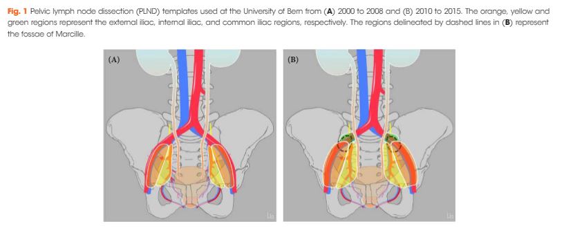
Article of the Week: More PLND template at RP detects metastases in the common iliac region and in the fossa of Marcille

Every Month, the Editor-in-Chief selects an Article of the Month from the current issue of BJUI. The abstract is reproduced below and you can click on the button to read the full article, which is freely available to all readers for at least 30 days from the time of this post.

In addition to the article itself, there is an accompanying editorial written by a prominent member of the urological community. This blog is intended to provoke comment and discussion and we invite you to use the comment tools at the bottom of each post to join the conversation.

If you only have time to read one article this week, it should be this one.

More extended lymph node dissection template at radical prostatectomy detects metastases in the common iliac region and in the fossa of Marcille


Lydia Maderthaner, Marc A. Furrer, Urs E. Studer, Fiona C. BurkhardGeorge N. Thalmann and Daniel P. Nguyen


Department of Urology, Inselspital, Bern University Hospital, University of Bern, Bern, Switzerland


Abstract

Objectives

To assess the effect of adding lymph nodes (LNs) located along the common iliac vessels and in the fossa of Marcille to the extended pelvic LN dissection (PLND) template at radical prostatectomy (RP).

Patients and Methods

A total of 485 patients underwent RP and PLND at a referral centre between 2000 and 2008 (historical cohort: classic extended PLND template) and a total of 268 patients between 2010 and 2015 (contemporary cohort: extended PLND template including LNs located along the common iliac vessels and in the fossa of Marcille). Descriptive analyses were used to compare baseline, pathological, complication and functional data between the two cohorts. A logistic regression model was used to assess the template’s effect on the probability of detecting LN metastases.

Results

Of 80 patients in the historical cohort with pN+ disease, the sole location of metastasis was the external iliac/obturator fossa in 23 (29%), and the internal iliac in 18 (23%), while 39 patients (49%) had metastases in both locations. Of 72 patients in the contemporary cohort with pN+ disease, the sole location of metastasis was the external iliac/obturator fossa in 17 patients (24%), the internal iliac in 24 patients (33%), and the common iliac in one patient (1%), while 30 patients (42%) had metastases in >1 location (including fossa of Marcille in five patients). Among all 46 patients in the contemporary cohort with ≤2 metastases, three had one or both metastases in the common iliac region or the fossa of Marcille. The adjusted probability of detecting LN metastases was higher, but not significantly so, in the contemporary cohort. There were no differences between the two cohorts in complication rates and functional outcomes.

Conclusion

A more extended template detects LN metastases in the common iliac region and the fossa of Marcille and is not associated with a higher risk of complications; however, the overall probability of detecting LN metastases was not significantly higher.


Editorial: PLND during RP for PCa: extending the template in the right patients without increasing complications

It took long time and consistent evidence to endorse the staging role of extended pelvic lymph node dissection (PLND) in prostate cancer (PCa). The poor performance of both conventional and functional imaging in identifying preoperative nodal status has contributed to making extended PLND the most accurate nodal staging procedure in PCa 1.

Current available guidelines recommend a standard extended PLND template that includes external, internal iliac and obturator lymph nodes 24; however, where does the need for a more extended template originate? Observational data suggest that a standard extended PLND template intercepts ~75% of all anatomical landing sites 4. Extending the anatomical template by adding nearby nodal stations would further minimize the risk of missing positive lymph nodes; however, it has previously been shown that a more accurate staging (i.e. a more extended template) might come at the price of longer operating time and a higher risk of procedure‐related complications 1.

According to the study by Maderthaner et al5, in the current issue of BJUI, an experienced academic surgical team is able to further extend the PLND template (including common iliac and the fossa of Marcille lymph nodes) without significantly increasing the risk of complications. In their study, 17% and 7% of the included men with pN+ disease had positive common iliac and fossa of Marcille lymph nodes, respectively.

Before celebrating this super‐extended template as safe and effective, however, at least three points need to be considered. First, these results were obtained by a group of skilled surgeons with longstanding experience in anatomical pelvic nodal dissection. It should not be taken for granted that this template in the hands of other surgeons would result in no additional complications, especially during the learning curve.

Second, >80% of men submitted to the super‐extended template did not have positive nodes outside the standard extended template boundaries, indicating possible overtreatment in a substantial proportion of men. Notably, extended PLND in this study was offered apparently without upfront preoperative lymph node invasion risk stratification.

Third, as a consequence, patient selection has a role to play. In other words, is super‐extended PLND appropriate for every patient? The use of available risk stratification tools in everyday clinical practice allows a more accurate decision process; this is the case for the Briganti nomogram concerning the need to perform an extended PLND 6. Could a similar approach be used in the setting of a super‐extended template to identify the best candidates? Recently, Gandaglia et al. 7 analysed data from 471 men with high‐risk PCa treated with radical prostatectomy and a super‐extended PLND including common iliac and pre‐sacral nodes in order to identify those men who really require such a super‐extended PLND. Interestingly, although not specifically designed for this task, the Briganti nomogram was able to provide a patient selection strategy: only 5% of patients with a nomogram‐derived N+ risk of <30% had positive common iliac and pre‐sacral nodes, indicating that the super‐extended PLND template should perhaps be considered exclusively in men with an N+ risk ≥30%.

In conclusion, a critical assessment of super‐extended staging PLND template would be welcome, allowing selection of the proper candidates, and a proper balance between accurate staging and the risk of treatment‐related complications.


Eugenio Vent imiglia, *Alberto Briganti,*† and Francesco Montorsi,*
*University Vita-Salute San Raffaele, Milan, Italy and Division of Experimental Oncology/Unit of Urology, URI, IRCCS Ospedale San Raffaele, Milan, Italy


References


  • Fossati N, Willemse P‐PM, Van den Broeck T et al. The benefits and harms of different extents of lymph node dissection during radical prostatectomy for prostate cancer: a systematic reviewEur Urol 201772: 84–109


  • Santis D, Henry A, Joniau S et al. Prostate Cancer EAU ESTRO SIOG Guidelines on 2017.


  • Mattei A, Fuechsel FG, Bhatta Dhar N et al. The template of the primary lymphatic landing sites of the prostate should be revisited: results of a multimodality mapping studyEur Urol 200853: 118–25


  • Maderthaner L, Furrer MA, Studer UE, Burkhard FC, Thalmann GN, Nguyen DP. More extended lymph node dissection template at radical prostatectomy detects metastases in the common iliac region and in the fossa of MarcilleBJU Int 2018121: 725–31


  • Briganti A, Larcher A, Abdollah F et al. Updated nomogram predicting lymph node invasion in patients with prostate cancer undergoing extended pelvic lymph node dissection: the essential importance of percentage of positive coresEur Urol 201261: 480–7


  • Gandaglia G, Zaffuto E, Fossati N et al. Identifying the candidate for super extended staging pelvic lymph node dissection among patients with high‐risk prostate cancerBJU Int 2018121: 421–7


Article of the Week: Profiling microRNA from nephrectomy and biopsy specimens

Every Week the Editor-in-Chief selects an Article of the Week from the current issue of BJUI. The abstract is reproduced below and you can click on the button to read the full article, which is freely available to all readers for at least 30 days from the time of this post.

In addition to the article itself, there is an accompanying editorial written by a prominent member of the urological community. This blog is intended to provoke comment and discussion and we invite you to use the comment tools at the bottom of each post to join the conversation.

If you only have time to read one article this week, it should be this one.

Profiling microRNA from nephrectomy and biopsy specimens: predictors of progression and survival in clear cell renal cell carcinoma


Casey G. Kowalik*, Drew A. Palmer*, Travis B. Sullivan, Patrick A. TeebagyJohn M. Dugan, John A. Libertino*, Eric J. Burks, David Canes* and Kimberly M. Rieger-Christ


Departments of *Urology, Translational Research Ian C. Summerhayes Cell and Molecular Biology Laboratory, and Pathology, Lahey Hospital and Medical Center, Burlington, MA, USA


Read the full article

Abstract

Objective

To identify microRNA (miRNA) characteristic of metastatic clear cell renal cell carcinoma (ccRCC) and those indicative of cancer-specific survival (CSS) in nephrectomy and biopsy specimens. We also sought to determine if a miRNA panel could differentiate benign from ccRCC tissue.

Materials and Methods

RNA was isolated from nephrectomy and kidney biopsy specimens (n = 156 and n = 46, respectively). Samples were grouped: benign, non-progressive, and progressive ccRCC. MiRNAs were profiled by microarray and validated by quantitative reverse transcription-polymerase chain reaction. Biomarker signatures were developed to predict cancer status in nephrectomy and biopsy specimens. CSS was examined using Kaplan–Meier and Cox proportional hazards analyses.

Results

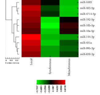
Microarray analysis revealed 20 differentially expressed miRNAs comparing non-progressive with progressive tumours. A biomarker signature validated in nephrectomy specimens had a sensitivity of 86.7% and a specificity of 92.9% for differentiating benign and ccRCC specimens. A second signature differentiated non-progressive vs progressive ccRCC with a sensitivity of 93.8% and a specificity of 83.3%. These biomarkers also discriminated cancer status in biopsy specimens. Levels of miR-10a-5p, -10b-5p, and -223-3p were associated with CSS.

Conclusion

This study identified miRNAs differentially expressed in ccRCC samples; as well as those correlating with CSS. Biomarkers identified in this study have the potential to identify patients who are likely to have progressive ccRCC, and although preliminary, these results may aid in differentiating aggressive and indolent ccRCC based on biopsy specimens.

Editorial: The utility of microRNAs as biomarkers in predicting progression and survival in patients with clear-cell renal cell carcinoma

RCC constitutes a diverse group of malignancies, yet the clear-cell subtype comprises ~80% of all diagnosed RCC cases [1]. The widespread use of abdominal imaging and subsequent stage migration has resulted in improved RCC 5-year cancer-specific survival. However, the overall mortality of RCC remains largely unchanged [2] and one-third of the patients have metastatic disease at the time of presentation [3]. Accordingly, the ability to precisely predict patient outcome has become an increasingly significant question in the management of these patients with RCC.

Accruing evidence suggests that changes in various biomarkers and their consequent downstream pathways affect cancer initiation and progression. Therefore, accurate prediction of the outcome and prognosis after treatment is necessary [4]. MicroRNAs (miRNAs) are small non-coding RNA molecules that can have significant functions in tumorigenesis [5]. Because of their ability in post-transcriptional regulation of gene expression, tumour-specific genetic defects in miRNA biogenesis and production correlate with development of human cancers. Thus, the differential expression of specific miRNA signatures in different tumours might become an important tool to help in directing cancer diagnosis and treatment [5].

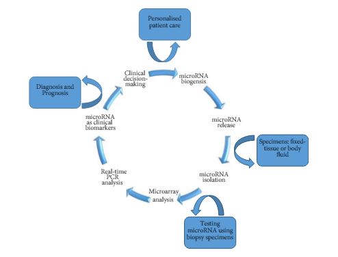
As such, Kowalik et al. [6] report on profiling miRNA to identify biomarker signatures predictive of clear-cell RCC (ccRCC) progression and survival. The authors used 202 formalin-fixed paraffin-embedded samples to isolate RNA from nephrectomy and biopsy specimens (n = 156 and n = 46, respectively) (Fig. 1).

Figure 1. Schematic diagram of miRNA-based biomarkers and potential utility in clinical decision-making approach for targeted therapy and RCC personalised treatment.

The primary analysis of their study [6] focused on the identification of miRNA signatures capable of differentiating between benign and ccRCC, as well as discerning those patients with a non-progressive ccRCC from a progressive clear-cell subtype. The secondary outcome examined the association of miRNA profiles discovered on cancer-specific survival.

In their initial microarray screening 20 differentially expressed miRNAs, comparing non-progressive with progressive tumours, were identified. The authors found four miRNA panels (10a-5p, 10b-5p, 106a-5p, and 142-5p) as a potential biomarker signature. This model was validated in nephrectomy specimens and resulted in a sensitivity of 86.7%, a specificity of 92.9%, and an area under the curve (AUC) of 0.930 for detecting ccRCC. Further analysis revealed a second signature of two biomarkers (miR-10a-5p and -223-3p) with 93.8% sensitivity, 83.3% specificity, and an AUC of 0.932 when validated for detecting progressive ccRCC. Similarly, the differential expression of these biomarkers could delineate cancer status in biopsy specimens. For correlation of miRNA expression levels with cancer-specific survival, higher expression levels of (miR-10a-5p and miR-10b-5p) and a lower expression level of (miR-223-3p) were significantly associated with survival (P< 0.001), and the median survival times were not reached.

In conclusion, the lack of precise prediction tools has led the authors to explore the potential utility of miRNAs as biomarkers to detect disease presence, biological aggressiveness, and prognosis in ccRCC. However, until future multicentre large prospective studies validate the results of the present work, the transition of miRNA from bench to bedside is emerging on the horizon and has encouraged urologists and scientists to pursue intense translational research in the field. The ability to use miRNAs as biomarkers might be promising for diagnostic and prognostic purposes. These biomarkers may exemplify different aspects of RCC pathogenesis and may potentially have important therapeutic implications to help in a clinical decision-making approach for targeted therapy and RCC personalised treatment.

Firas G. Petrosand Christopher J.D. Wallis†‡
*Department of Urology, The University of Texas MD Anderson Cancer Center, Houston, TX, USADivision of Urology, Department of Surgery, University of Toronto, Toronto, ON, Canada and Division of Urology, Department of Surgery, Sunnybrook Health Sciences Centre, Toronto, ON, Canada


Read the full article


References


1 Reuter VE, Presti JC Jr. Contemporary approach to the classication of renal epithelial tumors. Semin Oncol 2000; 27: 12437


2 Hollingsworth JM, Miller DC, Daignault S, Hollenbeck BK. Rising incidence of small renal masses: a need to reassess treatment effect. J Natl Cancer Inst 2006; 98: 13314


3 Gupta K, Miller JD , Li JZ, Russell MW, Charbonneau C. Epidemiologic and socioeconomic burden of metastatic renal cell carcinoma (mRCC): literature review. Cancer Treat Rev 2008; 34: 193205


5 Esquela-Kerscher A, Slack FJ. Oncomirs microRNAs with a role in cancer. Nat Rev Cancer 2006; 6: 25969


Article of the Week: Evaluation of Sig24, a 24-gene signature

Every week the Editor-in-Chief selects an Article of the Week from the current issue of BJUI. The abstract is reproduced below and you can click on the button to read the full article, which is freely available to all readers for at least 30 days from the time of this post.

In addition to the article itself, there is an accompanying editorial written by a prominent member of the urological community. This blog is intended to provoke comment and discussion and we invite you to use the comment tools at the bottom of each post to join the conversation.

Finally, the third post under the Article of the Week heading on the homepage will consist of additional material or media. This week we feature a video discussing the paper.

If you only have time to read one article this week, it should be this one.

Evaluation of a 24-gene signature for prognosis of metastatic events and prostate cancer-specific mortality

Kathryn L. Pellegrini*, Martin G. Sanda*, Dattatraya Patil*, Qi Long†‡§, MarıSantiago-Jimenez, Mandeep Takhar, Nicholas Erho, Kasra Youse, Elai DavicioniEric A. Klein**, Robert B. Jenkins††, R. Jeffrey Karnes‡‡ and Carlos S. Moreno§§§


*Department of Urology, Emory University School of Medicine, Winship Cancer Institute, Atlanta, GA‡ Department of Biostatistics and Epidemiology and Abramson Cancer Center, University of Pennsylvania Perelman School of Medicine, Philadelphia, PA, §Department of Biomedical Informatics, Emory University School of Medicine, Atlanta, GA, USA, GenomeDx Biosciences, Vancouver, BC, Canada, **Glickman Urological and Kidney Institute, Cleveland Clinic, Cleveland, OH, ††Department of Pathology and Laboratory Medicine, ‡‡Department of Urology, Mayo Clinic, Rochester, MN, and §§Department of Pathology and Laboratory Medicine, Emory University School of Medicine, Atlanta, GA, USA
Read the full article

How to Cite

Pellegrini, K. L., Sanda, M. G., Patil, D., Long, Q., Santiago-Jiménez, M., Takhar, M., Erho, N., Yousefi, K., Davicioni, E., Klein, E. A., Jenkins, R. B., Karnes, R. J. and Moreno, C. S. (2017), Evaluation of a 24-gene signature for prognosis of metastatic events and prostate cancer-specific mortality. BJU International, 119: 961–967. doi: 10.1111/bju.13779

Abstract

Objectives

To determine the prognostic potential of a 24-gene signature, Sig24, for identifying patients with prostate cancer who are at risk of developing metastases or of prostate cancer-specific mortality (PCSM) after radical prostatectomy (RP).

Patients and Methods

Sig24 scores were calculated from previously collected gene expression microarray data from the Cleveland Clinic and Mayo Clinic (I and II). The performance of Sig24 was determined using time-dependent c-index analysis, Cox proportional hazards regression and Kaplan–Meier survival analysis.

aotw-jun-3-results

Results

Higher Sig24 scores were significantly associated with higher pathological Gleason scores in all three cohorts. Analysis of the Mayo Clinic II cohort, which included time-to-event information, indicated that patients with high Sig24 scores also had a higher risk of developing metastasis (hazard ratio [HR] 3.78, 95% confidence interval [CI]: 1.96–7.29; P < 0.001) or of PCSM (HR 6.54, 95% CI: 2.16–19.83; P < 0.001).

Conclusions

The findings of the present study show the applicability of Sig24 for the prognosis of metastasis or PCSM after RP. Future studies investigating the combination of Sig24 with available prognostic tests may provide new approaches to improve risk stratification for patients with prostate cancer.

Editorial: Predicting outcome: role of gene signatures

Pathological assessments, such as Gleason grading, which is a strong clinical predictor of prostate cancer progression [1], have a role to play in predicting the outcome of a patient’s response to therapy. The addition of PSA and TNM staging are further used to inform appropriate treatment strategies in patients who are at low, intermediate and high risk of disease progression. Unfortunately, approximately 30% of men with intermediate-risk prostate cancer will fail to be cured by surgery or radiation therapy approaches. This is not surprising as primary prostate cancer represents a complex heterogeneous disease that is clearly not fully explained by the current clinical prognostic factors, and further molecular characterization is required. There is now significant emerging evidence that the molecular characterization of tumours is important to enable us to stratify prostate cancer patients by their response to primary therapy and to identify the next appropriate steps in their treatment pathway.

Long et al. [2] have identified gene signatures that can define different genomic subtypes of prostate cancer and are predictive of biochemical recurrence. Erho et al. [3] discovered and validated a 22-gene signature, which they termed a genomic classifier for the prediction of early metastasis after radical prostatectomy, while Penny et al. [4] have identified an mRNA expression signature of Gleason grade which is predictive of lethal prostate cancer.

Long et al. [2] identified a 24-gene signature, which they discovered through RNA sequencing analysis of 100 formalin-fixed paraffin-embedded prostatectomy samples. They went on to validate their findings in a publically available independent gene expression microarray dataset of 140 patients. This 24-gene signature forms the basis of the present study by Pellegrini et al. [5], who refer to this gene signature as Sig24. In their study, they firstly undertook to determine if Sig24 was associated with pathological Gleason score as a marker of tumour aggressiveness, and then if it had prognostic value for the identification of patients at risk of metastasis or prostate cancer-specific mortality after radical prostatectomy. The Gleason score association study was carried out using the data from three independent case–control sets, including 182 patients from the Cleveland Clinic and two cohorts (cohort I, n = 545; cohort II, n = 235) from the Mayo Clinic, for which gene expression analysis had previously been conducted by Genome Dx using the Affymetrix Human 1.0 ST Genechip platform. The Sig24 score was calculated for each patient and higher Gleason score was associated with significantly higher Sig24 scores. The association studies for metastatic disease and prostate cancer-specific mortality, however, were only carried out in the Mayo Clinic cohort II. For both clinical endpoints the Sig24 score combined with the clinical model outperformed the clinical model alone of PSA, Gleason score and tumour stage. For metastatic disease the area under the curve for the clinical model alone was 0.69 (0.62–0.77) compared with 0.73 (0.66–0.78) for the clinical model combined with Sig24. For the prostate cancer-specific mortality endpoint, the area under the curve for the clinical model alone was 0.69 (0.67–0.87) compared with 0.74 (0.63–0.85) for the clinical model combined with Sig24.

These gene signatures have significant potential for predicting the progression of disease rather than waiting for PSA relapse, which is currently used to identify disease recurrence, with interval to biochemical failure being the best univariate factor predicting prostate cancer mortality and overall survival [6]. This would allow the initiation of additional therapies before recurrence and a better outcome for the patient. This concept has been demonstrated in a study in which the use of the Decipher gene signature was shown to improve the identification of patients who could benefit from adjuvant radiotherapy and thus only these patients were targeted for therapy [7].

Incorporating these gene signatures in robust clinical assays and integrating them into clinical decision-making is the next essential step in order for these strategies to have an impact on patient outcomes.

Read the full article
Ronald W. Watson
UCD School of Medicine, University College Dublin, Dublin, Ireland

References

1 Gleason DF. Classication of prostatic carcinomas. Cancer Chemother
Rep 1966; 50: 1258


4 Penny KL, Sinnott JA, Fall K et al. mRNA expression signature of Gleason grade predicts lethal prostate cancer. J Clin Oncol 2011; 29:23916


6 Buyyounouski MK, Pickles T, Kestin LL, Allison R, Williams SGValidating the interval to biochemical failure for identication of potentially lethal prostate cancer. J Clin Oncol 2012; 30: 185763


Video: Evaluation of a 24-gene signature for prognosis of metastatic events and PCa-specific mortality

Evaluation of a 24-gene signature for prognosis of metastatic events and prostate cancer-specific mortality

Kathryn L. Pellegrini*, Martin G. Sanda*, Dattatraya Patil*, Qi Long†‡§, MarıSantiago-Jimenez, Mandeep Takhar, Nicholas Erho, Kasra Youse, Elai DavicioniEric A. Klein**, Robert B. Jenkins††, R. Jeffrey Karnes
‡‡ and Carlos S. Moreno§§§


*Department of Urology, Emory University School of Medicine, Winship Cancer Institute, Atlanta, GA‡ Department of Biostatistics and Epidemiology and Abramson Cancer Center, University of Pennsylvania Perelman School of Medicine, Philadelphia, PA, §Department of Biomedical Informatics, Emory University School of Medicine, Atlanta, GA, USA, GenomeDx Biosciences, Vancouver, BC, Canada, **Glickman Urological and Kidney Institute, Cleveland Clinic, Cleveland, OH, ††Department of Pathology and Laboratory Medicine, ‡‡Department of Urology, Mayo Clinic, Rochester, MN, and §§Department of Pathology and Laboratory Medicine, Emory University School of Medicine, Atlanta, GA, USA
Read the full article

How to Cite

Pellegrini, K. L., Sanda, M. G., Patil, D., Long, Q., Santiago-Jiménez, M., Takhar, M., Erho, N., Yousefi, K., Davicioni, E., Klein, E. A., Jenkins, R. B., Karnes, R. J. and Moreno, C. S. (2017), Evaluation of a 24-gene signature for prognosis of metastatic events and prostate cancer-specific mortality. BJU International, 119: 961–967. doi: 10.1111/bju.13779

Abstract

Objectives

To determine the prognostic potential of a 24-gene signature, Sig24, for identifying patients with prostate cancer who are at risk of developing metastases or of prostate cancer-specific mortality (PCSM) after radical prostatectomy (RP).

Patients and Methods

Sig24 scores were calculated from previously collected gene expression microarray data from the Cleveland Clinic and Mayo Clinic (I and II). The performance of Sig24 was determined using time-dependent c-index analysis, Cox proportional hazards regression and Kaplan–Meier survival analysis.

aotw-jun-3-results

Results

Higher Sig24 scores were significantly associated with higher pathological Gleason scores in all three cohorts. Analysis of the Mayo Clinic II cohort, which included time-to-event information, indicated that patients with high Sig24 scores also had a higher risk of developing metastasis (hazard ratio [HR] 3.78, 95% confidence interval [CI]: 1.96–7.29; P < 0.001) or of PCSM (HR 6.54, 95% CI: 2.16–19.83; P < 0.001).

Conclusions

The findings of the present study show the applicability of Sig24 for the prognosis of metastasis or PCSM after RP. Future studies investigating the combination of Sig24 with available prognostic tests may provide new approaches to improve risk stratification for patients with prostate cancer.

Sailing into “UnCHAARTED” waters

Chemotherapy comes alive for prostate cancer!

staff-chowdhury1Systemic therapy for metastatic prostate cancer has radically changed in the last 10 years with the introduction of several novel agents that have shown significant improvements in progression free and overall survival. These have all been studied in metastatic castrate refractory prostate cancer (mCRPC) and have improved overall survival but in each case by less than 6 months. (The latest major breakthrough is the introduction of a relatively old drug, docetaxel chemotherapy, earlier in the disease for hormone sensitive patients).

In this week’s New England Journal of Medicine we see the eagerly awaited results from the CHAARTED study from Christopher Sweeney and colleagues. This novel study aimed to improve treatment for men with newly diagnosed hormone sensitive metastatic prostate cancer by adding docetaxel chemotherapy to androgen deprivation therapy (ADT).

790 men with newly diagnosed metastatic prostate cancer were randomised to ADT plus docetaxel (6 cycles at 75mg/m2) or ADT alone. The addition of docetaxel to ADT was shown to significantly improve overall survival by 13.6 months (57.6 months vs. 44.0 months; p<0.001). The clinical benefit was greatest in the subgroup with high volume disease where the improvement in overall survival was 17 months (49.2 months versus 32.2 months). High volume disease was defined as the presence of visceral metastases and/or 4 or more bone metastases with at least one beyond the vertebral bodies or pelvis. The combination was well tolerated with approximately 6% of patients having neutropenic fever and one death possibly related to docetaxel.

The results from this study are truly practice changing. Supporting evidence from the UK STAMPEDE study (Systemic Therapy in Advancing or Metastatic Prostate Cancer: Evaluation of Drug Efficacy) was presented at this year’s American Society of Clinical Oncology (ASCO) meeting. STAMPEDE showed that for men with metastatic hormone sensitive prostate cancer 6 cycles of docetaxel in addition to ADT improved median overall survival by 22 months (43 versus 65 months).

Chemotherapy for metastatic prostate cancer has had a checkered past with a lack of enthusiasm and nihilism from clinicians and patients. The results from CHAARTED and STAMPEDE are already changing those views. The prostate cancer community needs to react to these results and look to make this treatment available to all suitable men. There are issues with regards to costs of chemotherapy (although docetaxel is now generic), workload, sequence, patient selection, toxicity management, etc. The CHAARTED and STAMPEDE investigators must also use this opportunity to interrogate the tumour samples from these studies to see if they can identify biomarkers that predict docetaxel activity. We will not get this opportunity again as docetaxel + ADT will be be standard of care for future studies.

The clinical benefit from the addition of docetaxel to ADT is one of the largest seen in any oncology study. All men presenting with newly diagnosed metastatic prostate cancer should be considered for 6 cycles of docetaxel in addition to ADT.


Simon Chowdhury is a Consultant Medical Oncologist at Guy’s, King’s and St Thomas’ Hospitals, London. He is actively involved in clinical trial research into urological cancers.


Ewing’s Sarcoma with Isolated Bladder Metastasis

We describe a case of an isolated bladder metastasis in EWS, which, to our knowledge, has not been previously reported.


Authors: Alexander Yeates MBBS, Peter Campbell FRACS, Queen Elizabeth II Jubilee Hospital, Brisbane, Australia
 
Corresponding Author: Alexander Yeates MBBS, Queen Elizabeth II Jubilee Hospital, Brisbane, Australia. Email: [email protected], [email protected]


Abstract
Ewing’s sarcoma (EWS) can occur in almost any bone or soft tissue, however cases involving the bladder are exceedingly rare. We describe a case of an isolated bladder metastasis in EWS, which, to our knowledge, has not been previously reported.
A twelve year old boy was initially diagnosed with primary EWS of the skull.  He was treated with local surgical resection, radiotherapy and chemotherapy.  He had no recurrence until four years later when he presented with painless haematuria.  Urinary cytology revealed small, round atypical cells, and he was referred for a urological opinion.  An intravenous pyelogram was normal.  Rigid cystoscopy revealed a 20 x 15mm lesion that was resected and histology confirmed recurrence of EWS.  After careful consideration, a partial cystectomy was performed with good post-operative recovery and subsequently he completed a course of adjuvant chemotherapy.  40 months of follow-up including blood tests, rigid cystoscopy, CXR, CT and USS have not revealed further recurrence.
The prognosis following recurrence of Ewing’s sarcoma is usually guarded, however features specific to this case, such as the prolonged interval to recurrence and presence of a distant, isolated recurrence are relatively reassuring.


Introduction
Ewing’s sarcoma (EWS) was initially described by James Ewing in 1921 as an undifferentiated, small, round cell tumour involving the diaphysis of long bones [1].  More recently, EWS, primitive neuroectodermal tumour and Askin’s tumour have all been classified under the common term of the Ewing’s sarcoma family of tumours following identification of the common translocation t(11;22)(q24;q12) resulting in the formation of the EWS-ETS fusion gene [2,3].  These tumours can occur in almost any bone or soft tissue, however cases of EWS involving the bladder are exceedingly rare [4,5] and a case of an isolated recurrence in the bladder has not previously been reported.


Case Report
An otherwise healthy twelve year old boy was diagnosed with EWS of the skull in 2003.  He was initially treated with local excision, radiotherapy (50.4Gy) and chemotherapy (vincristine 15mg/m2, cyclophosphamide 17,400mg/m2, doxorubicin 480mg/m2, ifosfamide 39,000mg/m2, carboplatin 3,000mg/m2 and etoposide 1,950mg/m2) followed by stem cell rescue.  Four years later he presented to an Accident and Emergency Department following a single episode of frank haematuria.  Abdominal and pelvic ultrasound scan demonstrated normal upper urinary tracts with a large amount of echogenic material consistent with clot within the bladder.  Urine microscopy showed clusters of small round atypical cells, lymphocytes and erythrocytes. An intravenous pyelogram was normal.
Following referral to a Urologist, rigid cystoscopy identified a 20x15mm solid necrotic lesion on the left lateral wall of the bladder (Figure 1).


Figure 1.Rigid cystoscopy showing bladder lesion.


The lesion was excised with a 26 Fr resectoscope.  Microscopic examination of the lesion revealed sheets of small, undifferentiated malignant cells invading into muscle (Figure 2).


Figure 2. Undifferentiated malignant tumour of bladder. (H&E stained section, original magnification x400)


Immunohistochemical testing showed the cells were strongly positive for CD99 in a membranous pattern with scattered cells positive for NFP and synaptophysin.  They were negative for S100, Desmin, Myo D1 and SMA.  Fluorescence in-situ hybridisation (FISH) using the Vysis [6] EWSR1 (22q12) dual colour break apart rearrangement probe showed rearrangement of the EWSR1 gene region confirming the diagnosis of metastatic EWS (Figure 3).


Figure 3. Fluorescence in-situ hybridisation of the bladder lesion using the Vysis EWRS1 (22q12) dual break apart probe shows rearrangement of the EWSR1 gene region, confirming the diagnosis of metastatic EWS.


Staging investigations (whole body bone scan and CT abdomen and pelvis) showed no evidence of disease elsewhere in the body.
Given that this appeared to represent an isolated tumour recurrence, the decision was made to perform a partial cystectomy in October 2007.  The lesion was identified and excised with a four centimetre margin (Figure 4).


Figure 4. Partial cystectomy specimen showing central area of ulceration.


Histology of the excised specimen showed an area of ulceration that was clear of the margins.  There was no evidence of residual tumour.  Post-operative recovery was uneventful.
Following partial cystectomy, the patient received adjuvant chemotherapy of topotecan (36mg/m2) and cyclophosphamide (18,000mg/m2).  This was adequately tolerated.  The patient was considered for sperm storage but was found to be azoospermic.
Follow-up has involved surveillance rigid cystoscopy and examination under anaesthesia at three-monthly intervals with interim outpatient reviews with surveillance full blood count, electrolytes and liver function blood tests, chest x-ray, abdominal and pelvic CT and ultrasound scans to exclude further metastatic disease.  Surveillance for over 40 months has so far revealed no evidence of local recurrence or further metastases.


Discussion
Rare cases of primary EWS of the bladder have been reported [4,5] and EWS has also been described as arising in the bladder as a second tumour in paediatric patients with a previous haematological malignancy [7].   To our knowledge this case represents the first report of primary skeletal EWS with an isolated bladder metastasis.    Given that the five-year survival rate for patients with recurrent EWS has been reported to be as low as 13% [8], the initial prognosis for this patient was guarded.  Significant risk factors for death following recurrence in Leavey’s study [9] included recurrence at combined local and distant sites, elevated LDH at initial diagnosis and initial recurrence less than two years following diagnosis.  In contrast, this patient had recurrence at a single distant site four years after the primary diagnosis of EWS and is now disease free four years after excision of the recurrent lesion.  This is relatively reassuring when considering the long-term prognosis.
It is also worth noting that this adolescent patient presented clinically with an episode of macroscopic haematuria.  This symptom cannot be ignored and should lead to consideration of cystoscopic examination of the bladder particularly in patients with a significant previous medical history [10].


References
1.  Ewing J.  Diffuse endothelioma of bone.  Proc NY Pathol Soc 1921;21:17-24.
2.  Aurias A, Rimbaut C, Buffe D, Zucker JM, Mazabraud A.  Translocation involving chromosome 22 in Ewing’s sarcoma: a cytogenic study of four fresh tumors.  Cancer Genet Cytogenet 1984;12:21-25.
3.  Whang-Peng J, Triche TJ, Knutsen T, Miser J, Douglass EC, Israel MA.  Chromosomal translocation in peripheral neuroepithelioma. N Engl J Med 1984;311:584-585.
4.  Gousse AE, Roth DR, Popek EJ, Cooley LD, Horowitz ME.  Primary Ewing’s sarcoma of the bladder associated with an elevated antinuclear antibody titer.  J. Urol.  1997;158:2265-2266.
5.  Okada Y, Kamata S, Akashi T, Kurata M, Nakamura T, Kihara K.  Primitive neuroectodermal tumor/Ewing’s sarcoma of the urinary bladder: a case report and its molecular diagnosis.  Int J Clin Oncol.  2010 Nov 10. [Epub ahead of print].
6.  Vysis package insert.  Invitrogen Spot-Light tissue Pre-treatment Kit package insert. KOJI,T, Molecular Histochemical Techniques, Springer- Verlag, Tokyo, 2000.
7.  Osone S, Hosoi H, Tanaka K, Tsuchiya K, Iehara T, Morimoto A, Hashida T, Yamashita M, Kawabata K, Nishijo K, Toguchida J, Hata J, Sugimoto T.  A case of a Ewing sarcoma family tumor in the urinary bladder after treatment for acute lymphoblastic leukemia.  J Pediatr Hematol Oncol. 2007 Dec;29(12):841-4.
8.  Bacci G, Ferrari S, Longhi A, Donati D, De Paolis M, Forni C, Versari M, Setola E, Briccoli A, Barbieri E.  Therapy and survival after recurrence of Ewing’s tumours: the Rizzoli experience in 195 patients treated with adjuvant and neo-adjuvant chemotherapy from 1979 to 1997.  Ann. Oncol 2003;14:1654-1659.
9.  Leavey PJ, Mascarenhas L, Marina N, Chen Z, Krailo M, Miser J, Brown K, Tarbell N, Bernstein ML, Granowetter L, Gebhardt M, Grier HE.  Prognostic Factors for Patients with Ewing sarcoma (EWS) at First Recurrence Following Multimodality Therapy – A Report from the Children’s Oncology Group.    Pediatr Blood Cancer. 2008 September ; 51(3): 334–338.
10.  Gordon C, Stapleton FB. Hematuria in adolescents.  Adolesc Med Clin. 2005;16:229-39.


Date added to bjui.org: 06/09/2011 


DOI: 10.1002/BJUIw-2011-028-web


© 2024 BJU International. All Rights Reserved.