Tag Archive for: #KidneyCancer

Posts

Editorial: Nephrometry scoring systems: valuable research tools, but can they be applied in daily clinical practice?

In this issue of BJUI Schiavina et al. [1] report on the RENAL and PADUA nephrometry scoring systems in predicting peri-operative outcomes, including warm ischaemia time and postoperative complications, in a multi-institutional cohort of patients undergoing robot-assisted partial nephrectomy. The authors showed that tumours classified as being of intermediate and high complexity on the PADUA score and high complexity on the RENAL score were associated with a nearly threefold higher risk of longer warm ischaemia times (>20 min). In addition, more complex tumours carried a higher risk of grade 3–4 postoperative complications (most commonly bleeding requiring angioembolization and urine leak requiring a ureteric stent). Notably, the two scoring systems were found to be similar predictors of these peri-operative outcomes on receiver-operating curve (ROC) analyses [1].

This represents the first large, multicentre study to evaluate the accuracy of these scoring systems in a cohort of patients who purely underwent robot-assisted surgery. A recent study by Borgmann et al. [2] found that, among the reported scoring systems, the RENAL nephrometry score correlated best with achieving negative surgical margins, shorter ischaemia times, and low postoperative complication rates; however, only 9% of patients underwent robot-assisted surgery. Another contemporary series showed concordance between the RENAL and PADUA scoring systems in predicting ischaemia times and complication rates, albeit in patients who only underwent open surgery [3].

Current guidelines recognize nephron-sparing approaches to small renal masses as the standard of care in well-selected patients, with the robot-assisted platform being predominantly adopted in clinical practice where available. Certainly, these nephrometry scores are valuable for urologists in counselling patients on the potential risk of complications specific to the surgical anatomy of the tumour. In addition, the RENAL and PADUA scores (and others) provide a quantitative, objective method for comparing data from different studies and different institutions.

As nephrometry scoring systems continue to be critically evaluated in the robotic surgery era, the question that naturally arises is: which system is best? With regard to this question, the data in the present study do not necessarily favour one or the other for the prediction of clinically relevant peri-operative outcomes. One must recognize, however, that several other anatomy-based scoring systems exist and were not examined in this manuscript [4-6]. While these are very valuable research and patient counselling tools, one must caution against using these nephrometry tools to make clinical decisions; for example, attempting to predict benign vs malignant histology (without a biopsy), attempting to predict high vs low grade tumours, or deciding on whether to perform a radical vs partial nephrectomy, or an open vs minimally invasive approach. After all, one must keep in mind that the area under the curve for these tools is in the range of 0.58–0.63 (0.50 being equivalent to toss of a coin).

It would have been interesting to include clinical size only in the present multivariate analysis (as was done for RENAL and PADUA scoring) and ROC analysis to compare this simple variable with the studied nephrometry scores. Future research should examine additional confounders that could potentially affect postoperative complication rates, such as BMI, adherent perinephric fat, experience of the surgeon actually performing the partial nephrectomy, technique of resection used (e.g. enucleation or resection) among others. This may help to distinguish a single system as the optimum model for use in research and in patient counselling regarding potential postoperative complications.

Matthew A. Meissner and Jose A. Karam


Department of Urology, University of Texas MD Andersonn Cancer Center, Houston, TX, USA


References


3 Kriegmair MC, Mandel P, Moses A et al. Dening Renal Masses: comprehensive Comparison of RENAL, PADUA, NePhRO, and C-Index Score. Clin Genitourin Cancer 2016; [Epub ahead of print]. doi: 10.1016/ j.clgc.2016.07.029.


5 Hakky TS, Baumgarten AS, Allen B, Lin HY, Ercole CE, Sexton WJSpiess PE et al. Zonal NePhRO scoring system: a superior renal tumor complexity classication model. Clin Genitourin Cancer 2014; 12: e138


6 Simmons MN, Ching CB, Samplaski MK, Park CH, Gill IS et al. Kidney tumor location measurement using the C index method. J Urol 2010; 183: 170813


Article of the Week: LCA – EuRECA study

Every week the Editor-in-Chief selects an Article of the Week from the current issue of BJUI. The abstract is reproduced below and you can click on the button to read the full article, which is freely available to all readers for at least 30 days from the time of this post.

In addition to the article itself, there is an accompanying editorial written by a prominent member of the urological community. This blog is intended to provoke comment and discussion and we invite you to use the comment tools at the bottom of each post to join the conversation.

If you only have time to read one article this week, it should be this one.

Oncological outcomes and complication rates after laparoscopic-assisted cryoablation: a European Registry for Renal Cryoablation (EuRECA) multi-institutional study

Tommy K. Nielsen*, Brunolf W. Lagerveld, Francis Keeley, Giovanni Lughezzani§Seshadri Sriprasad, Neil J. Barber**, Lars U. Hansen*,††, Nicole M. Buf§Giorgio Guazzoni§, Johan A. van der Zee, Mohamed Ismail, Khaled Farrag,Amr M. Emara**,‡‡, Lars Lund††,§§, Øyvind Østraat* and Michael Borre*


*Department of Urology, Aarhus University Hospital, Aarhus, Denmark, Department of Urology, Onze Lieve Vrouwe Gasthuis, Amsterdam, the Netherlands, Bristol Urological Institute, Bristol, UK, §Department of Urology, Istituto Clinico Humanitas IRCCS, Clinical and Research Hospital, Milano, Rozzano, Italy, Department of Urology, Darent Vally Hospital, Dartford, **Department of Urology, Frimley Park Hospital, Camberley, UK, ††Department of Urology, Odense University Hospital, Odense, Denmark, ‡‡Department of Urology, Ain Shams University, Cairo, Egypt, and §§Department of Urology, Viborg Regional Hospital, Viborg, Denmark


Read the full article

Abstract

Objective

To assess complication rates and intermediate oncological outcomes of laparoscopic-assisted cryoablation (LCA) in patients with small renal masses (SRMs).

Patients and Methods

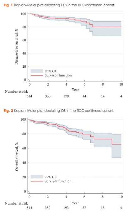
A retrospective review of 808 patients treated with LCA for T1a SRMs from 2005 to 2015 at eight European institutions. Complications were analysed according to the Clavien–Dindo classification. Kaplan–Meier analyses were used to estimate 5- and 10-year disease-free survival (DFS) and overall survival (OS).

aotw-mar-2-2017

Results

The median [interquartile (IQR)] age was 67 (58–74) years. The median (IQR) tumour size was 25 (19–30) mm. The transperitoneal approach was used in 77.7% of the patients. The median postoperative hospital stay was 2 days. In all, 514 patients with a biopsy-confirmed renal cell carcinoma (RCC) were available for survival analyses. The median (IQR) follow-up for the RCC-cohort was 36 (14–56) months. A total of 32 patients (6.2%) were diagnosed with treatment failure. The 5-/10-year DFS was 90.4%/80.0% and 5-/10-year OS was 83.2%/64.4%, respectively. A total of 134 postoperative complications (16.6%) were reported, with severe complications (grade ≥III) in 26 patients (3.2%). An American Society of Anesthesiologists score of 3 was associated with an increased risk of overall complications (odds ratio 2.85, 95% confidence interval 1.32–6.20; P = 0.005).

Conclusions

This large series of LCA demonstrates satisfactory long-term oncological outcomes for SRMs. However, although LCA is considered a minimally invasive procedure, risk of complications should be considered when counselling patients.

Read more articles of the week

Editorial: Laparoscopic renal mass cryoablation: an operation in search of an indication

In this issue of BJUI, Nielsen et al. [1] report the oncological and surgical outcomes from a multi-institutional cohort of patients receiving laparoscopic cryoablation (LCA) as primary therapy for solitary renal masses <4 cm in size (cT1a). This work represents the latest addition to a growing body of literature in an important oncological space that lacks prospective/randomized evidence to guide practitioners counselling patients with kidney cancer. Although the article does not advance the discussion toward higher levels of evidence, the results are nonetheless provocative and several strengths and weaknesses deserve comment.

While nephron-sparing surgery has become the recognized standard of care for cT1a renal lesions [2, 3], the reality remains that certain patients carry unacceptable risk profiles for partial nephrectomy, making less invasive options preferable. Such indications might include being elderly or frail, having hereditary kidney cancer syndromes prone to metachronous renal tumours, or having a solitary kidney. For such patients, focal renal mass ablative techniques have emerged as a safe alternative to extirpation that avoids the permanent nephron loss associated with radical nephrectomy. From an oncological perspective, however, cryotherapy, radiofrequency ablation and microwave ablation (by any approach) all have yet to be studied against partial nephrectomy in a prospective fashion. Numerous retrospective analyses have attempted to fill the void [4], yet the general consensus among most academic kidney surgeons is that renal mass ablation offers acceptable but inferior cancer control compared with surgery [5].

In this retrospective analysis by Nielsen et al., 808 patients underwent LCA between 2005 and 2015, 514 (63.4%) of whom had pre-procedural biopsy-proven RCC. The principal findings described in the present study include not only 5- and 10-year disease-free and overall survival, but also morbidity and mortality outcomes after LCA. The authors should be commended for the structure of their design, which included a high proportion of patients with available preoperative biopsy data. Additionally, clear definitions of treatment success, ‘residual unablated tumour’ and ‘local tumour progression’ are provided, and consistent follow-up imaging protocols were employed by the institutions involved. In each of these ways, Nielsen et al. overcome many of the pitfalls that have clouded the interpretation of results from previous reports.

Nevertheless, the oncological outcomes reported in this study, which are on a par with those for partial nephrectomy as well as other ablative techniques, must be approached with a degree of skepticism. As there is no alternative treatment cohort included in the study, omission of anatomical complexity data (in the form of nephrometry scoring) prohibits any meaningful comparison with patients having undergone ablative procedures or partial nephrectomy from other series. Availability of these data is essential for the reader to gauge the influence of selection bias in the interpretation of the results.

From a morbidity and mortality standpoint, the reported 16% overall complication rate, 3% rate of severe complications (defined as Clavien III–V) and three deaths within 30 days of the procedure might have been strengthened by the missing nephrometry data, as the rate of complications would be expected to increase with the complexity of the renal mass [6]. Also noticeably absent from the analysis are granular comorbidity and previous surgery data, both of which intuitively predispose patients to complications when undergoing minimally invasive surgery.

With these limitations in mind, the experienced kidney surgeon is not likely to see LCA as an equally effective or safer alternative to minimally invasive partial nephrectomy, which can be performed with similar complication rates and length of hospital stay without sacrificing oncological efficacy in most patients. Similarly, the question of why the practitioner should assume the risks of LCA when percutaneous cryoablation is readily available at many contemporary kidney cancer centres is unanswered by the present study. Indeed, with increasingly complex renal masses being managed via minimally invasive nephron-sparing surgery, and active surveillance of small renal masses gaining traction in the appropriate patient population, cryoablation via a laparoscopic approach unfortunately may represent another urological application without a well-defined indication going forward. We hope that the results presented by Nielsen et al. in this issue of BJUI encourage investigators to enroll patients in prospective trials aimed at comparing available ablative techniques or partial nephrectomy in matched cohorts to identify the ideal patient population for this operation and further clarify the oncological and clinical outcomes compared with surgical excision.

Daniel C. Parker and Brian W. Cross


Department of Urologic Oncology, University of Oklahoma Stephenson Cancer Center, Oklahoma City, OK, USA


Read the full article


References


1 NielsenT, Lagerveld B, Keeley F et al. Oncologic outcomes and complication rates after laparoscopic-assisted cryoablation: a EuRECA multi-institutional study. BJU Int 2016. [Epub ahead of print].


2 Campbell SC, Novick AC, Belldegrun A et al. Guideline for management of the clinical T1 renal mass. J Urol 2009; 182: 12719


3 Ljungberg B, Bensalah K, Caneld S et al. EAU guidelines on renal cell carcinoma: 2014 update. Eur Urol 2015; 67: 91324


4 Wagstaff P, Ingels A, Zondervan P et al. Thermal ablation in renal cell carcinoma management: a comprehensive review. Curr Opin Urol 2014; 24: 47482


5 Kutikov A, Smaldone MC, Uzzo RG. Focal therapy for treatment of the small renal mass: dealers choice or a therapeutic gamble? Eur Urol 2015; 67: 2601


Article of the Week: Combining Nanotech Drug Delivery and Thermoablation in an in vivo mouse model of RCC

Every week the Editor-in-Chief selects an Article of the Week from the current issue of BJUI. The abstract is reproduced below and you can click on the button to read the full article, which is freely available to all readers for at least 30 days from the time of this post.

In addition to the article itself, there is an accompanying editorial written by a prominent member of the urological community. This blog is intended to provoke comment and discussion and we invite you to use the comment tools at the bottom of each post to join the conversation.

If you only have time to read one article this week, it should be this one.

Nanotechnology combined therapy: tyrosine kinase-bound gold nanorod and laser thermal ablation produce a synergistic higher treatment response of renal cell carcinoma in a murine model

James Liu*, Caleb Abshire*, Connor Carry*, Andrew B. Sholl, Sree Harsha Mandava*, Amrita Datta*, Manish Ranjan*, Cameron Callaghan*, Donna V. Peralta, Kristen S. Williams, Weil R. Lai*, Asim B. Abdel-Mageed*, Matthew Tarr and Benjamin R. Lee§


Departments of *Urology, Pathology, Tulane University School of Medicine, Department of Chemistry, University of New Orleans, New Orleans, LA, and §Division of Urology, University of Arizona College of Medicine, Tucson, AZ, USA


Read the full article

Abstract

Objectives

To investigate tyrosine kinase inhibitors (TKI) and gold nanorods (AuNRs) paired with photothermal ablation in a human metastatic clear cell renal cell carcinoma (RCC) mouse model. Nanoparticles have been successful as a platform for targeted drug delivery in the treatment of urological cancers. Likewise, the use of nanoparticles in photothermal tumour ablation, although early in its development, has provided promising results. Our previous in vitro studies of nanoparticles loaded with both TKI and AuNRs and activated with photothermal ablation have shown significant synergistic cell kill greater than each individual arm alone. This study is a translation of our initial findings to an in vivo model.

Materials and Methods

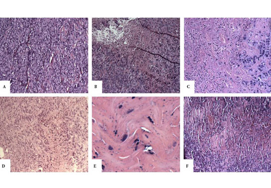
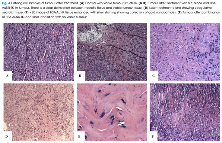
Immunologically naïve nude mice (athymic nude-Foxn1nu) were injected subcutaneously bilaterally in both flanks (n = 36) with 2.5 × 10⁶ cells of a human metastatic renal cell carcinoma cell line (RCC 786-O). Subcutaneous xenograft tumours developed into 1-cm palpable nodules. AuNRs encapsulated in human serum albumin protein (HSA) nanoparticles were synthesised with or without a TKI and injected directly into the tumour nodule. Irradiation was administered with an 808-nm light-emitting diode laser for 6 min. Mice were humanely killed 14 days after irradiation; tumours were excised, formalin fixed, paraffin embedded, and evaluated for size and the percentage of necrosis by a genitourinary pathologist. The untreated contralateral flank tumours were used as controls.

Results

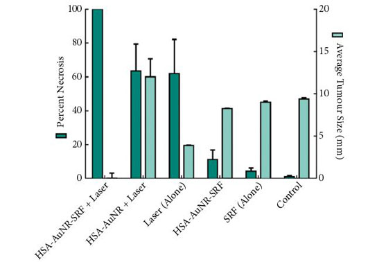
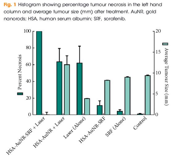
In mice that did not receive irradiation, TKI alone yielded 4.2% tumour necrosis on the injected side and administration of HSA-AuNR-TKI alone yielded 11.1% necrosis. In the laser-ablation models, laser ablation alone yielded 62% necrosis and when paired with HSA-AuNR there was 63.4% necrosis. The combination of laser irradiation and HSA-AuNR-TKI had cell kill rate of 100%.

aotw-22-2-17

Conclusions

In the absence of laser irradiation, TKI treatment alone or when delivered via nanoparticles produced moderate necrosis. Irradiation with and without gold particles alone also improves tumour necrosis. However, when irradiation is paired with gold particles and drug-loaded nanoparticles, the combined therapy showed the most significant and synergistic complete tumour necrosis of 100% (P < 0.05). This study illustrates the potential of combination nanotechnology as a new approach in the treatment of urological cancers.


Read more articles of the week


Editorial: Synergistic effects in combinational drug delivery and thermal ablation using nanotechnology

Chemotherapy, the dominant therapeutic approach to the treatment of a wide variety of cancers, is intrinsically inefficient because its drug delivery is non-specific. This leads to a trade-off between undesirable cytotoxic effects in healthy cells and associated side effects and lower efficacy in killing cancerous cells at lower concentrations.

The study in the present issue of BJUI by Lui et al. [1], from the University of Arizona College of Medicine, attempts to circumvent these undesirable side effects by employing nanoparticles as drug delivery vectors, isolating chemotherapeutic agents from the systemic environment while allowing them to accumulate in solid tumours with leaky vasculature and impaired lymphatics, and improving cellular uptake both passively and through targeted therapy. In recent years, this therapeutic approach has been extended by combining targeted drug delivery via nanoparticles with temperature-based treatments. In the combined therapy, either the nanoparticles exhibit strong absorption in the human tissue transparency window in the near infra-red, enabling laser excitation, or alternatively, via the absorption of ultrasound. Synergy implies more than a simple linear addition of chemotherapeutic agents and high temperature ablation, and it has been suggested previously that the efficacy of chemotherapeutic agents is improved at elevated temperatures [2].

A wide variety of nanoparticle vectors has been investigated, but gold nanoparticles have been shown to be biocompatible and elementally stable, while possessing the ability to bind various compounds for immune system evasion, and are a malleable structure for further design considerations [3] as well as exhibiting a strong and wavelength-tunable absorption resonance for near infra-red laser excitation.

In genitourinary oncology, the use of nanotechnology as a carrier for drug delivery, laser ablation, gene therapy and imaging has grown rapidly in the past decade. The work reported in the present study follows one of the first studies on synergistic therapeutic treatments, which was conducted by Stern et al. [4] from the University of Texas Southwestern. They used a combination of gold and tyrosine kinase inhibitor (TKI) nanotechnology with laser ablation. Their work was performed in an in vivo metastatic RCC mouse model [4] and shows that gold nanoparticles improve heat delivery to cancer while both sparing local benign tissues and also significantly improving tumour necrosis.

A previous study by Lui’s group [5] has already demonstrated the efficacy of using gold nanorods loaded with human serum albumin (HSA) and a TKI (sorafenib) as effective drug delivery vectors, as well as gold nanorods (AuNR) for tumour ablation. The purpose of the study was to explore the synergistic effects when gold ablation is also paired with chemotherapeutics; therefore, in each individual experimental arm low amounts of HSA-AuNR and HSA-AuNR-TKI were used to further magnify any co-acting effect when combined with thermal ablation.

For that study, immunologically naïve nude mice (Athymic Nude-Foxn1nu) were injected bilaterally on the flanks (= 36) with 2.5 × 106 cells of a human metastatic RCC cell line (RCC 786-O). Subcutaneous xenograft tumours developed 1-cm palpable nodules. AuNR encapsulated in HSA protein nanoparticles were synthesized with or without a TKI and injected directly into the tumour nodule. Once tumours reached an appropriate size, they were directly injected with 0.1 mL of 10 mM sorafenib solution in PBS, 0.1 mL suspension of HSA-AuNR stock, or 0.1 mL of HSA-AuNR-TKI stock. In the treatment groups, laser ablation was performed 24 h later to allow the cellular uptake of the particle with irradiation, administered with an 808-nm LED diode laser for 6 min. The mice were killed 14 days after irradiation. The tumours were then excised, formalin-fixed, paraffin-embedded and evaluated for size and percent necrosis by a genitourinary pathologist. Untreated contralateral flank tumours were used as controls. The area of laser treatment was 1 cm in diameter, completely covering the tumours. The mice were anaesthetized during laser treatment with continuous isoflurane.

The results of the final percentage tumour necrosis and average tumour size are shown in Fig. 1. To be able to infer synergistic effects, a careful study design was used, such that neither HSA-AuNR vs sorafenib alone, nor HSA-AuNR vs laser treatment alone showed statistically significant differences, which showed that nanoparticles concentrations were insufficient in each individual experimental arm to result in any statistical improvement when compared with control settings (Fig. 1). In mice that did not receive irradiation, a TKI alone yielded 4.2% tumour necrosis on the injected side and administration of HSA-AuNR-TKI alone yielded 11.1% necrosis. In laser ablation models, laser ablation alone yielded 62% necrosis and, when paired with HSA-AuNR, yielded 63.4% necrosis; however, the combination of laser irradiation and HSA-AuNR-TKI had cell kill of 100%. The clear conclusion is that in the absence of laser irradiation, TKI treatment alone or when delivered via nanoparticle produced moderate necrosis. Irradiation with and without gold particles alone also improves tumour necrosis. When irradiation is paired with gold particle and drug-loaded nanoparticle treatment, however, the combination therapy showed the most significant and synergistic complete tumour necrosis of 100% (P < 0.05). The overwhelming tumour necrosis of combinational nanotechnology shows synergistic kill rather than simple additive effects of each treatment method.

aotw-ed-22-2-17

This significantly improved efficacy of combination nanotechnology can be explained by the complementary mechanisms of action for each individual arm. By combining AuNR and sorafenib in an albumin vehicle, better delivery of both chemotherapeutic drug and thermal ablative particle to the tumour site is possible. Likewise, when tumour cells are activated by laser there is not only cell lysis, attributable to rapid temperature increase, but also cells that do survive upregulate heat shock proteins, such as FasL which mediate apoptosis [6]. Studies have found that TKIs play a critical part in intracellular pathways that enhance this effect, possibly by upregulating FasR and thereby accelerating apoptosis [7]. A laser-induced temperature increase may disrupt albumin construction and facilitates intracellular drug delivery. The verification of synergistic tumour necrosis from combination nanotechnology is an exciting springboard for future experiments, while the translation of this effective in vitro model into a murine tumour model illustrates that nanotechnology is a reliable platform demanding future clinical evaluation.

The present study beautifully illustrates the enormous potential of combination nanotechnology as a new approach in the treatment of urological cancers. The next step in pursuing more effective combination nanotechnology is to better calibrate factors such as drug load, AuNR load and laser settings. In particular, narrow size distribution of the gold nanoparticles, fully optimizing their absorption resonance optimized at the irradiation wavelength will enable lower nanoparticle loading and either lower irradiation thresholds or deeper tissue activation. These studies will not only help find new ways to eradicate tumours but will also add to the precision of minimally invasive surgical technology.

Because of the hazardous nature of chemotherapeutics, a specialized CTSD pharmacy must handle, prepare, and dispense these kinds of medications.

Read the full article

Wayne Dickson
Department of Physics, King’s College London, London, UK

References


2 Zhang W, Guo Z, Huang D et al. Synergistic effect of chemo- photothermal therapy using PEGylated graphene oxide. Biomaterials 2011; 32: 855561


3 Stern JM, Staneld J, Lotan Y, Park S, Hsieh JT, Cadeddu JA. Efcacy of laseractivated gold nanoshells in ablating prostate cancer cells in vitro. J Endourol 2007; 8: 93


Immunotherapy in urological malignancies: can you take your knowledge to the next level?

bju13648-fig-0001In this month’s issue of the BJUI, we highlight the evolving era of immunotherapy in solid tumour therapy. As urological surgeons, we spend a large portion of our time working with anatomy, instruments, and robots – things we can see, touch, and control. Immunotherapy is a different conversation – cartoon pathways, process blockades, molecular expression levels, combination therapies, and treatment resistance. Curing a patient with a successful operation is a major draw to our field, but we know our limitations when faced with lethal variant prostate cancer, and high-grade/high-stage bladder and kidney cancers in particular. Systemic therapies have been around for decades and are part of our guidelines – so why all the excitement over immunotherapy?

Our highlighted articles are a review from Mataraza and Gotwals [1] and a comment from Elhage et al. [2]. I urge you to start with the review article [1] and give it a full line-by-line read. You may need to pull out pen and paper, and practice spelling and pronouncing a number of new compounds. They may sound as awkward as abiraterone did the first time you heard of it years ago but will eventually become familiar and attached to yet another catchy trade name from pharma. Here is a quick list/homework assignment: ipilimumab, nivolumab, pembrolizumab, pidilizumab, atezolizumab. Another 20 or more are in development. Challenge yourself to write out their pathways, and you may re-learn a thing or two about familiar agents like sipuleucel-T, interferon α, and interleukin 2.

A major theme in both articles is the experience with immunotherapy in advanced melanoma. The enticing message is that a cohort of patients with metastatic melanoma treated with ipilimumab survived 3 years and the Kaplan–Meier curves plateau out to 10 years. This observation sparks different possible futures such as immune ‘memory’, durable response, and ultimately the word we like to use in surgery – cure.

The picture in urological cancers is not entirely as rosy as the melanoma Kaplan–Meier curve. Multiple trials are highlighted by our review with familiar themes of single agent trials, combination immunotherapies, and combined immunotherapy plus anti-angiogenesis agents. Many trials enrol heavily pre-treated populations with limited remaining options. Many endpoints still observe responses followed by resistance patterns. An important theme to follow is the coupling of biomarkers that link expression to treatment response (i.e. predictive vs prognostic), and the USA Food and Drug Administration (FDA) has approved such a biomarker for nivolumab response. However, even this story line is perplexing, as drug response is not always linked to the marker, and immune cell expression may be ‘inducible and dynamic’ [1].

Last step – re-read the review and comment articles and see if you can write down some key agenda items for future immunotherapy. How are checkpoint inhibitors different from vaccines? How do we generate a durable immune response? What is the ‘abscopal effect’? What are three major areas of research and development in immunotherapy?

If you can spend the time on these articles and ponder these challenging questions, you will move up to the next level of understanding and enjoy a greater appreciation of the next abstract you hear at a major meeting. In closing, I am reminded of the oft-repeated words from the hit television show Game of Thrones (based on the novels of George R.R. Martin) from the House Stark: ‘Winter is Coming’. In urological oncology, ‘Immunotherapy is Coming’, so be prepared!


John W. Davis

BJUI Associate Editor Urological Oncology


References

1 Mataraza JM, Gotwals P. Recent advances in immuno-oncology and its application to urological cancers. BJU Int 2016; 118: 50614


2 Elhage O, Galustian C , Dasgupta P. Immune checkpoint blockade treatment for urological cancers? BJU Int 2016; 118: 498505


Further Randomised Controlled Trials are needed….No! say something original.

Capture“As we all know, prostate/kidney/bladder cancer is a common disease…” aaargh!!! Of course it is, that’s why you are writing about it and trying to get this piece of work into this journal and why everyone who reads it might be interested; because it is so important and common! If we all know it anyway why are you bothering to tell us this whilst wasting time and your word count and not getting on with presenting the actual research? Anyone who doesn’t know that prostate cancer is pretty common isn’t a doctor let alone a urologist. This is found more often than I can stand and got me thinking about all the other scientific catchphrases and tactics that serve more to irritate than inform.

Common1Common2Common3

As the BJUI associate editor for Innovation and one of the triage editors, I read around 600 BJUI submissions each year as part of my role. This is not to mention the additional manuscripts I formally review for this journal and others and there are certain phrases and statements that really just make my blood boil. Time and time again the same statements come up that are put into medical papers seemingly without any thought and which add nothing other than serving to irritate the editor, reviewer and reader.

The throwaway statement that “further randomised trials are needed” is often added to the end of limited observational and cohort studies, presumably by young researchers and almost never adds anything. Anyone who has ever been involved with a surgical RCT will know how challenging it is to set one up and run one, let alone recruit to one which is why so few exist and why so many have failed. Just saying more RCTs are needed without thought to why they haven’t already been carried out just frustrates the reader and shows a lack of true comprehension of the subject. Suggesting an valid alternative to an RCT however might actually get people thinking.

Further1

So what else is in the wastebucket of things that cause journal irritation? Well conclusions that have no basis in the results that have been shown; such as XXX is a safe and generally acceptable procedure after 3 cases, of which one had a 2 litre blood loss; or we advise everyone to switch to our technique on the basis of this uncontrolled retrospective cohort. Another is YY is the “Gold Standard” even though this is just opinion that is usually very outdated and this way of doing things was really only the standard approach 20 years ago!

Failure to acknowledge the study limitations is another area that particularly winds me up especially when the authors did a procedure one way 500 times then subsequently did it 50 times in a subtly different way and state that the second is better without mentioning that they might have learnt a fair bit from the previous huge number of cases!

So please let me know what irritates you in a paper so I can watch out for it and makes sure never to use it myself


Ben Challacombe
Associate Editor, BJUI 


Editorial: Regulatory T cells in renal cell carcinoma: additional fuel to the bonfire of debate

In the developing immune system, all T cells are positively selected in the neonatal thymus for the ability to recognize self-antigens, the major histocompatibility complex (MHC) proteins. Thus, the mature T-cell repertoire is trained to ‘see’ foreign pathogens ‘complexed’ with those self-antigens (‘MHC-presentation’). Fundamentally, this requirement predisposes mammalian systems to the development of autoimmune diseases, as all T cells are self-reactive. That such diseases are the exception rather than the rule is attributable to a small population (∼2–5%) of circulating T cells, termed ‘regulatory T cells’ (Tregs), that suppress the activation and function of many other immune cells. The fine balance between Tregs and other pro-inflammatory cells is essential for maintaining self-tolerance while allowing immunological reactivity against danger signals such as foreign antigens (mostly pathogens) and malignant cells. Many pathogens co-evolving alongside the mammalian immune system have learned to ‘hijack’ this balance to propagate disease or to inhibit their own clearance, notably Leishmaniasis, malaria, tuberculosis, HIV, hepatitis C virus and Helicobacter pylori. In these scenarios, an excess of Tregs induced by the pathogens prevents their clearance and establishes infective chronicity.

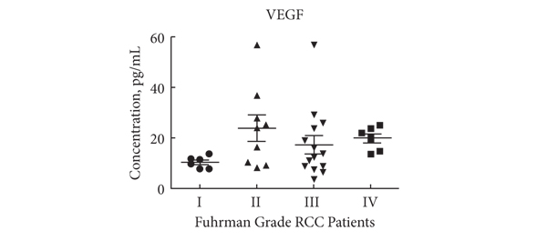
Likewise, in malignant diseases, such as pancreatic and ovarian cancer, an excess of Tregs is thought to contribute to failure of the immune system to clear neoplastic cells. Whether the tumour environment appropriates the regulatory function of Tregs to propagate its own survival in a manner akin to infectious agents, or whether Tregs infiltrate larger tumours in which there is more chronic inflammation is unclear. Nevertheless, a correlation between higher Treg numbers and poorer outcomes is a common feature of malignancies. In this issue of the BJUI Polimeno et al. add evidence to the debate over whether Treg numbers in RCC are associated with worse outcomes. While previous publications both support (Cancer Immunol Immunother 2007, BJU Int 2009) and refute (Clin Cancer Res 2007) this assertion, the data presented by Polimeno et al. identify not only increased circulating Treg numbers in patients with RCC but also find an association betweenTreg numbers, especially those that express the naïve T-cell marker CD45RA, and both larger tumour load and worse prognosis. In the same dataset, as expected, the authors also find that a shorter disease-free survival was evident in patients with lower numbers of tumoricidal natural killer cells. In the serum, patients with RCC had higher concentrations of soluble factors involved in cell growth and movement, such as epidermal growth factor, hepatocyte growth factor, vascular endothelial growth factor and interferon γ-induced protein 10 (also known as CXCL10), and markers of active inflammation, such as interleukins 6 and 8.

These observations suggest several broad possibilities: (i) that the ‘Tregs’ identified in the tumour environment and circulation are not Tregs but are in fact other activated T cells that temporarily express the same surface markers as bona fide Tregs; (ii) that Tregs in the context of RCC are unable to control tumour-associated inflammation; (iii) that Tregs contribute to tumour survival by inhibiting clearance of neoplasms by other immune cells, resulting in chronic inflammation; and/or (iv) that Tregs are actively contributing to the inflammation by converting to pro-inflammatory phenotypes, as has been demonstrated by several groups. These possibilities can be differentiated by isolating Tregs from the tumour environment or local draining lymph nodes and testing their functional characteristics in vitro; however, the fact that CD45RA+ Tregs were independently associated with worse outcomes makes (i) and (ii) less likely, as such cells are less likely to be recently activated and are inherently less plastic than other populations of Tregs.

In our opinion, the clinical value of the data presented in this paper and those of others, even if the underlying biology is poorly understood, should next be determined in a prospective study to see whether the immunological ‘fingerprint’ in peripheral blood can correctly identify those patients who are more likely to do poorly, targeting them for closer monitoring and/or more aggressive therapy.

Behdad Afzali and Giovanna Lombardi
Medical Research Council Centre for Transplantation, King’s College London, King’s Health Partners, Guy’s Hospital, and National Institute for Health Research Biomedical Research Centre at Guy’s and St Thomas’ NHS Foundation Trust and King’s College London, Guy’s Hospital, London, UK

Read the full article
© 2024 BJU International. All Rights Reserved.