Tag Archive for: ERG

Posts

Article of the Week: Immunocytochemical expression of ERG in urine identifies prostate cancer

Every Week the Editor-in-Chief selects an Article of the Week from the current issue of BJUI. The abstract is reproduced below and you can click on the button to read the full article, which is freely available to all readers for at least 30 days from the time of this post.

In addition to the article itself, there is an accompanying editorial written by a prominent member of the urological community. This blog is intended to provoke comment and discussion and we invite you to use the comment tools at the bottom of each post to join the conversation.

If you only have time to read one article this week, it should be this one.

Immunocytochemical detection of ERG expression in exfoliated urinary cells identifies with high specificity patients with prostate cancer

Raj P. Pal*, Roger C. Kockelbergh, John Howard Pringl e*, Lara Cresswell, Roger
Hew§, John P. Dormer§, Colin Cooper, John Kilian Mellon, Julian G. Barwell** and Edward J. Hollox**


*Department of Cancer Studies and Molecular Medicine, University of Leicester, Department of Urology, Department of Cytogenetics, §Department of Cellular Pathology, University Hospitals of Leicester NHS Trust, Leicester, Department of Cancer Genetics, University of East Anglia, Norwich, and **Department of Genetics, University of Leicester, Leicester, UK


Objectives

To evaluate the immunocytochemical detection of ERG protein in exfoliated cells as a means of identifying patients with prostate cancer (PCa) before prostate biopsy.

Materials and Methods

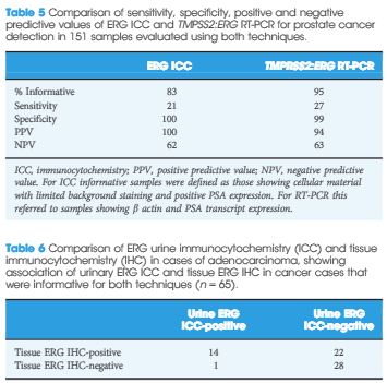
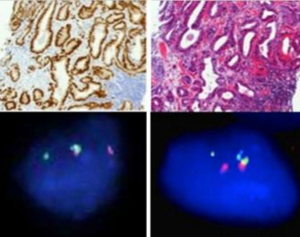
Urine samples (30 mL) were collected after digital rectal examination (DRE) from 159 patients with an elevated age-specific prostate-specific antigen (PSA) and/or an abnormal DRE who underwent prostate biopsy. In all cases, exfoliated urinary cells from half of the urine sample underwent immunocytochemical assessment for ERG protein expression. Exfoliated cells in the remaining half underwent assessment ofTMPRSS2:ERG status using either nested reverse-transcriptase (RT)-PCR (151 cases) or fluorescence in situ hybridization (FISH; eight cases). Corresponding tissue samples were evaluated using FISH to determine chromosomal gene fusion tissue status and immunohistochemistry (IHC) to determine ERG protein expression. Results were correlated with clinicopathological variables.

Results

The sensitivity and specificity of urinary ERG immunocytochemistry (ICC) for PCa were 22.7 and 100%, respectively. ERG ICC results correlated with advanced tumour grade, stage and higher serum PSA. In comparison, urine TMPRSS2:ERG transcript analysis had 27% sensitivity and 98% specificity for PCa detection. On tissue IHC, ERG staining was highly specific for PCa. In all, 52% of cancers harboured foci of ERG staining; however, only 46% of cancers that were found to have ERG overexpression were positive on urine ICC. The ERG ICC results showed strong concordance with urinary RT-PCR and FISH, and tissue IHC and FISH.

ew

Conclusion

This is the first study to show that cytological gene fusion detection using ICC is feasible and identifies patients with adverse disease markers. ERG ICC was highly specific, but this technique was less sensitive than RT-PCR.

Editorial: New possibilities for urinary molecular diagnostics

Fusion of androgen-driven serine protease transmembrane protease (TMPRSS) and oncogenic erythroblast transformation-specific-related gene (ERG) genes is a specific alteration in human prostate cancer and many studies have been performed in order to analyse its role as a biomarker or, even more interestingly, as a tumour promoter [1]. Recently, Nguyen et al. [1] demonstrated that ERG activates the Yes-associated protein 1 (YAP1) transcriptional programme thus inducing prostate carcinogenesis. YAP is a transcriptional coactivator involved in regulation of many biological processes. Thus, there is increasing evidence showing that ERG is causally involved in prostate cancer development. Since the original publication by Tomlins et al. [2], in which the gene fusion was discovered, researchers have addressed numerous scientific questions in order to reveal importance of the TMPRSS:ERG fusion in prostate cancer. This is particularly important because several other proteins have been proposed as prostate tumour markers; however, many of them cannot be used in clinical practice. The reasons for that are related to differences in sampling of tissues and methodologies. Thus, biomarker research will in future probably be restructured on the basis of standardisation of experimental procedures. Biomarker research work related to the TMPRSS:ERG fusion appears to be more advanced. In addition to tissue diagnostics, studies on detection of ERG may be performed in urine as evidenced in the paper by Pal et al. [3]. Moreover, the authors for the first time show that exfoliated urinary cells can be used to identify patients with prostate cancer by detection of positive ERG samples. The authors used nested PCR and urinary immunocytochemistry and fluorescence in situ hybridisation in this study. As the TMPRSS:ERG fusion is detectable in ≈50% of prostate cancer specimens, the data presented in the study published in this issue of BJUI are of considerable interest.

Combining the use of TMPRSS:ERG detection and other markers in urine should further improve diagnostics of prostate cancer. Another tumour marker, prostate cancer antigen 3 (PCA3), is frequently used in prostate cancer diagnostics. In urine, TMPRSS:ERG urinary transcript detection is superior to PSA and PCA3 for identifying patients with cancer [4]. The results support data recently published according to which the determination of urine TMPRSS2:ERG together with PCA3 may be used for a more individualised prostate cancer risk assessment [5].

As the authors state in the paper [3], differences between results of some marker studies on TMPRSS:ERG detection may be a consequence of the appearance of less common isoforms, which are not detectable by all assays.

In summary, Pal et al. [3] show for the first time that patients with prostate cancer could be identified by immunocytochemistry on the basis of detection of ERG in exfoliated urine cells. In addition, the authors [3] also show that the appearance of ERG in exfoliated urine cells is associated with a bad prognosis. Indirectly, the present paper [3], supports the concept that the presence of TMPRSS:ERG regulates various cellular events leading to increased migration and proliferation of prostate cancer cells [6]. It is expected that further improvements in urinary molecular diagnostics based on the presence of TMPRSS:ERG will be achieved in the near future. However, clinicians should also be aware that, despite its high specificity, there are limitations of methodology used in this paper, in particular a large proportion of tumours may remain undetected and not all cancers exfoliate into the urine.

Zoran Culig
Experimental Urology, Department of Urology, Medical University of Innsbruck, Anichstrasse 35, Innsbruck, A-6020, Austria


References


1 Nguyen LT, Tretiakova MS, Silvis MR et al. ERG activates the YAP1 transcriptional program and induces the development of age-related prostate tumors. Cancer Cell 2015; 27: 797808


2 Tomlins SA, Rhodes DR, Perner S et al. Recurrent fusion of TMPRSS2 and ETS transcription factor genes in prostate cancer. Science 2005; 310: 6448


5 Tomlins SA, Day JR, Lonigro RJ et al. Urine TMPRSS2:ERG plus PCA3 for individualized prostate cancer risk assessment. Eur Urol 2015; [Epub ahead of print] DOI: 10.1016/j.eururo.2015.04.039


© 2024 BJU International. All Rights Reserved.