Tag Archive for: Editorial

Posts

Editorial: The Advanced Prostate Cancer Consensus on a regional level – what can we learn?

In this issue of BJUI Chiong et al [1] present the results of the Asia Pacific (APAC) Advanced Prostate Cancer Consensus Conference (APCCC) 2018, during which the implications of the APCCC 2017 findings were discussed in the context of the APAC region. For background, it is important to understand the concept of the original APCCC and why it was initiated [1,2,3].

The consensus conference aims to target areas of controversy in the clinical management of advanced prostate cancer where evidence is either limited or lacking or where interpretation of evidence is controversial. The expert consensus aims to complement existing clinical practice guidelines that are mostly based on high‐level evidence. The APCCC’s most prominent aim is knowledge translation, in the sense of improving care of men with advanced prostate cancer worldwide who are treated outside of centres of excellence. During the original APCCC in St Gallen, where 61 prostate cancer experts and scientists were assembled, the majority of the consensus questions were discussed; these had been prepared prior to the conference under the idealistic assumption that all diagnostic procedures and treatments (including expertise in their interpretation and application) mentioned were readily available. These assumptions have been specifically chosen, because availability of systemic treatment options for advanced prostate cancer, access to next‐generation imaging (whole‐body MRI and positron‐emission tomography [PET]) and expertise in molecular techniques and interpretation of results vary widely across the world. The original global APCCC did not generally address regional or country‐specific situations, but APCCC 2017 did have a special session and also voting questions for treatment options in countries with limited resources. Importantly, consensus recommendations may even inform and influence regulatory authorities, for example, if a specific treatment is considered to be the best option by the majority of experts and availability in a certain country is lacking.

The APAC APCCC 2018 consisted of 20 experts (mostly urologists) from 15 countries and discussed the findings and voting results of five of the 10 APCCC 2017 topics. Whether or not Turkey should be considered an APAC country is unclear. The most relevant observations were as set out below:

  • There is huge variation in access to drugs used for treatment of advanced prostate cancer in the APAC region. Australia and Hong Kong have access to almost all treatment options (notably cabazitaxel is not mentioned) compared with countries such as Vietnam or the Philippines, where there is limited availability of many compounds. Regarding imaging technologies (standard CT is not mentioned), there seems to be wide availability of next‐generation imaging such as whole‐body MRI and choline‐ or PSMA‐PET technologies; however, these imaging methods are often not reimbursed.
  • Pharmaco‐ethnic issues have so far not been considered by the original APCCC and the APAC report clearly highlights the need to address such issues. The higher toxicity of docetaxel in Asian men may influence treatment recommendations, especially in situations such as low‐volume metastatic castration‐naïve prostate cancer, where the role of early addition of docetaxel to androgen deprivation therapy is less clear.
  • The authors of the APAC meeting state that ketoconazole and bicalutamide are still widely used despite the proven superiority of enzalutamide vs bicalutamide. A possible reason for this is the lack of reimbursement in some APAC countries.
  • There is an obvious need for clinical trials in the APAC region because of variations in genetics, genomics, epidemiology and pharmaco‐ethnicity. Such trials may answer questions about toxicity/tolerability and also optimal use of resources in the context of economic limitations.

In summary, the APAC APCCC 2018 is an excellent example of how the global APCCC findings should be discussed and integrated on a regional or even country‐specific level. The authors are therefore to be congratulated for their efforts and for writing up the discussions. The next APCCC  (2019; apccc.org) will take up a number of points raised by the APAC meeting, namely, more panel experts from APAC countries and pharmaco‐ethnic topics.

References

  1. Edmund C, Declan GM, Hideyuki A et al. Management of patients with advanced prostate cancer in the Asia Pacific region: ‘real‐world’ consideration of results from the Advanced Prostate Cancer Consensus Conference (APCCC) 2017. BJU Int 2019; 123: 22–34
  2. Gillessen S, Omlin A, Attard G et al. Management of patients with advanced prostate cancer: recommendations of the St Gallen Advanced Prostate Cancer Consensus Conference (APCCC) 2015. Ann Oncol 2015; 26: 1589–604
  3. Gillessen S, Attard G, Beer TM et al. Management of patients with advanced prostate cancer: the report of the Advanced Prostate Cancer Consensus Conference APCCC 2017. Eur Urol 2018; 73: 178–211


Editorial: Retzius‐sparing robot‐assisted radical prostatectomy

In their commentary in the current issue of BJUI, Stonier et al. [1] examine the potential technical pitfalls and published results of the Retzius‐sparing technique of robotic radical prostatectomy. The authors reviewed three studies from three different groups [2,3], including a study by our group [4], and raised three specific concerns: the oncological efficacy of the procedure; the long learning curve; and the generalizability of the technique to challenging surgical scenarios. We offer a few clarifications and comments.

The first study on Retzius‐sparing robot‐assisted radical prostatectomy came from the Bocciardi group [2]. This was a prospective, single‐arm study of 200 patients. The authors reported a 14‐day continence rate of 90–92%, a 1‐year potency rate of 71–81% (in preoperatively potent patients undergoing bilateral intrafascial nerve‐sparing) and a positive surgical margin rate of 25.5%. The positive surgical margin rate improved in patients with pT2 disease, from 22% to 9% (P = 0.04) over the course of the study (initial 100 vs subsequent 100 patients), while in patients with pT3 disease, it remained stable at ~45%. Lim et al. [3] also noted an improvement in their overall positive surgical margin rate from 20% to 8% when comparing the initial 25 patients with the subsequent 25 patients. In that study, a standard robot‐assisted radical prostatectomy comparator arm was included and there were no differences in overall positive surgical margin rates (14% in both arms), while continence was better with the Retzius‐sparing approach.

Recognizing the potentially technically challenging nature of the Bocciardi approach, we performed a randomized controlled trial to objectively evaluate the technique. Randomized controlled trials are typically designed to answer a single question. Our trial was designed to determine whether there were differences in the rate of return of urinary continence, the primary benefit that previous non‐controlled studies had reported. This our study clearly showed [4].

Once the trial was completed, post hoc analysis of secondary outcomes was performed [5]. One of these outcomes was the positive surgical margin rate. In our trial, we noted an overall positive surgical margin rate of 25% in the Retzius‐sparing arm vs 13% in the control arm, a difference that did not achieve statistical significance (P = 0.11). Stonier et al. [1] suggested that if the sample size of our trial were doubled, then the positive surgical margin rate in each group would be doubled as well, leading to significance. This conclusion is problematic. The likelihood that doubling the sample size would result in the exact doubling of numbers in all four cells of a 2 × 2 contingency table is estimated at <5% using Fisher’s exact test (this calculation is different from the P value). Furthermore, the surgical margins depend as much on the pathological stage as on surgical approach. In our trial, patients were matched preoperatively for risk in the best manner possible for a pragmatic randomized trial. However, it is impossible to predict and control for the final pathological characteristics. Pathological analysis showed that patients undergoing Retzius‐sparing surgery did have significantly more aggressive disease: ≥pT3 disease in 45% vs 23.3% of patients (P = 0.04) [4, 5]. This, by itself, could account for a substantial difference in surgical margin rates.

In writing our paper, we made no judgements as to whether the Bocciardi or posterior technique is fundamentally superior to an anterior or Menon approach, whether it is easier to perform, how generalizable it is [6], or what the learning curve may be. That is best left to the individual surgeon’s training and judgement. We do suggest, however, that surgical margins be interpreted as a function of pathological variables, and not in isolation, and that it is simplistic to assume that identical results will be obtained by doubling sample size. We suggest that such conclusions are hypothesis‐generating, and should best be explored through a separate, purpose‐designed randomized trial.

Authors: Akshay Sood, Firas Abdollah and Mani Menon

References

  1. Stonier T, Simson N, Davis J, Challacombe B. Retzius‐sparing robot‐assisted radical prostatectomy (RS‐RARP) vs standard RARP: it’s time for critical appraisal. BJU Int 2019; 123: 5–10
  2. Galfano A, Di Trapani D, Sozzi F et al. Beyond the learning curve of the Retzius‐sparing approach for robot‐assisted laparoscopic radical prostatectomy: oncologic and functional results of the first 200 patients with >/= 1 year of follow‐up. Eur Urol 2013; 64: 974–80
  3. Lim SK, Kim KH, Shin TY et al. Retzius‐sparing robot‐assisted laparoscopic radical prostatectomy: combining the best of retropubic and perineal approaches. BJU Int 2014; 114: 236–44
  4. Dalela D, Jeong W, Prasad MA et al. A pragmatic randomized controlled trial examining the impact of the Retzius‐sparing approach on early urinary continence recovery after robot‐assisted radical prostatectomy. Eur Urol 2017; 72: 677–85
  5. Menon M, Dalela D, Jamil M et al. Functional recovery, oncologic outcomes and postoperative complications after robot‐assisted radical prostatectomy: an evidence‐based analysis comparing the Retzius sparing and standard approaches. J Urol 2018; 199: 1210–7
  6. Galfano A, Secco S, Bocciardi AM. Will Retzius‐sparing prostatectomy be the future of prostate cancer surgery? Eur Urol 2017; 72: 686–8


Editorial: Reply: RS-RARP vs standard RARP

Since the introduction of robotic surgery in the treatment of patients with prostate cancer (PCa), different surgical innovations have been implemented in order to preserve postoperative functional outcomes while maintaining oncological safety. Sparing the Retzius space during robot‐assisted radical prostatectomy (RARP) was introduced early this decade by Galfano et al [1]. Interestingly, 90% and 96% of patients treated with Retzius‐sparing RARP (RS‐RARP) were continent (no pad/safety pad) at 1 week and 1 year, respectively. Similarly, our group reported a 70% continence rate (no pad) at 1 month after RS‐RARP [2].

The fast urinary continence recovery after RS‐RARP is related to several anatomical factors: the anterior Retzius space is kept intact; the urinary bladder is not dropped; the endopelvic fascia and puboprostatic ligaments are preserved; and there is minimal distortion of the supporting urethral tissues. A recent study reported [3] that less bladder neck descent was observed during postoperative cystogram in patients treated with RS‐RARP than in those treated with standard RARP.

In a recent randomized controlled study, the postoperative continence rate at 1 week was 48% in standard RARP compared with 71% in RS‐RARP (P = 0.01), and this difference was maintained at 3 months (86% standard RARP vs 95% RS‐RARP; P = 0.02). At 1 year, however, the effect on urinary continence difference was muted (93.3% standard RARP vs 98.3% RS‐RARP; P = 0.09) [4]. Similarly, Chang et al. [3] found that the higher continence rate at 1 week (73.3% RS‐RARP vs 26.7% standard RARP; P = 0.000) had vanished at 1 year (100% vs 93.3%; P = 0.15). By contrast, a large recent prospective series showed that the superiority of RS‐RARP in terms of higher early urinary continence was maintained at 1 year (97.5% RS‐RARP vs 68.5% standard RARP) [5].

In addition to a higher early continence rate, RS‐RARP has a lower incidence of postoperative inguinal hernia occurrence compared with standard RARP [6]. Theoretically, RS‐RARP may provide several other potential advantages. It may be advantageous if patients require future surgery necessitating access to the Retzius space and dropping of the bladder, such as an artificial urinary sphincter implantation, an inflatable penile prosthesis insertion, or kidney transplantation. In addition, in patients with previous inguinal hernia repair using mesh, it enables the avoidance of anterior adhesions by accessing the prostate directly from the Douglas pouch. Notably, large‐size glands and/or middle‐lobe, advanced/high‐risk PCa, and patients with previous prostatic surgeries can be managed safely with RS‐RARP in experienced hands.

Undoubtedly, oncological safety is our main concern in treating cancer. To determine the effectiveness of new treatment methods, long‐term follow‐up is warranted. Biochemical recurrence (BCR) is widely used as a primary oncological outcome to assess PCa treatment success. To our knowledge, after radical prostatectomy, ~35% of patients are at risk of developing BCR in the next 10 years. Currently, there are insufficient data regarding the oncological outcomes of RS‐RARP. Only four articles have compared early oncological outcomes between RS‐RARP and standard RARP, and there was no significant difference (Table 1).

More recently, we reported on the mid‐term oncological outcomes of 359 patients who underwent RS‐RARP. The median follow‐up was 26 months. Although this period is not long enough to reach a meaningful conclusion on the oncological safety of RS‐RARP, it is the longest follow‐up period reported in literature. Overall, the positive surgical margin (PSM) rate was 30.6% (14.6% in pT2 and 40.8% in pT3a disease) and the BCR rate was 14.8%. In terms of functional outcomes, the urinary continence rate at 1 year was 93.9% [7]. Interestingly, 164 patients (45.7%) of our cohort had high‐risk PCa. In these patients, the PSM rate was 41.2%, the BCR rate was 22%, and the 3‐year BCR‐free survival (BCRFS) rate was 72%. We compared our results with those in patients with high‐risk PCa treated with standard RARP in the literature. In studies that used the D’Amico criteria the median follow‐up ranged from 12.5 to 37.3 months, the PSM rates were 20.5% to 53.3%, the BCR rates were 17.4% to 31% and the 3‐year BCRFS rates were 41.4% to 86%. In studies that used the National Comprehensive Cancer Network criteria, the median follow‐up ranged from 23.6 to 27 months, the PSM rates were 29% to 38%, the BCR rates were 9.4% to 33%, and the 3‐year BCRFS rates were 55% to 66% [7].

In summary, RS‐RARP is a novel surgical approach which is associated with better urinary continence recovery in the first few months compared with standard RARP [2,3,4,5]. This superiority might be maintained [5] or equalized at 1 year [3,4]. A few studies have compared the early oncological results between RS‐RARP and standard RARP and no significant difference was found [2,3,4,5]. Recently, our group reported the mid‐term oncological outcomes of patients with high‐risk PCa treated with RS‐RARP and these were similar to those of large studies of conventional RARP. This confirms effective and safe mid‐term BCR control after RS‐RARP, while the long‐term oncological results are awaited [7]. Currently, >4 000 cases of RS‐RARP are performed worldwide and more centres are beginning to use and converting to Retzius‐sparing surgery. All centres are experiencing faster recovery of continence. Thanks are due to Drs Galfano and Bocciardi for exploring and sharing this surgical frontier.


References

  1. Galfano A, Di Trapani D, Sozzi F, et al. Beyond the learning curve of the Retzius‐sparing approach for robotassisted laparoscopic radical prostatectomy: oncologic and functional results of the first 200 patients with ? 1 year of follow‐up. Eur Urol 2013; 64: 974‐80
  2. Lim SK, Kim KH, Shin TY et al. Retzius‐sparing robot‐assisted laparoscopic radical prostatectomy: combining the best of retropubic and perineal approaches. BJU Int 2014; 114: 236–44
  3. Chang LW, Hung SC, Hu JC et al. Retzius‐sparing robotic‐assisted radical prostatectomy associated with less bladder neck descent and better early continence outcome. Anticancer Res 2018; 38: 345–51
  4. Menon M, Dalela D, Jamil M et al. Functional recovery, oncologic outcomes and postoperative complications after robot‐assisted radical prostatectomy: an evidence‐based analysis comparing the Retzius sparing and standard approaches. J Urol 2018; 199: 1210–7
  5. Sayyid RK, Simpson WG, Lu C et al. Retzius sparing robotic assisted laparoscopic radical prostatectomy: a safe surgical technique with superior continence outcomes. J Endourol 2017; 31: 1244–50
  6. Chang KD, Abdel Raheem A, Santok GDR et al. Anatomical Retzius‐space preservation is associated with lower incidence of postoperative inguinal hernia development after robot‐assisted radical prostatectomy. Hernia 2017; 21: 555–61
  7. Abdel Raheem A, Kidon C, Alenzi M et al. Predictors of biochemical recurrence after retzius‐sparing robot‐assisted radical prostatectomy: analysis of 359 cases with a median follow‐up of 26 months. Int J Urol 2018; 25: 1006–14


Editorial: Can machine‐learning algorithms replace conventional statistics?

Wong et al. [1] evaluate 19 clinical variables (training data) and three supervised machine‐learning algorithms to predict early biochemical recurrence after robot‐assisted prostatectomy. They further compare the areas under the curve (AUCs) resulting from these algorithms with the AUC of a conventional Cox regression model and conclude that the machine‐learning algorithms can produce accurate disease prognosis, perhaps better than a traditional Cox regression model. As the authors state, predictive models have the potential to better individualize care to patients at highest risk of prostate cancer recurrence and progression.

The authors should be commended for their adoption of machine‐learning algorithms to better interpret the vast volumes of clinical data and assess prognosis after robot‐assisted prostatectomy. This should represent another step forward for the management of prostate cancer, where tailored treatment is now largely based on the clinical risk stratification of the disease [2]. Incidentally, we are also in an era where we are seeing aspects of artificial intelligence (machine learning being a subset of it) vastly transform how we view and process data in everyday life. This has been true in medicine as well, particularly for prostate cancer [3].

While our own research group has also evaluated machine‐learning algorithms to process surgeon performance metrics and predict clinical outcomes after robot‐assisted prostatectomy [4], I want to express a word of caution. Utilization of machine learning does not in itself imply automatic superiority over conventional statistics [5] despite literature that has demonstrated so [3]. The success of predictive models in machine learning still relies on the quality of data introduced and careful execution of the analysis. In our experience, it works best when highly experienced clinicians and data scientists are working hand in hand.

Furthermore, I would argue that the results of this present study do not necessarily show that machine learning is superior to conventional statistics, but rather it highlights an inherent advantage of machine learning. While traditional analyses require the a priori selection of a model based on the available data, machine learning has more flexibility for model fitting [6]. Additionally, inclusion of variables in traditional analyses is constrained by the sample size. In contrast, by design, machine learning models thrive on their ability to consider many variables concurrently, and as such, have the potential to detect underlying patterns that may otherwise be undetectable when data are examined effectively in individual silos.

We look forward to the external validation of the methodology described in the present article. Big and diverse data are critical requirements of machine learning. A multi‐institutional, multi‐surgeon cohort is necessary to confirm the findings in this report. A further step from there is the adoption of such prediction models into clinical use. The ultimate question is how improved prognostic data may influence surgeon and patient decisions.

Conflict of Interest

Dr Hung reports personal fees from Ethicon, Inc, outside the submitted work.

References

  1. Wong NC, Lam C, Patterson L, Shayegan B. Use of machine learning to predict early biochemical recurrence following robotic prostatectomy. BJU Int 2019; 123: 51–7
  2. D’Amico AV, Whittington R, Malkowicz SB et al. Biochemical outcome after radical prostatectomy, external beam radiation therapy or interstitial radiation therapy for clinically localized prostate cancer. JAMA 1998; 280: 969–74
  3. Hung AJ, Chen J, Che Z et al. Utilizing machine learning and automated performance metrics to evaluate robot‐assisted radical prostatectomy performance and predict outcomes. J Endourol 2018; 32: 438–445
  4. Kattan MW. Comparison of Cox regression with other methods for determining prediction models and nomograms. J Urol 2003; 170 (6 Pt 2): S6–9
  5. Hung AJ, Chen J, Gill IS. Automated performance metrics and machine learning algorithms to measure surgeon performance and anticipate clinical outcomes in robotic surgery. JAMA Surg 2018; 153: 770–1

Editorial: Mental imagery: ‘you can observe a lot by watching!’

Urethrovesical anastomosis (UVA), like any other surgical anastomosis, is a key example of motor muscle memory, where aligning two hollow structures should result in a watertight anastomosis defining its success and help avoid complications. The authors [1] investigated the importance of cognitive training during UVA, which has been shown to be a promising supplement to skill‐based training. The authors utilised the Global Evaluative Assessment of Robotic Skills (GEARS), which has been validated for assessment of general robotic rather than procedure‐specific skills. As the authors chose UVA to evaluate training, they could have used the Robotic Anastomosis Competency Evaluation (RACE), which has been developed and validated for specific evaluation of UVA [2]. However, the study eloquently revealed higher scores, using the validated movement imagery questionnaire modified for robot‐assisted surgery whilst evaluating mental imagery.

Motor imagery utilises imagining action without its physical execution and this leads to eliciting activity in regions of brain normally activated during performance. Motor imagery has shown significant neural activity in important brain area involved in somatosensory perception, especially kinesthetic information from motor perception and muscle spindles. Such areas become active when a motor illusion is induced that ultimately share the same basis with areas active during executing movement. Mental imagery also yields more benefits if its sessions are interposed between periods of training [3]. Unfortunately, the ability to imagine more complex tasks is less accurate when utilising mental imagery [4]. In future, studies using procedure‐specific evaluation, such as RACE, may help us understand in depth the role of mental imagery during various steps of complex task, such as UVA. The hypothesis of improvement of skills whilst utilising supplemental cognitive training is reasonable; future studies will benefit from utilising an elaborate cognitive assessment. Metrics such as electroencephalograms (EEGs) and eye tracking, or even less sophisticated tools like the National Aeronautics and Space Administration Task Load Index (NASA‐TLX) self‐assessment questionnaires, have previously been used for assessment of cognitive load [5]. Objective feedback provided by a brain–computer interface (BCI) can increase the brain activation levels produced during motor imagery and thereby help in improving performance [6].

Motor imagery has been used as a popular input for BCI and in future could be used as a link to establishing instruction to semi‐autonomous robotic systems [6]. Meanwhile, a motor imagery BCI using EEG is utilising intention recognition through decoding brain activity, which ultimately could allow for intuitive control of devices like robotic systems.

References

  1. Raison N, Ahmed K, Abe T et al. Cognitive training for technical and non‐technical skills in robotic surgery: a randomised controlled trial. BJU Int 2018; 122: 1075–81
  2. Raza SJ, Field E, Jay C et al. Surgical competency for urethrovesical anastomosis during robot‐assisted radical prostatectomy: development and validation of the robotic anastomosis competency evaluation. Urology 2015; 85: 27–32
  3. Nicholson VP, Keogh JW, Low Choy NL. Can a single session of motor imagery promote motor learning of locomotion in older adults? A randomized controlled trial. Clin Interv Aging 2018; 13: 713–22
  4. Kalicinski M, Kempe M, Bock O. Motor imagery: effects of age, task complexity, and task setting. Exp Aging Res 2015; 41: 25–3
  5. Besharat Shafiei S, Hussein AA, Ahmed Y, Guru K. Can eye tracking help explain an expert surgeon’s brain performance during robot‐assisted surgery? J Urol 2018; 199 (Suppl.): e1–2
  6. Batula AM, Kim YE, Ayaz H. Virtual and actual humanoid robot control with four‐class motor‐imagery‐based optical brain‐computer interface. Biomed Res Int 2017; 2017: 1463512.


Editorial: A novel nomogram for predicting ECE of prostate cancer

We read with great interest the publication on the side‐specific multiparametric magnetic resonance imaging (mpMRI)‐based nomogram from Martini et al. [1].

The prediction of extracapsular extension (ECE) of prostate cancer is of utmost importance to inform accurate surgical planning before radical prostatectomy (RP).

Today, surgical strategy is tailored to the patient’s characteristics, and the need for a correct prediction of ECE is of paramount importance to guarantee oncological safety, as well as optimal functional outcome. The most up‐to‐date guidelines suggest referring to nomograms to decide whether or not to perform nerve‐sparing (NS) surgery. Since the first version of the Partin Tables in 1993, several models have been developed based on PSA, Gleason score at prostate biopsy, and clinical staging, as the most used covariates.

Furthermore, mpMRI is increasingly used in the diagnostic pathway of prostate cancer to aid prostate biopsy targeting and to attain a more accurate diagnosis of clinically significant prostate cancer. Despite its recognised role in the detection of cancer, the accuracy for local staging is poor, providing a low and heterogeneous sensitivity for the detection of ECE [2].

Given this limitation, the addition of MRI to clinically derived nomograms might result in an improved assessment of preoperative local staging. In a retrospective analysis of 501 patients who underwent RP, MRI + clinical models outperformed clinical‐based models alone for all staging outcomes, with better discrimination in predicting ECE with MRI + Partin Tables and MRI + Cancer of the Prostate Risk Assessment (CAPRA) score than nomograms alone [3].

In the current article, Martini et al. [1] suggest a novel nomogram for predicting ECE that includes the presence of a ‘documented definite ECE at mpMRI’ as an additional variable beyond PSA, Gleason score, and maximum percentage of tumour in the biopsy core with the highest Gleason score. Readers should recognise that this is the first model integrating side‐specific MRI findings together with side‐specific biopsy data to provide a ‘MRI‐based side‐specific prediction of ECE’, in an effort to support the surgical decision for a uni‐ or bilateral NS approach.

However, given the frail generalisability of nomograms in different datasets even after external validation [4], a predictive tool has to be built on a rigorous methodology with clear reproducibility of all steps the covariates derive from.

In this respect, the current model raises some concerns.

The schedule of preoperative MRI assessment is arbitrary, with imaging being performed either before (23.9%) or after systematic biopsy (76.1%), and amongst patients with a MRI prior to biopsy, only 94 of 134 patients underwent additional targeted sampling. As a result, MRI is applied by chance in three different ways: before prostate biopsy without targeted sampling, before prostate biopsy with targeted sampling, and after prostate biopsy.

Based upon this heterogeneous MRI timing, the performance of such a model in a novel population may be biased depending on the diagnostic pathway applied at each institution.

The choice of the variables included represents another point of concern. The output of two out of four covariates, ECE depiction at mpMRI and the percentage of tumour in the biopsy core, have been deliberately dichotomised, without taking into account the continuous trend intrinsic to both variables.

Actually, local staging in the European Society of Urogenital Radiology (ESUR) guidelines has been scored on a 1–5 point scale to grade the likelihood of an ECE event. The authors deliberately dichotomised mpMRI findings, considering ‘the loss of prostate capsule and its irregularity’ as suggestive of ECE and ‘broad capsular contact, abutment or bulge without gross ECE’ evocative of organ‐confined disease. As a result, the included MRI covariate may account for a gross prediction of ECE, maintaining the inaccurate and inter‐reader subjective interpretation of local staging intrinsic to MRI.

Beyond those methodological concerns and the moderate sample size that may limit the reproducibility of the model, we wonder if such a prediction really assists the surgeon’s capability to perform a tailored surgery.

The ‘all or none’ era of NS surgery is over, and we are currently able to grade NS according to different approaches reported in the literature. Particularly, Tewari et al. [5] proposed a NS approach based on four grades of dissection, with the veins on the lateral aspect as vascular landmarks to gain the correct dissection planes. Patel et al. [6] described a five‐grade scale of dissection, using the arterial periprostatic vasculature as a landmark to the same purpose.

If we are able to grade a NS surgery, the prediction of ECE should be graded as well and should answer the prerequisite of knowing the amount of prostate cancer extent outside the capsule. How does a surgeon make the decision to follow a more or less conservative dissection otherwise?

We tried to address this issue by using a tool aimed at predicting the amount of ECE [the Predicting ExtraCapsular Extension in Prostate cancer tool] [6] and supporting the choice of the correct plane of dissection with a suggested decision rule. In our study, developed on a large sample of nearly 12 000 prostatic lobes and several combined clinicopathological variables, the absence of imaging characterization was the major point of weakness.

To date, the ideal predictive tool has yet to be described. However, in the modern era of precision surgery, we think that a model should encompass the surgical knowledge and techniques currently available.

Future developments will probably include three‐dimensional surgical navigation models displayed on the TilePro™ function of the robotic console (Intuitive Surgical Inc., Sunnyvale, CA, USA), based on the integration of MRI (for the number, size and location of disease) and predictive tools (to define the amount of ECE).


References

  1. Martini A, Gupta A, Lewis SC et al. Development and internal validation of a side‐specific, multiparametric magnetic resonance imaging‐based nomogram for the prediction of extracapsular extension of prostate cancer. BJU Int 2018; 122: 1025–33
  2. de Rooij M, Hamoen EH, Witjes JA, Barentsz JO, Rovers MM. Accuracy of magnetic resonance imaging for local staging of prostate cancer: a diagnostic meta‐analysis. Eur Urol 2016; 70: 233–45
  3. Morlacco A, Sharma V, Viers BR et al. The incremental role of magnetic resonance imaging for prostate cancer staging before radical prostatectomy. Eur Urol 2017; 71: 701–4
  4. Bleeker SE, Moll HA, Steyerberg EW et al. External validation is necessary in prediction research: a clinical example. J Clin Epidemiol 2003; 56: 826–32
  5. Tewari AK, Srivastava A, Huang MW et al. Anatomical grades of nerve sparing: a risk‐stratified approach to neural‐hammock sparing during robot‐assisted radical prostatectomy (RARP). BJU Int 2011; 108: 984–92
  6. Patel VR, Sandri M, Grasso AA et al. A novel tool for predicting extracapsular extension during graded partial nerve sparing in radical prostatectomy. BJU Int 2018; 121: 373–82


Editorial: Multi-parametric MRI: an important tool to improve risk stratification for active surveillance in prostate cancer

Multiparametric MRI (mpMRI) has become an important adjunct in the management of localized prostate cancer (PCa), particularly in the active surveillance (AS) setting. Current guideline recommendations [1,2] have recommended incorporation of mpMRI into AS protocols to improve patient stratification and reclassification.

Bryant et al. [3], based on updated National Institute of Health and Care Excellence (NICE) guidelines [1], report on the effect of mpMRI incorporation into their institution’s AS protocols, specifically focusing on the time to treatment and number of biopsies required to trigger treatment. In 2014, they replaced protocol‐driven biannual prostate biopsies (PBs) with mpMRI ± cognitive targeted biopsy and systematic biopsy (TB). With a median follow‐up of 2.4 years, they found that more men who underwent TB progressed to treatment than men who underwent PB alone (44% vs 37%; P = 0.003). The median number of biopsies (beyond the original diagnostic biopsy) required to trigger intervention was 1.55. Based on these results, the authors conclude that mpMRI‐driven TB increases reclassification compared with protocol‐driven PB.

This is consistent with increasing evidence that mpMRI enhances, and sometimes, exceeds detection of clinically significant PCa over TRUS‐guided prostate biopsy alone. The PROMIS study [4], a multicentre paired validation study that compared mpMRI to TRUS‐guided biopsy in the diagnostic setting, found that mpMRI had better sensitivity (93% vs 43%; P < 0.001) and negative predictive value (89% vs 74%; P < 0.001) than TRUS‐guided biopsy in detecting clinically significant cancer (defined as Gleason grade ≥4 + 3). While the concerns about foregoing a systematic biopsy at the time of targeted biopsy in that study were warranted, there was consensus that prebiopsy mpMRI increased the yield for clinically significant PCa.

In the AS setting, unfortunately, randomized data are lacking; however, retrospective series and systematic reviews provide some guidance. In a systematic review, Schoots et al. [5] found that a positive mpMRI in the AS setting was associated with a higher risk of upgrading at the time of radical prostatectomy and a higher risk of reclassification at the time of confirmatory biopsy. Yet, a negative mpMRI did not preclude reclassification and upgrading, indicating the continued need for systematic biopsy. Recabal et al. [6] confirmed these conclusions in their retrospective assessment of an institutionally maintained prospective dataset. While MRI‐targeted biopsies detected higher grade cancer in 23% of men, they missed higher grade clinically significant cancers in 17%, 12% and 10% of patients with mpMRI scores of 3, 4 and 5, respectively. This suggests that both targeted and systematic biopsy should be used for the optimal detection of clinically significant PCa in men on AS.

The present study by Bryant et al. [3] reaffirms the value of mpMRI in the AS paradigm. Yet, some concerns about their study cohort and methodology should be noted. First, as the authors clearly note as a limitation, despite completing a targeted and systematic biopsy, all the samples were sent as a single specimen, precluding the ability to distinguish between targeted biopsy and systematic biopsy cores. As the absolute difference in the rate of progression to treatment between the PB and TB arms was only 7%, it is uncertain how much of that was attributable to the addition of targeted biopsy alone.

Additionally, in a closer analysis of their study population, it should be noted that 35% of the patients had Gleason Grade Group 2 disease or higher at the time of inclusion, representing a higher‐risk AS patient population than guideline recommendations. This may account for the higher rate of progression to treatment in this study cohort independent of grade progression – 24% of patients progressed to treatment based on PSA progression alone and an additional 10% were based on mpMRI findings alone.

Lastly, the median number of biopsies required to trigger intervention was 1.55 and, for the majority of patients, this was just one additional biopsy beyond the original diagnostic biopsy. Guideline recommendations indicate the importance of a confirmatory biopsy to exclude Gleason sampling error [2]; however, by definition, many of these patients were essentially upstaged or redirected to active treatment after a confirmatory biopsy. With 59% of the entire AS population never receiving a confirmatory biopsy beyond their original diagnostic biopsy and many progressing to treatment after a confirmatory biopsy, this study population may not reflect a well‐selected low‐risk PCa patient population for AS.

Despite these limitations, the work by Bryant et al. [3] adds to the growing body of evidence supporting the use of mpMRI‐targeted biopsies in addition to systematic biopsy to more accurately risk stratify men for AS, particularly at the time of diagnosis. It remains unknown how we can use mpMRI to individually tailor surveillance strategies or if mpMRI may ultimately replace surveillance biopsies over time.

References

  1. Graham J, Kirkbride P, Cann K, Hasler E, Prettyjohns M. Prostate cancer: summary of updated NICE guidance. BMJ (Clinical research ed.). 2014348: f7524
  2. Mottet N, Bellmunt J, Bolla M et al. EAU‐ESTRO‐SIOG Guidelines on Prostate Cancer. Part 1: screening, diagnosis, and local treatment with curative intentEur Urol 201771: 618–29


Editorial: Contemporary quality‐of‐life scores provide a key foundation for high‐quality cancer research

Prostate cancer is the most common male malignancy in many countries, including the UK/Northern Ireland. Given excellent oncological outcomes for appropriately treated localised cancer, there is an increasing focus on understanding the quality‐of‐life implications of different treatment options.

As Donnelly et al. [1] emphasise, contemporary cohorts of untreated men can provide useful comparisons for inferring the impact of treatment. Specifically, updated population‐level observations of urinary, bowel, and sexual dysfunction are needed to provide a baseline for such discussions. Surveys should focus on particular populations (e.g. geographic), utilise prostate cancer‐specific questionnaires, and ensure age‐matched cohorts. Such baseline characteristics are essential to teasing apart the impact of prostate cancer and its treatment from ageing and comorbidities.

Donnelly et al. [1] sampled 10 000 men in Northern Ireland aged >40 years, using the EuroQoL five Dimensions five Levels (EQ‐5D‐5L) survey to assess a general health baseline and Extended Prostate Cancer Composite (EPIC) questionnaire to determine bladder, bowel, and sexual function more specifically. In all, 2 955 men responded, although ultimately only men aged >60 years were analysed to better match the age distribution of patients with prostate cancer. Strikingly, they found that nearly two out of five men reported at least one urinary, bowel, or sexual issue. A third of men reported some degree of urinary leakage, 26% had some degree of bowel problems, and as much as 57.9% of respondents had some problem with sexual function [1].

Nearly two decades ago, Litwin [2] published a health‐related quality of life control sample of older men in the USA without prostate cancer using the University of California Los Angeles Prostate Cancer Index (UCLA‐PCI, a precursor to EPIC). He found ageing subjects had diminished urinary continence, bowel function, and sexual potency, with similar rates to the Northern Ireland study: a third reported urinary leakage, a third had bowel complaints, and nearly two‐thirds claimed to have erectile dysfunction (ED).

In contrast, patient‐reported outcomes in the Prostate Testing for Cancer and Treatment (ProtecT) trial showed low levels of urinary incontinence and bowel symptoms, and one‐third of men had sexual dysfunction [3]. The difference here in ED when compared to Donnelly et al. [1] may be attributed to the age distribution differences between the cohorts, as ProtecT included men aged 50–69 years and the Northern Ireland group looked only at men aged >60 years. This highlights the importance of ensuring age‐matched cohorts when using population‐based surveys as baselines for assessment counselling.

Furthermore, Resnick et al. [4] evaluated the change in patient‐reported urinary incontinence and ED over time in two cohorts of patients enrolled almost 20 years apart. They compared patients enrolled in 1994–1995 in the Prostate Cancer Outcomes Study (PCOS) vs those enrolled in 2011–2012 in the Comparative Effectiveness Analysis of Surgery and Radiation (CEASAR) study. Men in PCOS were surveyed using UCLA‐PCI, and those in CEASAR completed EPIC‐26. They found that self‐reported urinary incontinence was more common in CEASAR than in PCOS (7.7% vs 4.7%), as was ED (44.7% vs 24%). These differences could be due to rising rates of comorbidities associated with ED and urinary incontinence or they may reflect an increase in social awareness and disclosure of these issues.

Taken together, these self‐reported rates of pretreatment urinary and sexual function underscore the potential for significant variation in reporting of patient quality‐of‐life outcomes in prostate cancer.

This does not mean that patient‐reported outcomes should be ignored. Rather the takeaway is that we must invest in tools to ensure that reporting is appropriate, standardised, and accurate [5]. And regardless of whether these data are collected prospectively, or retrospectively, it is vital to use appropriate statistical methods and scientific principles to account for bias and to ensure that causal inferences are valid [6].

As prostate cancer survival and mortality rates improve, patients and clinicians must weigh treatment‐specific short‐ and long‐term effects on quality of life. Patient‐reported outcome measures are vital to assessing these major impacts. Contemporary, population‐based cohorts such as that provided by Donnelly et al. [1], provide a key tool for better interpreting and understanding these results.

References

  1. Donnelly DW, Donnelly C, Kearney T et al. Urinary, bowel and sexual health in older men from Northern Ireland. BJU Int 2018; 122: 845–57
  2. Litwin MS. Health related quality of life in older men without prostate cancer. J Urol 1999; 161: 1180–4


Editorial: The opioid epidemic: a wake‐up call for us all

The article in this issue of BJUI by Theisen et al. [1] is a timely reminder of the duty of all prescribers (including surgeons) to be mindful of the potential unintended consequences and off‐target effects of medicines.

Although some of the factors that have led to serious opioid‐related problems are particularly related to the US setting, we in Europe and other continents should not be complacent [2, 3].

The US Department of Health and Human Services (HHS) stated that, in 2016, opioid deaths had risen to > 42 000 deaths, of which an estimated 40% involved a prescription opioid [4].

The underlying reasons for this opioid epidemic are multiple and complex.

The prevalence of pain in the population is high, as are patients expectations and demands for treatment. The ageing population, living with multiple painful conditions, including cancer survivors and patients with persisting post‐surgery chronic pain, has further increased the demand for analgesics.

Meanwhile, the WHO’s drive over the last 30 years to eradicate ‘opiophobia’ and ensure that opioids are available for cancer pain, together with the advent of potent prolonged‐release opioid formulations, led to a transfer of this therapeutic experience to non‐cancer pain. A few questioned the wisdom of this strategy, but reassurance was drawn from an 11‐line letter in the New England Journal of Medicine in 1980, oft cited and misquoted as evidence that addiction was rare with long‐term opioids [5]. Subsequently, the journal has added a note warning readers that ‘the letter has been heavily and uncritically cited by sources using it to suggest opioids are not addictive.’ In fact, the authors surveyed the files of inpatients who were administered predominantly short‐term opioids in hospital, including patients who had only received one dose, and concluded that in this population, development of new addiction was rare.

Add to these factors, the well‐intentioned drive to assess and treat pain with initiatives such as ‘Pain – the fifth vital sign’, and pharmaceutical company promotion of their new opioid formulations, and the scene was set for greatly increased opioid initiation, escalation of dosage and repeat prescribing without regular patient review. In addition to these factors, it was also identified that a proportion of patients continue to receive opioids long after their surgery [6].

By 2017, year‐on‐year increases in long‐term opioid prescribing compounded by the diversion of the medicines, illicit manufacture and importing of compounds, such as fentanyl analogues, culminated in the staggering US mortality data and the HHS declaring a public health emergency with a five‐point strategy to combat the opioid crisis.

What strategies can we adopt during and after surgery? Better multimodal acute inpatient analgesia and working closely with our acute pain colleagues will surely assist in achieving less need for subsequent opioid prescribing on discharge. Using enhanced recovery pathways encourages the use of opioid‐sparing local and regional anaesthetic blocks, together with simple analgesia rather than prolonged use of high‐dose opioids. The goal must be to discharge patients on less potent analgesics and for a shorter duration. The analogy is with antibiotic prescribing where only a limited supply is dispensed. We need to develop pain discharge plans which can be communicated to the primary care physician incorporating tapering, patient education and emphasis on avoiding the repeat prescribing of opioids. Where pain persists, the patient should be referred back to the surgical or pain management team sooner in order to review progress. We should be wary of prescribing modified‐release preparations of a drug such as morphine or oxycodone because these contain a high dose, which can be extracted from the slow‐release preparation for abuse purposes. Similarly, the use of opioid‐based patches encourages extended use of opioid drugs, sometimes without a full understanding of the hourly or daily morphine equivalent dosage. Looking forward, there is the promise of new non‐opioid analgesics for chronic pain on the horizon, in particular long‐acting, prolonged‐release local anaesthetics for use in the wound or for nerve blocks. We need to adopt strategies for the regular review of pain medication rather than the all too often ‘automatic’ repeat prescription.

In urology, we have seen a significant reduction in the use of opioids on discharge through the use of less invasive, endoscopic/robotic techniques, local anaesthetic blocks such as the transversus abdominis block, which is so valuable in abdominal procedures, wound local anaesthetic infusion catheters and the use of regular simple analgesics given by the clock, providing excellent opioid‐sparing background analgesia.

Less opioid drug prescribing in the community is the way forward as Theisen et al. describe. As peri‐operative physicians, we must respond to this challenge if we are to avert a similar crisis to that seen in the USA. In peri‐operative practice, responsible and appropriate opioid‐prescribing remains an essential part of good pain management, while we strive to reduce both dose and duration of therapy. These strategies serve both wider society and the individual patient, for whom the benefit is reduced dose‐dependent opioid side effects. In the modern era where specialist advice is available through multidisciplinary team working, we need to minimize repeat prescribing and ensure that a specific opioid tapering plan is in place. The latter relies on good communication, teamwork and partnership, the essential ‘Domain 3’ of General Medical Council Good Medical Practice [7].

References

  1. Theisen K, Jacobs B, Macleod L, Davies B. The United States opioid epidemic: a review of the surgeon’s contribution and health policy initiatives. BJU Int 2018; 122: 754–9
  2. Stannard C. Opioids in the UK: what’s the problem? BMJ 2013; 347: f5108
  3. Weisberg DF, Becker WC, Fiellin DA, Stannard C. Prescription opioid misuse in the United States and the United Kingdom: cautionary lessons. Int J Drug Policy 2014; 358: 1124–30
  4. US Department of Health and Human Services. What is the U.S. Opioid Epidemic? Available at: https://www.hhs.gov/opioids/about-the-epidemic/index.html. Accessed October 2018
  5. Porter J, Jick H. Addiction rare in patients treated with narcotics. N Engl J Med 1980; 302: 123
  6. Clarke H, Soneji N, Ko TD, Yun L, Wijeysundera DN. Rates and risk factors for prolonged opioid use after major surgery: population based cohort study. BMJ 2014; 348: g1251
  7. GMC Good Medical Practice, 2013. Available at: www.gmc-uk.org/guidance Accessed October 2018


Editorial: Close surgical margins after RP: how to make a complex story even more complex

Surgical margin (SM) status after radical prostatectomy (RP) for clinically localized prostate cancer (PCa) is a measure of surgical quality and retains some prognostic value. Positive SMs (PSMs) have long been considered an adverse oncological outcome because they were repeatedly found to be associated with a higher risk of biochemical recurrence (BCR), and are still among the factors guiding the decision to deliver adjuvant treatments; however, the long‐term impact of PSMs on survival remains uncertain because it is largely affected by other concurrent risk modifiers [1,2,3].

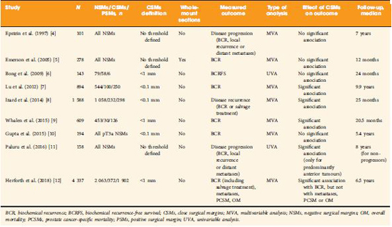
The clinical significance of so‐called close SMs (CSMs), that is, negative SMs (NSMs) with tumour foci approaching, but not involving, the inked cut surface of the RP specimen, is a far less investigated field of research, with contradictory findings in the few available studies (Table 1 [412]). Some studies showed a significant association with risk of disease progression (mainly measured with BCR), while others did not.


The study by Herforth et al. [12] published in this issue of BJUI further adds to the debate on CSMs, with an analysis of the largest series reported to date. The authors assessed the impact of CSMs vs NSMs vs PSMs after RP on BCR, PCa‐specific and overall survival in ~4 300 men included in the Shared Equal Access Regional Cancer Hospital cohort. CSMs were defined as cancer foci within 1 mm from the inked specimen surface, and were found in 372 patients (9%). The median follow‐up was 6.5 years. On multivariable analysis accounting for several established prognostic factors, CSMs were significantly associated with a higher BCR risk compared with NSMs, but a lower risk compared with PSMs. Notably, SM status alone did not influence PCa‐specific or overall survival. Major limitations to this retrospective analysis were lack of central pathology review and inadequate follow‐up length to assess survival.

The main question yet to answer is whether CSMs entail a biological entity that is distinct from both negative (but not close) SMs and PSMs. Advances in this area cannot be made without taking into consideration the knowledge of PSMs that has accumulated over the past years. We suggest, therefore, that the following principles be adhered to in order to ascertain the true significance of CSMs.

    1. Uniform definition
      Some of the available studies used an arbitrary threshold (0.1 or 1 mm) to designate CSMs, but distance between tumour and SMs should be ideally evaluated as a continuous variable before attempting to categorize it.
    2. Accurate pathology examination
      It has been hypothesized that CSMs could be the expression of occult PSMs that are present in different close planes of resection missed by standard sectioning as a result of block sampling bias 11. Encountering CSMs should, then, probably prompt further specimen processing that requires standardization.
    3. Correct prognostic assessment
      It is now accepted that PSMs per se are not sufficient to confer a dismal prognosis, rather it is the concomitant effect of other pathological risk factors (such as stage, tumour volume, Gleason score at SMs, location and extent of PSMs) that determines the aggressive tumour behaviour. The same could apply to CSMs; therefore, their prognostic effect should be investigated by adding ‘interaction terms’ to classic multivariable models that account for a putative synergistic biological effect. It might well be, in fact, that the simultaneous presence of CSMs and extracapsular disease (or higher Gleason score, greater tumour volume, perineural/lymphovascular invasion) results in a final risk of detrimental outcome exceeding the additive combination of the individual risks.
    4. Adequate follow‐up
      At least a decade is required to appropriately test the association of CSMs in patients undergoing RP with endpoints of meaningful interest.

The truth about SMs after RP is still hard to reach, and the issue of CSMs possibly complicates this scenario. While we await further characterization of PCa facilitated by advances in genetic profiling, we recommend that future clinical research in the field does not run into the methodological obstacles of the past.

Gianluca Giannarini, Alessandro Crestani and Claudio Valotto

Urology Unit, Academic Medical Centre ‘Santa Maria della Misericordia’, Udine, Italy


References

  1. Yossepowitch O, Bjartell A, Eastham JA et al. Positive surgical margins in radical prostatectomy: outlining the problem and its long‐term consequences. Eur Urol 2009; 55: 87–99
  2. Yossepowitch O, Briganti A, Eastham JA et al. Positive surgical margins after radical prostatectomy: a systematic review and contemporary update. Eur Urol 2014; 65: 303–13
  3. Stephenson AJ, Eggener SE, Hernandez AV et al. Do margins matter? The influence of positive surgical margins on prostate cancer‐specific mortality. Eur Urol 2014; 65: 675–80
  4. Epstein JI, Sauvageot J. Do close but negative margins in radical prostatectomy specimens increase the risk of postoperative progression? J Urol 1997; 157: 2413
  5. Emerson RE, Koch MO, Daggy JK, Cheng L. Closest distance between tumor and resection margin in radical prostatectomy specimens: lack of prognostic significance. Am J Surg Pathol 2005; 29: 225–9
  6. Bong GW, Ritenour CW, Osunkoya AO, Smith MT, Keane TE. Evaluation of modern pathological criteria for positive margins in radical prostatectomy specimens and their use for predicting biochemical recurrence. BJU Int 2009; 103: 327–31 
  7. Lu J, Wirth GJ, Wu S et al. A close surgical margin after radical prostatectomy is an independent predictor of recurrence. J Urol 2012; 188: 91–7
  8. Izard JP, True LD, May P et al. Prostate cancer that is within 0.1 mm of the surgical margin of a radical prostatectomy predicts greater likelihood of recurrence. Am J Surg Pathol 2014; 38: 333–8
  9. Whalen MJ, Shapiro EY, Rothberg MB et al. Close surgical margins after radical prostatectomy mimic biochemical recurrence rates of positive margins. Urol Oncol 2015;33:494.e9–14
  10. Gupta R, O’Connell R, Haynes AM et al. Extraprostatic extension (EPE) of prostatic carcinoma: is its proximity to the surgical margin or Gleason score important? BJU Int 2015; 116: 343–50
  11. Paluru S, Epstein JI. Does the distance between tumor and margin in radical prostatectomy specimens correlate with prognosis: relation to tumor location. Hum Pathol 2016; 56: 11–15 Erratum in: Hum Pathol 2017; 60: 212
  12. Herforth C, Stroup SP, Chen Z et al. Radical prostatectomy and the effect of close surgical margins: results from the SEARCH database. BJU Int 2018; 122: 592–8


© 2024 BJU International. All Rights Reserved.