Tag Archive for: Editorial

Posts

Editorial: RPLND – Open Surgery’s Next Challenger Is Ready To Enter The Ring

By Tim Dudderidge

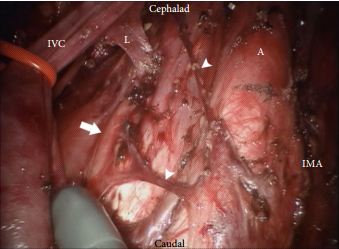
The da Vinci surgical system delivers the benefits of laparoscopic surgery with an easier and more precise human–tissue interface than conventional laparoscopic instruments. Nearly all major uro-oncological procedures are being performed robotically. In this issue of BJUI, Cheney et al. [1] present their technique and initial experience of robot-assisted retroperitoneal lymph node dissection (RA-RPLND) for patients with primary and post-chemotherapy non-seminoma germ cell tumours. Quality indicators for RA-RPLND include adequate clearance of the desired surgical field, satisfactory lymph node yield, acceptable perioperative morbidity and length of stay, as well as longer-term functional and oncological outcomes. So how well does RA-RPLND stand up to scrutiny?

The technique employed by Cheney et al., placing the robot at the head of the patient, is unfamiliar to most urologists I suspect. It appears to offer excellent access to the retroperitoneum, but still requires a re-docking when performing full bilateral dissections. Whether this technique is superior to the lateral approach that I and others have used for modified dissections requires further study [2,3]. The lymph node yield was lower than that previously reported for open RPLND and while Cheney et al. [1] observe this may be due to the use of a modified template where appropriate, the absence of any in-field recurrences at a median of 22 months is perhaps the more reliable sign that there is oncological equivalence. Concerns that a true template dissection cannot be completed with a robot-assisted laparoscopic approach are probably unjustified in my opinion. The description of surgical technique by Cheney et al., including suture ligation and division of lumbar vessels, confirms that if a surgeon is minded to do so, a complete bilateral or modified template clearance can be completed.

The absence of significant complications in this series is impressive; however, there were three out of 18 conversions to open surgery. The mean length of stay of 2.4 days is close to the 3–4 days stay I would expect after an uncomplicated open RPLND in a young fit man. However, 1–2 night stays were seen in their later cases as they gained experience. Perhaps more importantly in a group of working age men, return to full physical activity within 3 weeks is possible [2].

As highlighted by Cheney et al. [1], minimally invasive primary RPLND has been previously reported both by laparoscopic and robotic approaches. Their larger series provides an important demonstration that the robotic approach facilitates the more complex undertaking of post-chemotherapy RPLND. Furthermore they show that except for operative time, all other outcomes were similar in primary and post-chemotherapy cases.

As an enthusiast for minimally invasive therapies, I of course welcome these results and think that along with other published and presented series, they provide sufficient evidence to consider a more formal evaluation of this approach. However, how feasible is the wider introduction of RA-RPLND? Despite having experience of robotics and working in a team performing around 30 RPLNDs a year, I was only able to identify five cases during a 1-year period suitable for a robotic approach. With experience this could have been a higher proportion, but it is fair to conclude that suitable cases in typical cancer centres would be limited in number. This is particularly so for the UK and other European countries, where primary RPLND is not used. Cheney et al. [1] had similarly low numbers each year and recruited their cohort of 18 cases over 5 years.

An international multicentre registry is arguably the best way to gather more information on the safety and completeness of template dissection RPLND. Existing registries, e.g. the BAUS complex operations database, have already provided valuable insights into the results of RPLND in the UK [4] and could be combined with other international RA-RPLND databases already being compiled (Erik Castle MD personal communication). Partnership of testicular cancer surgeons without robotic experience with experienced robotic surgeons may also facilitate the development of additional centres for development of this procedure. They will also aid optimal patient selection and help avoid incomplete template dissections, which may compromise the excellent cancer control we are now used to.

There are clear potential advantages with a minimally invasive approach to RPLND, not least of which are the avoidance of a laparotomy scar, the reduction of complications and an earlier return to normal activity. Cheney et al. [1] have shown that their technique is feasible, safe and effective in the medium term and their results justify wider consideration of the procedure for further study and improvement.

Tim Dudderidge

University Hospital Southampton, Southampton, UK

References

1 Cheney SM, Andrews PE, Leibovich BC, Castle EP. Robot-assisted retroperitoneal lymph node dissection: technique and initial case series of 18 patients. BJU Int 2015; 115: 114–20

2 Dudderidge T, Pandian S, Nott D. Technique and outcomes for robotic assisted post-chemotherapy retroperitoneal lymph node dissection (RPLND) in Stage 2 non-seminomatous germ cell tumour (NSGCT). BJU Int 2012; 110: 97

3 Dogra PN, Singh P, Saini AK, Regmi KS, Singh BG, Nayak B. Robot assisted laparoscopic retroperitoneal lymph node dissection in testicular tumor. Urol Ann 2013; 5: 223–6

4 Hayes M, O’Brien T, Fowler S, BAUS RPLND Group. Contemporary retroperitoneal lymph node dissection (RPLND) for testis cancer in the UK – a national study. J Urol 2014; 191 (Suppl.): e89–90


Editorial: Bone Metastases in Prostate Cancer: Which Scan?

In this issue of BJUI, Poulsen et al. [1] present a prospective comparison of 18F-fluoride (NaF) and 18F-choline (FCH) positron emission tomography (PET)/CT with planar whole-body bone scintigraphy (WBS) using spinal MRI, including short tau inversion recovery (STIR), T1 and T2 sequences, as the reference standard in 50 hormone-naïve patients with confirmed bone metastases on WBS. They found that both PET/CT methods were significantly more sensitive and accurate than WBS and that FCH PET/CT was more specific than NaF PET/CT.

It has become increasingly recognised that planar WBS is no longer the most accurate method of assessing the skeleton for metastases and that novel imaging methods, including PET/CT, single-photon emission CT (SPECT)/CT and whole-body MRI offer advantages [2].

What is surprising in the presented results is that NaF PET/CT shows poor specificity (54%), a result that is discordant with previous literature [3, 4]. Compared with PET alone, using the CT component of hybrid PET/CT reduces false-positive interpretation of NaF uptake in benign lesions [3]. This raises the question as to whether the CT component of the PET/CT acquisition was used to full effect in the present study. The use of spinal MRI as a reference standard is also a possible limitation that is recognised by the authors, as this limits the comparison to only the spine, and MRI in itself is a method with known limitations. All patients had abnormal WBS for entry into the trial and whilst the PET methods were more sensitive on a lesion basis, a patient-based comparison was therefore not possible; however, the results imply that PET methods may identify metastatic disease in patients with normal WBS, as has been previously reported [3, 5].

Nevertheless, the authors should be congratulated in reporting valuable data from a prospective study where all imaging was performed in hormone-naïve patients, minimising confounding treatment-related effects, and within a small time window of 30 days; however, some questions remain. WBS is no longer state of the art for imaging the skeleton with radiolabelled bisphosphonates, such as 99mTc-methylene diphosphonate (MDP). Although NaF PET/CT has been shown to be superior to planar WBS augmented with SPECT [3], there have not been head-to-head comparisons with 99mTc-MDP SPECT/CT, where the potential advantages of the pharmacokinetics of NaF and the superior spatial resolution of PET compared with SPECT may not be as great. This may be particularly important given the difference in costs and availability of the two methods.

Despite the results from the present study, which show superiority of FCH PET/CT compared with NaF PET/CT with regard to specificity, taking the available literature as a whole, it remains unresolved as to what the best test for staging the skeleton in patients with high-risk prostate cancer should be at diagnosis. The different mechanisms of uptake of the PET tracers should be noted. NaF uptake reflects the local bone osteoblastic reaction to tumour within the bone marrow, whereas FCH uptake reflects metabolic activity within the tumour cells themselves. In prostate cancer, where the predominant effect is an increase in osteoblastic activity in the adjacent bone, the bone-specific tracers such as 99mTc-MDP and NaF have shown high sensitivity; however, direct imaging of tumour cell metabolism, such as increased choline kinase activity and cell membrane synthesis with FCH, may be advantageous in detecting metastases in the bone marrow before an osteoblastic reaction has occurred [6]. It is possible that both PET tracers may be required to provide optimum diagnostic accuracy and of course FCH PET/CT also provides valuable data on nodal and visceral metastatic disease. In patients with recurrent disease, better specificity has been reported with FCH [4], NaF possibly being limited by non-specific treatment-related effects such as osteoblastic flare. For similar reasons it may be that the more tumour-specific imaging methods, such as FCH PET/CT or diffusion-weighted MRI, may be better in assessing the treatment response of skeletal metastases. Questions therefore remain as to the best imaging test at different times in the management of patients with metastatic prostate cancer. 99mTc-MDP SPECT/CT deserves a full assessment, but perhaps the recent advent of PET/MRI and the potential synergies available from this hybrid technique may help resolve some of the remaining issues.

Read the full article

Gary Cook*† and Vicky Goh*‡

*Division of Imaging Sciences and Biomedical Engineering, King’s College London, † Clinical PET Centre, and ‡ Department of Radiology, Guy’s and St Thomas’ Hospitals NHS Foundation Trust, London, UK

References

1 Poulsen MH, Petersen H, Høilund-Carlsen PF et al. Spine metastases in prostate cancer: comparison of [99mTc]MDP wholebody bone scintigraphy, [18F]choline PET/CT, and [18F]NaF PET/CT. BJU Int 2014; 114: 818–23

2 Fogelman I, Blake GM, Cook GJ. The isotope bone scan: we can do better. Eur J Nucl Med Mol Imaging 2013; 40: 1139–40

3 Even-Sapir E, Metser U, Mishani E et al. The detection of bone metastases in patients with high-risk prostate cancer: 99mTc-MDP Planar bone scintigraphy, single- and multi-field-of-view SPECT, 18F-fluoride PET, and 18F-fluoride PET/CT. J Nucl Med 2006; 47: 287–974

4 Langsteger W, Balogova S, Huchet V et al. Fluorocholine (18F) and sodium fluoride (18F) PET/CT in the detection of prostate cancer: prospective comparison of diagnostic performance determined by masked reading. Q J Nucl Med Mol Imaging 2011; 55: 448–57

5 Kjölhede H, Ahlgren G, Almquist H et al. Combined 18F-fluorocholine and 18F-fluoride positron emission tomography/computed tomography imaging for staging of high-risk prostate cancer. BJU Int 2012; 110: 1501–6

6 Beheshti M, Vali R, Waldenberger P et al. Detection of bone metastases in patients with prostate cancer by 18F fluorocholine and 18F fluoride PET-CT: a comparative study. Eur J Nucl Med Mol Imaging 2008; 35: 1766–74


Editorial: Complex tumours, partial nephrectomy and functional outcomes

In the paper by Volpe et al. [1], excellent renal functional outcomes are associated with partial nephrectomy in patients with high PADUA score cancers. The study is notable because it shows that, even in patients who are typically considered candidates for radical nephrectomy, partial nephrectomy can maintain excellent estimated GFR (eGFR) and outcomes; however, because we perform nephron-sparing procedures on patients who may also be candidates for radical nephrectomy, we must consider the varied nature of some of the data on partial nephrectomy.

The literature on renal ischaemia and functional outcomes is heterogeneous and highly debated [2]. There have been several contradictory studies and changes over time in the literature based on technology, surgeon, centre, measurement and, now, correlation with parenchyma-sparing.

A study conducted by the European Organisation for the Research and Treatment of Cancer (EORTC) compared radical nephrectomy (essentially an ischaemic time of infinity) and partial nephrectomy, reporting a 10-year overall survival benefit for patients treated with radical nephrectomy [3]. Nevertheless, this oft-criticized randomised trial also showed better eGFR in partial nephrectomy. The survival benefit reported in that study is countered by population-based studies suggesting that partial nephrectomy may still be a better option when feasible [4]. Unfortunately, these population-based studies may be considered to provide a lower level of evidence than a randomised study, and are also prone to several biases, the most notable being selection of both patients and centres. Surgeons may be more likely to perform nephron-sparing in patients in lower-risk groups. There are also other questions to consider. If a patient is more likely to be referred to a larger centre for partial nephrectomy, are they not also likely to be referred for their coronary artery bypass, aortic surgery, general medical care and even emergency care? Are these patients more likely to seek out second opinions for all of their medical care? Will this affect mortality? Are they more motivated and engaged in their own overall healthcare? These are just a few of the confounding factors that could influence outcomes and are difficult to control in population-based studies. Nevertheless, I am a firm believer in partial nephrectomy, and particularly in preserving renal function, as the better choice for the treatment of both straightforward and complex lesions. It will be difficult, however, to completely negate the implications of the EORTC trial.

Does reasonable ischaemic time affect eGFR outcome? The present study by Volpe et al. [1] would suggest that reasonable ischaemic times are completely acceptable. Several contradictory studies point out the benefits and risks of a limited or minimized clamp time for partial nephrectomy. Another separate paper by White et al. [5] is consistent with other studies that show that a clamped partial nephrectomy, even for high complexity masses, results in a minimal loss of renal function, if at all. Although there is also enthusiasm for a zero ischaemia technique, it is critical to point out that this may be surgeon-, patient-, technique- and institution-dependent. Ultimately, however, we are splitting hairs over a few points of eGFR. The real issue with long-term GFR outcomes in our patients is not only the impact of a few minutes of renal ischaemia, but also control of hypertension, diabetes and their role in medical renal disease. There is an absence of urological literature that controls for patients’ glycated haemoglobin levels or measures hypertension monthly and records the response to medical therapy. These critical pieces of information confound all eGFR and comparative measurements and make it difficult to compare published outcomes. Perhaps the best medical advice we can give patients is to diet, exercise and eat healthily for better overall health. In some sense, this advice may be far more important than the decision of partial vs radical nephrectomy for a complex mass.

What are the logical conclusions of these dilemmas? Clamped partial nephrectomy is possible in complex cases, and the procedure salvages eGFR. Further refinements are also interesting academically, including papers on parenchyma-sparing. Nevertheless, if we are serious about ‘healthy kidneys’, we might take a holistic approach and encourage our patients to pursue a healthier lifestyle so they can bolster lifelong preservation of renal function and general wellness. Would the effect be more profound than a few minutes of ischaemic time? I am betting it would.

Read the full article

Sam B. Bhayani 

Division of Urological Surgery, Washington University School of Medicine and Barnes-Jewish West County Hospital, St Louis, MO, USA

References

1 Volpe A, Garrou D, Amparore D et al. Perioperative and renal functional outcomes of elective robot-assisted partial nephrectomy for renal tumors with high surgical complexity. BJU Int 2014; 114: 903–9

2 Lane BR, Russo P, Uzzo RG et al. Comparison of cold and warm ischemia during partial nephrectomy in 660 solitary kidneys reveals predominant roles of nonmodifiable factors in determining ultimate renal function. J Urol 2011; 185: 421–7

3 Van Poppel H, Da Pozzo L, Albrecht W et al. A prospective, randomized EORTC intergroup phase 3 study comparing the oncologic outcome of elective nephron-sparing surgery and radical nephrectomy for low-stage renal cell carcinoma. Eur Urol 2011; 59: 543–52

4 Sun M, Trinh Q-D, Bianchi M et al. A non-cancer related survival benefit is associated with partial nephrectomy. Eur Urol 2012; 61: 725–31

5 White MA, Georges-Pascal H, Autorino R et al. Outcomes of robotic partial nephrectomy for renal masses with nephrometry score of ≥ 7. Urology 2011; 77: 809–13


Editorial: Histone deacetylase inhibition: a new target for Peyronie’s disease?

Peyronie’s disease is a chronic and progressive disease characterised by fibrotic plaque of the tunica albuginea of the penis that can cause deformity, pain during erection and erectile dysfunction. Fibrosis is the hallmark of the pathology of Peyronie’s disease and is known to be driven by fibroblasts and myofibroblasts, which produce excessive amounts of extracellular matrix proteins and, hence, disturb the architecture of the tunica albuginea.

In this issue of BJUI, Kwon et al. [1] have shown that selective inhibition of histone deacetylase isoform 2 (HDAC-2) using a small hairpin silencing RNA elicits reversal of plaque development in vivo and prevention of collagen production and myofibroblast transformation in vitro. Histone deacetylases (HDACs) are a group of enzymes that remove acetyl groups from lysine amino acid in histones, causing histones to wrap around the DNA tightly and, ultimately, affecting gene transcription. In addition HDACs can de-acetylate cytosolic proteins and hence alter their function. Because of their direct effect on cell growth and death, HDACs have recently been attractive targets for anti-cancer drug development. Currently, there are > 100 clinical trials recruiting patients to investigate the clinical efficacy of HDAC inhibitors, most of which are non-selective HDAC inhibitors, bearing in mind that there are 11 isoforms of HDACs.

HDAC inhibitors have been suggested to have anti-fibrotic effects in the lung, liver, kidney and skin. They have been shown to reduce myofibroblast transformation and fibroblast activation, and counteract TGF-β actions and extracellular matrix production [2]. Although the exact mode of action of HDAC inhibitors in fibrosis is not clear, it has been suggested that HDAC inhibitors might repress the TGF-β pathway and interfere with phosphorylation and activation of STAT3, a key transcription factor in inflammatory pathways. Among all the isoforms of HDAC, HDAC-2 has been implicated in pathogenesis of fibrosis, firstly in kidney fibrosis [3] and later in Peyronie’s disease [4]. Currently available small-molecule HDAC inhibitors target more than one isoform of HDAC; to our knowledge isoform-selective small-molecule inhibitors are not available yet. Kwon et al. [1] have solved this problem using small hairpin silencing RNA to target HDAC-2 specifically. Although the clinical feasibility of such a silencing RNA approach remains to be tested, their study nevertheless gives an important indication for HDAC-2 as a possible target for fibrotic diseases, such as Peyronie’s. No doubt further research and development will be required to validate this target and develop small-molecule inhibitors selective for HDAC-2.

Read the full article

Selim Cellek* and David J. Ralph*†

*Centre for Biomedical Engineering, Cranfield University, Cranfield, and † University College London Hospital, London, UK

References

1 Kwon K-D, Choi MJ, Park J-M et al. Silencing histone deacetylase 2 using small hairpin RNA induces regression of fibrotic plaque in a rat model of Peyronie’s disease. BJU Int 2014; 114: 926–36

2 Pang M, Zhuang S. Histone deacetylase: a potential therapeutic target for fibrotic disorders. J Pharmacol Exp Ther 2010; 335: 266–72

3 Noh H, Oh EY, Seo JY et al. Histone deacetylase-2 is a key regulator of diabetes- and transforming growth factor-beta1-induced renal injury. Am J Physiol Renal Physiol 2009; 297: F729–39

4 Ryu JK, Kim WJ, Choi MJ et al. Inhibition of histone deacetylase 2 mitigates profibrotic TGF-β1 responses in fibroblasts derived from Peyronie’s plaque. Asian J Androl 2013; 15: 640–5


Editorial: Robotic and conventional open radical cystectomy lead to similar postoperative health-related quality of life

In this month’s issue of BJU International, Messer et al. [1] devise a prospective randomised trial to compare postoperative health-related quality of life (HRQoL) after robot-assisted (RARC) vs conventional open radical cystectomy (ORC). The investigators evaluated 40 patients over a follow-up period of 1 year and found no significant difference in HRQoL between surgical approaches. Moreover, they showed that the postoperative decrease in HRQoL returns to baseline within 3 months of surgery.

RC is one of the most challenging and potentially mutilating surgical interventions in the urological field and represents the standard-of-care treatment for patients with muscle-invasive bladder cancer. It is associated with a non-negligible risk of morbidity and mortality [2]. With the advent of new technologies, such as the Da Vinci surgical robot, carefully designed studies are needed to weigh the potential benefits of a novel approach against the increased costs associated with such tools. While RARC holds the promise of combining the benefits of a minimally invasive intervention with the precise robotic translation of the surgeon’s movements, these claims remain to be definitely proven in the clinical setting. As such, further elucidating the effect of surgical approach on perioperative outcomes after RC is essential for treatment planning, patient counselling and informed decision-making before surgery.

QoL is increasingly used as a quantitative measure of treatment success [3, 4]. These measures are gaining considerable traction in the USA, as reimbursements will soon be tied to patient satisfaction. While previous retrospective studies suggest that RARC has comparable perioperative oncological outcomes with potentially lower morbidity relative to ORC [5], there is a scarcity of high-quality evidence on HRQoL outcomes of RARC vs ORC. The difficulties of conducting randomised trials in the surgical setting are reflected by the relatively few participants in the Messer et al. [1] trial. Nonetheless, in their pilot study, the authors demonstrated the feasibility of a HRQoL trial in RC patients. Furthermore, they deliver initial evidence on the impact of surgical approach on HRQoL after RC.

From a clinical perspective, the authors contribute interesting findings to the ongoing debate. Their results suggest that the potential benefits of robot-assisted surgery on HRQoL may be limited in patients undergoing complex oncological surgery such as RC. Several hypotheses may be pertinent to their conclusions. For example, performing an open urinary diversion after RARC that can take as much time as the actual extirpative RC may mitigate any potential benefit of the minimally invasive approach. Furthermore, the study findings may be largely influenced by the surgical skills of the participating surgeons. Maybe the correct interpretation of their study findings is that there was no significant difference in HRQoL outcomes between ORC and RARC, at the institution where the trial was performed.

Nonetheless, the authors suitably demonstrate the feasibility of performing a randomised trial in this field and pave the way towards adequately powered, randomised multicentre trials that can provide further evidence on what impact RARC may have on perioperative outcomes and beyond.

Read the full article

Julian Hanske, Florian Roghmann, Joachim Noldus and Quoc-Dien Trinh*

Department of Urology, Marien Hospital, Ruhr-University Bochum, Herne, Germany, and *Division of Urologic Surgery and Center for Surgery and Public Health, Brigham and Women’s Hospital, Harvard Medical School, Boston, MA, USA

References

1 Messer JC, Punnen S, Fitzgerald J, Svatek R, Parekh DJ. Health-related quality of life from a prospective randomised clinical trial of robot-assisted laparoscopic vs open radical cystectomy. BJU Int 2014; 114: 896–902

2 Roghmann F, Trinh QD, Braun K et al. Standardized assessment of complications in a contemporary series of European patients undergoing radical cystectomy. Int J Urol 2014; 21: 143–9

3 Cookson MS, Dutta SC, Chang SS, Clark T, Smith JA Jr, Wells N. Health related quality of life in patients treated with radical cystectomy and urinary diversion for urothelial carcinoma of the bladder: development and validation of a new disease specific questionnaire. J Urol 2003; 170: 1926–30

4 Loppenberg B, von Bodman C, Brock M, Roghmann F, Noldus J, Palisaar RJ. Effect of perioperative complications and functional outcomes on health-related quality of life after radical prostatectomy. Qual Life Res 2014. doi: 10.1007/s11136-014-0729-1

5 Kader AK, Richards KA, Krane LS, Pettus JA, Smith JJ, Hemal AK. Robot-assisted laparoscopic vs open radical cystectomy: comparison of complications and periopera


Editorial: Statins and biochemical recurrence after radical prostatectomy – who benefits?

In the present issue of the BJUI Allott et al. [1] report results from a study where they used the Shared Equal Access Regional Cancer Hospital (SEARCH) database to explore the risk of biochemical recurrence (BCR) after radical prostatectomy (RP) among men who used statins after RP. They report improved BCR-free survival among statin users, especially among men with high-risk disease at baseline. The results provide some new insights into the current discussion on statins and prostate cancer outcomes.

Statins have recently shown promise as chemotherapeutic agents against prostate cancer. There is conflicting evidence on the effect on overall prostate cancer risk, but most studies able to evaluate the risk by tumour stage have reported lowered risk of advanced prostate cancer among statin users compared with the non-users [2], and lowered prostate cancer-specific mortality [3].

Taken together, these epidemiological findings suggest that statins may not strongly lower the risk of initiation of prostate cancer, but may be able to slow down the progression of the most dangerous form of the disease. In vitro studies support this by reporting growth inhibition and lower metastatic activity of prostate cancer cells after statin treatment [4].

Despite this, there has been recent controversy on statins’ effect on BCR of prostate cancer after radical treatment. A recent meta-analysis concluded that statin users may have a lower risk of BCR after external beam radiation therapy, but not after RP [5]. This could be due to statins acting as radiation sensitizers. Reports of improved BCR-free survival in statin users after brachytherapy would support this [6].

However, there are also differences in the characteristics of patients managed with RP or radiation therapy. Men undergoing RP have localised disease, which usually means low- to medium-grade tumours (Gleason ≤7), as high-grade disease (Gleason 8–10) progresses early and is more often locally advanced or already metastatic at diagnosis, leading to the choice of radiation therapy with neoadjuvant androgen deprivation instead of RP if curative treatment is still deemed possible.

This leads to the question whether the differing association between statins and BCR by treatment method is explained by patient selection, and whether statins are most effective against progression of high-grade disease. The study reported by Allott et al. [1] in this issue of the BJUI certainly suggests so. They report lowered risk of BCR among men who used statins after RP. They were able to study the effect of statin usage occurring after RP, not just usage at the time of RP. When the analysis was stratified by tumour characteristics, the improvement in relapse-free survival was strongest among men with high-risk disease (Gleason score ≥4 + 3; positive surgical margins).

The present study [1] supports the notion that statins could target a mechanism that is essential for progression of high-risk prostate cancer. This would be in concordance with the previously reported lowered risk of advanced prostate cancer and decreased prostate cancer mortality among statin users, as high-grade/high-risk cancer is the type progressing into advanced and fatal stages. On the other hand, if statins do not affect low-grade prostate cancer, this could explain why many RP series have not observed differences in biochemical relapses by statin use, as patients in these studies often have low-grade disease.

As always, statins’ benefits against prostate cancer are not really proven until verified in randomised clinical trials properly designed and powered to detect a difference in cancer endpoints. Designers of such trials should consider targeting the statin intervention to men with high-grade and/or high-risk prostate cancer for efficient study design.

Read the full article

Teemu J. Murtola*†

*School of Medicine, University of Tampere, and † Department of Urology, Tampere University Hospital, Tampere, Finland

References

1 Allott EH, Howard LE, Cooperberg MR et al. Postoperative statin use and risk of biochemical recurrence following radical prostatectomy: results from the Shared Equal Access Regional Cancer Hospital (SEARCH) database. BJU Int 2014; 114: 661–6

2 Bansal D, Undela K, D’Cruz S, Schifano F. Statin use and risk of prostate cancer: a meta-analysis of observational studies. PLoS ONE 2012; 7:e46691

3 Yu O, Eberg M, Benayoun S et al. Use of statins and the risk of death in patients with prostate cancer. J Clin Oncol 2014; 32: 5–11

4 Brown M, Hart C, Tawadros T et al. The differential effects of statins on the metastatic behaviour of prostate cancer. Br J Cancer 2012; 106: 1689–96

5 Park HS, Schoenfeld JD, Mailhot RB et al. Statins and prostate cancer recurrence following radical prostatectomy or radiotherapy: a systematic review and meta-analysis. Ann Oncol 2013; 24: 1427–34

6 Moyad MA, Merrick GS, Butler WM et al. Statins, especially atorvastatin, may improve survival following brachytherapy for clinically localized prostate cancer. Urol Nurs 2006; 26: 298–303


Editorial: Evolution of extracorporeal shockwave lithotripsy (ESWL)

Much has changed since the introduction of extracorporeal shockwave lithotripsy (ESWL); however, in many ways the principles remain constant. This manuscript by Jagtap et al. [1] is a large series of patients over 25 years and encapsulates the changes in ESWL over that time. This paper has all the limitations inherent in a retrospective review but within this offers interesting data. In particular the use of two different machines and refinements in technique are eloquently described. This shows an improvement due to both the change in technology but also in the importance of modifications of technique. The particular factors improving stone-free rate (SFR) were; better localisation with ultrasonography and X-ray, better coupling and use of coupling gel, change in selection criteria for both the patient and stone, ramping up the power and a staff training programme. This emphasis on technique is especially pertinent in healthcare systems where mobile lithotripters are still in use. These are renowned to have lower SFRs than static machines, which may be due to the technical delivery of treatment as much as the efficacy of the lithotripter.

What is the future for ESWL? The paper reflects the perception globally that whilst the incidence of urolithiasis is increasing, the use of ESWL is not increasing at the same rate, particularly for ureteric stones, and they cite the potential factors for this. This has also been noted in the UK and our own recent review of Hospital Episode Statistics (HES) data even suggest the rate of ESWL has plateaued for both ureteric (3000/year) and renal (19 500/year) stones in the last 3 years [2, 3]. There has been discussion within the UK about centralising endourology services using the same model as for cancer, with provision of static lithotripters within those centres. This would potentially have the advantage of creating high-volume centres with quality being easier to standardise and monitor; however, this would have to be balanced against patients probably having to travel further to access ESWL. The use of Hounsfield units remains a topic of debate with conflicting data and limited clinical application [4, 5]. Optimising targeting to minimise tissue damage with maximal stone fragmentation remains a challenge and modifications to lithotripters with dual-imaging modalities, dual heads, alterations in shockwave delivery rate, control of respiratory effort and novel feedback devices have had limited success. Increasing levels of obesity within developed countries are a factor in the utilisation of ESWL, as there is a limit on focal distance. All of these factors along with the continued improvement in the optics, miniaturisation of ureteroscopes and advent of holmium laser have contributed to a surge in the use of ureteroscopy, despite publications and guidelines showing similar success rates [6].

Read the full article

Kay Thomas

Clinical lead for Urology, Honorary Senior Lecturer Kings College London, UK

References

1 Jagtap J, Mishra S, Bhattu A, Ganpule A, Sabnis R, Desai M. Evolution of shockwave lithotripsy (SWL) technique: a 25-year single centre experience of >5000 patients. BJU Int 2014; 114: 748–53

2 Turney BW, Reynard JM, Noble JG, Keoghane SR. Trends in urological disease. BJU Int 2011; 109: 1082–7

3 Withington J. Personal communication from Royal College of Surgeons. July 2014

4 Pareek G, Armenakas A, Fracchia JA. Hounsfield units on computerized tomography predict stone free rates after extracorporeal shock wave lithotripsy. J Urol 2012; 169: 1679–81

5 Foda K, Abdeldaeim H, Youssif M, Assem A. Calculating the number of shock waves, expulsion time and optimum stone parameters based on noncontrast computerized tomography characteristics. Urology 2013; 82: 1026–31

6 Türk C, Knoll T, Petrik A et al. EAU Guidelines on Urolithiasis, 2014. Available at: https://www.uroweb.org/gls/pdf/22%20Urolithiasis_LR.pdf. Accessed July 2014


Editorial: ‘Discontent is the first necessity of progress’, Thomas A. Edison

This study from Kaag et al. [1] investigates predictors of renal functional decline after radical nephroureterectomy (RNU) in patients with upper tract urothelial carcinoma (UTUC). They evaluate early (2 months) and late (6 months) predictors of renal functional decline, finding that on a multivariable model only age at surgery and preoperative renal function were independently associated with early postoperative function. This is an intuitive finding whereby we expect older patients and those with lower renal function to have a more dramatic decrease in renal function after RNU.

Age, preoperative renal function, and Charlson score were associated with late functional recovery. The latter is a counterintuitive finding, as higher Charlson score was associated with less decrease in renal function. Charlson comorbidity was not significant on univariate analyses. Why it would become significant on multivariate is unclear. Whether it is an artifact related to study methodology or is a real phenomenon will require further study.

Unquestionably, this study [1] adds to the growing discontent of our current management of UTUC. The authors cogently discuss the issues related to better risk stratification as a natural consequence of instituting a neoadjuvant chemotherapy paradigm in those with high-risk disease. Multiple retrospective studies have failed to show a benefit of adjuvant chemotherapy, whereas now we have a matched-cohort study showing significant rates of downstaging and complete remission [2], and as well significantly improved 5-year survival, with institution of a neoadjuvant paradigm [3]. One cannot view the dismal outcomes of this disease without being discontent and wishing for progress. We need to continue getting out the message to not only urologists who reflexively institute RNU in patients with a risk-unstratified upper tract filling defect, but as well many medical oncologists who can only function based on guidance from level I data, which for this disease, will be a long time coming.

Read the full article

Surena F. Matin

Department of Urology, MD Anderson Cancer Center, Houston, TX, USA

References

1 Kaag M, Trost L, Thompson RH et al. Pre-operative predictors of renal function decline following radical nephroureterectomy for upper tract urothelial carcinoma. BJU Int 2014; 114: 674–9

2 Matin SF, Margulis V, Kamat A et al. Incidence of downstaging and complete remission after neoadjuvant chemotherapy for high-risk upper tract transitional cell carcinoma. Cancer 2010; 116: 3127–34

3 Porten S, Siefker-Radtke AO, Xiao L et al. Neoadjuvant chemotherapy improves survival of patients with upper tract urothelial carcinoma. Cancer 2014; 120: 1794–9

Editorial: Where next in ketamine uropathy? Dedicated management centres?

Tam et al. [1] in this month’s BJUI publish the largest prospective cohort to date on ketamine uropathy (KU). KU is a growing international problem since initial reports in 2007 from Canada and Hong Kong, where ketamine is second only to heroin in popularity amongst drug takers [2, 3]. Prevalence of KU may be higher than previously thought with up to a quarter of people misusing ketamine reporting urinary symptoms [4].

Importantly, the Tam et al. [1] paper demonstrates the benefit of stopping ketamine amongst those presenting with KU. Dose, frequency and dependency upon ketamine have been reported as risk factors for developing KU [1, 4]. Achieving cessation is not always straightforward following identification, assessment and urology input. Consistent with the Winstock et al. [4] recommendations a multi-disciplinary approach is required to assess symptoms and risk profile. The recommendation of Tam et al. of a one-stop clinic is thus appealing.

The key to diagnosing KU, is a focused history including specific drug use, performing non-invasive uroflowmetry investigations and upper tract imaging. Urologists need to be aware of motivational interviewing strategies, and incorporate them in their assessment. Presenting symptoms include dysuria, frequency, urgency and pain that may be consequent on the small contracted bladder that develops in KU. The diagnosis should exclude other bladder diseases and cystoscopy and biopsy is advised [5]. If left late, pain and bladder contraction can be so severe that bladder augmentation, cystectomy and neobladder or ileal conduit may be required [6]. It is strongly advised that ketamine use is stopped before, as ketamine metabolites will be readily absorbed through bowel and potentially lead to a fatal overdose.

In the Tam et al. [1] paper, renal ultrasonography (US, performed on a second visit) showed hydronephrosis in 8%. However, their client uptake for renal US was only 50%. Having a one-stop KU clinic with integrated US is more patient-friendly and consistent with our unit’s one-stop clinic approach [7]. Management of hydronephrosis and reversal of renal impairment is crucial and more definitive surgical management may be warranted. Renal failure secondary to KU may rise as the numbers of ketamine users continues to climb.

What makes KU interesting and difficult to manage is the stigmatising nature of illicit drug use that makes patients uncomfortable in disclosing ketamine use. Patients may not recognise the causal link between ketamine use and their discomfort. Instead symptoms may be attributed to other pathologies such as UTIs, sexually transmitted infections (common in high-risk drug use behaviour), excessive alcohol or caffeine consumption or be mistaken for ‘K cramps’, which may be a direct result of ketamine itself [8]. Pain team input may be required. The Bristol unit report managing KU pain with buprenorphine patches, co-codamol (combination of codeine phosphate and paracetamol) and amitriptyline [5], whereas the Tam et al. [1] unit prefer a combination of diclofenac, anti-cholinergics and opioids.

Promoting early treatment seeking will help reduce the time between symptom onset and assessment. However, due to the nature of ketamine patients, their history may be unreliable, follow-up intermittent and compliance poor. These issues may lead to a delay in presentation and referral.

Ultimately, what is required is a raised awareness among users of the potential for ketamine to cause irreversible bladder and upper tract harm. While abstinence may be the most attractive option for clinicians this remains an unrealistic and unhelpful approach for many users including those most at risk. Consideration needs to be given to support users to reduce harm and to maintain abstinence once achieved. Stopping ketamine may require psychological, addiction and even psychiatric support.

Importantly, clinicians should accept that ketamine users are interested in their own health and wellbeing. They may appreciate learning strategies to minimise their harm risk. Harm reduction strategies as outlined in the Global Drug Survey Highway Code (stay well hydrated, have breaks between use periods, and avoid alcohol use) not only encourage safer use but can raise awareness of symptoms suggestive of KU [9].

Given the complexity of ketamine patients and the fact that users share information, provision of high-quality care from a dedicated understanding team has obvious advantages. An age-appropriate unit including a urologist, psychiatrist, pain management consultant and a sexual health expert provides a comprehensive approach. A one-stop clinic, as described by Tam et al., may expedite initial assessment but withdrawal from ketamine requires long-term investment to achieve overall improvements in KU outcomes.

The key message to get out to the ketamine-using community is that as a rule, marked improvement in function follows cessation of ketamine use. There is an increasing role for the urologist to be a source of credible information to ketamine users and healthcare professionals. Finally, dedicated management centres offering a holistic approach to the management of these patients seems ideal. This will concentrate exposure and understanding of KU, which we hope will help continue to improve management of this difficult condition.

Read the full article

Claire F. Taylor, Adam R. Winstock* and Jonathon Olsburgh

Young Onset Urology Clinic, Urology/Renal Unit, Guy’s and St Thomas’ Hospital, and *South London and Maudsley NHS Trust, London, UK

References

1 Tam YH, Ng CF, Pang KK et al. One-stop clinic for ketamine-associated uropathy: report on service delivery model, patients’ characteristics and non-invasive investigations at baseline by cross-sectional study in a prospective cohort of 318 teenagers and young adults. BJU Int 2014; 114: 754–60


2 Chu PS, Kwok SC, Lam KM et al. ‘Street ketamine’-associated bladder dysfunction: a report of ten cases. Hong Kong Med J 2007; 13: 311–3


3 Shahani R, Streutker C, Dickson B, Stewart RJ. Ketamine-associated ulcerative cystitis: a new clinical entity. Urology 2007; 69: 810–2


4 Winstock AR, Mitcheson L, Gillatt DA, Cottrell AM. The prevalence and natural history of urinary symptoms among recreational ketamine users. BJU Int 2012; 110: 1762–6


5 Wood D, Cottrell A, Baker SC et al. Recreational ketamine: from pleasure to pain. BJU Int 2011; 107: 1881–4


6 NgCF,ChiuPK,LiMLetal.Clinical outcomes of augmentation cystoplasty in patients suffering from ketamine-related bladder contractures. Int Urol Nephrol 2013; 45: 1245–51


7 Coull N, Rottenberg G, Rankin S et al. Assessing the feasibility of a one-stop approach to diagnosis for urological patients. AnnRCollSurg Engl 2009; 91: 305–9


8 Winstock AR, Mitcheson L. New recreational drugs in the primary care approach to patients who use them. BMJ 2012; 344: e288


9 Global Drug Survey Ltd. Global Drug Survey Highway Code. Available at: https://www.globaldrugsurvey.com/wp-content/uploads/2014/04/The -High-Way-Code_Ketamine.pdf. Accessed September 2014


Editorial: Extent of lymph node metastases

The role of prostatectomy in lymph node metastasized prostate cancer has been subject to changing opinions. Classically, a nodal dissection was performed as the initial step in the procedure and prostatectomy was avoided in men with cryosection-proven metastases. Biochemical recurrence during the first 3 years occurs in the majority of men with pN1 disease [1]. Early data from randomized trials shows only a 50% prostate cancer-specific survival 12 years after prostatectomy and nodal metastases without immediate adjuvant treatment [2]. Recently, Passoni et al. [3] showed a higher 10-year overall survival of 82.8% in men with nodal metastases, of whom the majority were treated with adjuvant androgen ablation and/or radiotherapy. This percentage is remarkably similar to the treatment arm of the earlier-mentioned study reported by Messing et al. [2], which showed a 10-year disease-specific survival of >80%. At 10 years about half the patients who died, did so from prostate cancer; therefore, although reasonable intermediate range survival can be obtained in men with nodal metastases of prostate cancer, the major cause of death remains prostate cancer when surgery is applied at the age of 65 years. Although adjuvant androgen ablation may improve survival, as suggested by the above-mentioned observations, some men may not experience recurrence after resection of nodal metastases and would experience the toxicity of androgen ablation unnecessarily. The identification of these men would reduce costs and toxicity.

Passoni et al. [3] presented a multicentre study on prognostic factors after prostatectomy for node-positive disease. The number of removed nodes (median 10) seems relatively low compared with the 17 reported in their earlier single-centre study, but may be a good reflection of urological practice in general. By comparison, the percentage of men who underwent adjuvant radiotherapy in the multicentre study was low (16%). Data from da Pozzo et al. [4] suggest that adjuvant radiotherapy may be of benefit in men with limited nodal metastases. It would be of interest to study whether men with a later biochemical recurrence would be those that did experience recurrence only locally and therefore would be those most likely to benefit from adjuvant (or salvage) radiotherapy.

In the current study by Passoni et al. [1] in the BJUI, the follow-up was relatively short (16 months). Earlier data from this author group showed that number of positive nodes and lymph node density were good predictors of cancer-specific survival after prostatectomy. This earlier observation is now confirmed in a multicentre analysis with a different endpoint: biochemical recurrence. What is notable is the fact that this confirmation was obtained in a series of patients with fewer nodes removed. The value of the marker ≤2 positive nodes becomes limited with the observation that this group contained 85% of men in their series. The second marker found, the size of the node, showed a more general distribution but as a single marker had no predictive value. The differences in Harrel’s c values from the base model containing other clinical characteristics are limited and reproducibility of measures needs attention. Still, the observation that extent of nodal metastases is of prognostic value after surgery is notable.

Ideally, markers could predict the absence of further disease progression in men after prostatectomy for nodal metastasized prostate cancer. None of the studied characteristics fulfill this need because at 36 months after prostatectomy the majority of men, even those in the best prognostic group, do experience biochemical recurrence that will result in prostate cancer-related death. Gleason score is a strong predictor of the presence of nodal metastases [5], and some have suggested that nodal Gleason grade is of prognostic value in men with pN+ disease. Until these markers have been further evaluated, it remains important to address the fact that reported cancer-specific survival in most men with pN+ disease is >10 years [6]. Although tempting to speculate that prostatectomy and (extended) lymph node dissection plays a role in this, the almost inevitable development of biochemical recurrence reported in the current study by Passoni et al. [1], even in patients in the best prognostic group, stresses the systemic nature of this disease which will require a multimodality approach in most men at some point.

Read the full article

Henk G. van der Poel

Department of Urology, Netherlands Cancer Institute, Amsterdam, The Netherlands

References

1 Passoni N, Fajkovic H, Xylinas E. Prognosis of patients with pelvic lymph node metastasis following radical prostatectomy: value of extranodal extension and size of the largest lymph node metastasis. BJU Int 2014; 114: 503–10

2 Messing EM, Manola J, Yao J et al. Immediate versus deferred androgen deprivation treatment in patients with node-positive prostate cancer after radical prostatectomy and pelvic lymphadenectomy. Lancet Oncol 2006; 7: 472–9

3 Passoni NM, Abdollah F, Suardi N et al. Head-to-head comparison of lymph node density and number of positive lymph nodes in stratifying the outcome of patients with lymph node-positive prostate cancer submitted to radical prostatectomy and extended lymph node dissection. Urol Oncol 2013; 29: 29.e21–8

4 Da Pozzo LF, Cozzarini C, Briganti A et al. Long-term follow-up of patients with prostate cancer and nodal metastases treated by pelvic lymphadenectomy and radical prostatectomy: the positive impact of adjuvant radiotherapy. Eur Urol 2009; 55: 1003–11

5 Ross HM, Kryvenko ON, Cowan JE, Simko JP, Wheeler TM, Epstein JI. Do adenocarcinomas of the prostate with Gleason score (GS)</=6 have the potential to metastasize to lymph nodes? Am J Surg Pathol 2012; 36: 1346–52

6 Touijer KA, Mazzola CR, Sjoberg DD, Scardino PT, Eastham JA. Long-term outcomes of patients with lymph node metastasis treated with radical prostatectomy without adjuvant androgen-deprivation therapy. Eur Urol 2013; 65: 20–5

© 2024 BJU International. All Rights Reserved.