Tag Archive for: Editorial

Posts

Capsaicin, resiniferatoxin and botulinum toxin-A – a trip down memory lane

Over 20 years ago, I went to work at Queen Square, the Mecca of Neurology, as Medical Research Council fellow to Prof. Clare Fowler, an international expert in the neurogenic bladder. She has now retired leaving a lasting legacy, which features in this edition of the BJUI.

I clearly remember my first meeting with Vijay Ramani (now Consultant Urologist in Manchester) and Dirk De Ridder (Associate Editor, BJUI), which led to a collaborative paper on the effects of capsaicin in refractory neurogenic detrusor overactivity (NDO) [1]. While we were busy studying suburothelial nerves in NDO, with many hours of computerised image analysis, a seminal paper describing the ‘capsaicin receptor’ appeared in Nature [2]. This was my first encounter with transient receptor potential (TRP) channels. They continue to excite urologists and neurologists alike as potential therapeutic targets in overactive and painful bladders [3].

Just like semisynthetic capsaicin, derived from chillies, which acted through TRP receptors, TRPV1 antagonists are effective but have numerous side-effects including hyperthermia. No surprises here But there are other subtypes, such as TRPV4 and TRPM8, which are generating a lot of interest in the field of drug discovery.

Life, of course, moved on. Capsaicin never received a license for NDO and was followed by resiniferatoxin (RTX), which also made a rapid exit as it adhered to the plastic bags that it was dispensed in as a solution. Botulinum toxin-A turned out to be the game changer [4]. After extensive trials and safety studies, it has changed the lives of many millions with incontinence secondary to DO, who have failed most other first-line treatments. It has a licence for clinical use and the science behind its mechanism of action has led to many fascinating discoveries.

So, are TRP inhibitors the next big thing in functional urology? After 20 years of fundamental research, they certainly have the potential. As with most eureka moments in translational research, only time will tell.


References


1 De Ridder D, Chandiramani V, Dasgupta P, Van Poppel H, Baert LFowler CJ. Intravesical capsaicin as a treatment for refractory detrusor hyperreexia: a dual center study with long-term followup. J Urol 1997; 158: 208792


2 Caterina MJ, Rosen TA, Tominaga M, Brake AJ, Julius D. A capsaicin- receptor homologue with a high threshold for noxious heat. Nature 1999; 398: 43641


Prokar Dasgupta @prokarurol
Editor-in-Chief, BJUI 


Editorial: Patients not p-values

A well powered study can attain statistical significance at a small effect size, but in real-life clinical practice, we do not routinely judge the success or failure of treatment based on the mean result for the hundreds of patients we have treated previously. Nor do we compare the response to treatment with what would have happened if we gave our patient a placebo; instead, clinical effectiveness is determined by the response of the individual patient seated across the desk in our clinic. In an ideal world, therefore, clinical significance, as well as statistical significance, should be built into study design and influence sample size and methodology in much the same way. In this way, we could attempt to assign objectivity to what is essentially a subjective metric: ‘did this treatment work for you?’

It is 25 years since the concept of ‘minimum clinically important difference’ (MCID) was first postulated [1] and almost 20 years since Barry et al. [2] applied this theory to LUTS and the IPSS in particular. MCID represents the smallest change as a result of treatment that is of clinical importance. In a measure such as blood pressure or diabetic control, this is the difference that makes a meaningful impact on complications, but in a quality-of-life field, such as measurement of urinary symptoms where we are predominantly treating the bother caused by the symptoms, the MCID is the smallest change that is noticeable to the patient. Barry et al. showed that a three-point improvement in IPSS is the minimum change required for a patient to notice a slight improvement in symptoms (five points correlating with a moderate improvement and eight points with marked improvement). For the IPSS quality-of-life item, the MCID is considered to be 0.5 points. This is based on two considerations: in other well studied questions with similar seven-point Likert scales, the MCIDs are usually ∼0.5, with the rule of thumb that the MCID is ∼0.5 of the standard deviation/one standard error of measurement. The 2010 National Institute for Health and Care Excellence LUTS in Men Guideline examined the concept of what constituted the MCID for flow rate changes; the evidence base is weak, but a change of 2 mL/s was taken as the MCID, based on the evidence available and expert opinion [3]. A change of three points in total IPSS, however, whilst noticeable, does not necessarily imply a significant improvement in overall or disease-specific quality of life. Furthermore, in a patient with severe symptoms, an improvement of three points may represent a much smaller change than in a patient with milder symptoms at baseline, and for this reason, an improvement in IPSS of ≥25% from baseline has also been proposed as a threshold for clinically meaningful improvement.

The study by Nickel et al. [4] is a rare example of an attempt to integrate the concept of MCID into LUTS trial reporting, analysing the proportion of men with LUTS/BPH, treated with tadalafil 5 mg once daily, who achieved a meaningful improvement in symptoms based on changes in both actual and percentage IPSS. This analysis again shows the power of placebo in LUTS treatment, with approximately half the patients in placebo arms of the four studies achieving the MCID on the IPSS. For those treated with tadalafil, a greater proportion achieved the MCID, with 71.1% seeing an improvement of ≥3 points on the IPSS, and 61.7% a ≥25% change in total IPSS. This benefit over placebo was greater when more demanding clinical thresholds were used, e.g. 50 or 75% improvement on IPSS.

It is encouraging to see a paper that reports clinical significance, but whilst of interest, the study is a post hoc analysis of four trials designed to test tadalafil vs tamsulosin or placebo, for licensing approval, and not a trial designed specifically to measure the clinical significance of changes in symptoms. It is a useful reminder to urologists, however, of the concept of MCID, which despite being well established is not widely known. MCID should be incorporated into the analysis of any results based on patient-reported outcomes [5] where the clinical significance of the results may not be immediately apparent to the clinician.

Read the full article
by Jonathan Rees


Backwell & Nailsea Medical Group, Nailsea, North Somerset, UK


References


Editorial: The impact of the surgical correction of Peyronie’s disease – a patient’s perspective

Peyronie’s disease (PD) is an acquired benign connective tissue disorder of the tunica albuginea of the penis that leads to the formation of fibrous inelastic plaques. As a result of pain, worsening quality of erections, penile shortening and deformity, the quality of life of both the patient and their partner may be significantly affected, and this may lead to depression, low self-esteem and relationship difficulties [1].

At present, surgery represents the ‘gold standard’ treatment when PD is stable, and should be offered to guarantee a penis straight and rigid enough to allow penetrative intercourse.

The flow chart in the 2010 guidelines on PD indicates the type of surgery that should be offered according to the preoperative quality of the erection, degree of deformity and penile length, but patient perception of preoperative penile shortening is not taken into consideration [2]. Penile shortening does play an important part, however, with regard to postoperative patient satisfaction, as confirmed by Akin-Olugbade et al. [3], whose series of patients with PD reported the lowest satisfaction rates after penile prosthesis implantation.

According to the present series by Kueronya et al. [4], in which patient-perceived pre- and postoperative penile length loss in patients with PD was evaluated, 79.1% of patients perceived a degree of length loss attributable to PD, and a subjective loss of length of >2.5 cm translated into reduced ability with regard to sexual intercourse. In particular, patients who underwent penile prosthesis implantation reported more significant perceived shortening. This is not surprising, as patients with larger plaques, more severe forms of PD and fibrosis are more likely to have erectile dysfunction and ultimately to require a penile prosthesis implantation. Among patients who did not undergo penile prosthesis implantation, those requiring Nesbit plication reported less preoperative shortening than those requiring plaque incision and grafting, as the latter group presented with more severe deformities.

Further penile length loss caused by the surgical correction leads to bother to the patients, irrespective of the magnitude of the loss. The message from the present series by Kueronya et al. is that, to achieve higher postoperative satisfaction rates in this unfortunate cohort of patients, the choice of the type of surgery should take into consideration patient’s perceived preoperative penile shortening and not be based solely on the 2010 PD guidelines algorithm, because ultimately patients wish to obtain full restoration of the shape and size of penis they had before the onset of PD [2].

As patient’s perceived penile length plays such an important role in a patient’s postoperative satisfaction and because patients undergoing penile prosthesis implantation are those who have lost more length, length restoration should be offered simultaneously with penile prosthesis implantation [5, 6].

Kueronya et al. should be congratulated for their work, which is the first series evaluating patient’s perceived penile shortening and may represent a significant step towards the restoration of an adequate sex life in patients with PD.

Read the full article
Giulio Garaffa and David J. Ralph


St Peters Andrology and the Institute of Urology, University College London Hospitals, London, UK


References


2 Ralph D, Gonzalez-Cadavid N , Mirone V et al. The management of Peyronies Disease: evidence-based 2010 guidelines. J Sex Med 2010; 7: 235974


3 Akin-Olugbade O, Parker M, Guhring P, Mulhall J. Determinants of patient satisfaction following penile prosthesis surgery. J Sex Med 2006; 3: 7438


6 Egydio PH, Kuehhas FE, Sansalone S. Penile girth and length restoration in severe Peyronies Disease using circular and longitudinal grafts. BJU Int 2013; 111 (4 Pt B): E2139


Editorial: Selecting the right α-blocker – is silodosin your best option?

A significant proportion of aging men will have bothersome LUTS and will eventually seek help for this problem. Various medical therapies are available to help aleviate these symptoms. Amongst the various treatments, α-blockers are some of the most widely used drugs. Novara et al. [1] recently published a report on the efficacy and safety of silodosin in a pooled analysis of individual patient data from three registrational randomized controlled trials comparing silodosin and placebo in patients with LUTS. Their study contributes pertinent information to aid the clinician in determining which α-blocker is best suited for specific patients with LUTS.

In the current study, patients were subdivided into groups in order to better understand which patient would benefit most from the use of silodosin [2]. In addition, the article examines the safety of silodosin in these same distinct patient groups. With regard to efficacy, silodosin was significantly more effective than placebo in improving all IPSS-related variables and maximum urinary flow rate, regardless of the patient’s age. When comparing the efficacy of silodosin in different age groups, no difference was observed for any of the IPSS variables, whereas patients aged <65 years had a statistically significantly greater maximum urinary flow rate.

With regard to safety, silodosin was associated with a significantly higher adverse event (AE) rate compared with placebo. When comparing the safety of silodosin in patients aged <65 years and >65 years, the overall AE rate, ejaculatory dysfunction and discontinuation rate attributable to AEs were all higher in the younger age group. Interestingly, in patients with concomitant use of antihypertensive drugs, the use of silodosin was not associated with a higher risk of either dizziness or orthostatic hypotension.

In a previous study by the same authors, no clinically relevant or statistically significant differences with regard to diastolic blood pressure, systolic blood pressure or heart rate in patients taking silodosin as compared to placebo were found [3]; however, a minor statistically significant difference vs placebo was observed with tamsulosin. The present study by Novara et al. [2] further supports the belief that silodosin is a safe drug from a cardiovascular standpoint.

From a sexual standpoint, silodosin does not seem to perform as well. In the present study, patients in the silodosin group had significantly more adverse events as compared with the placebo group. Retrograde ejaculation was by far the most common side effect affecting 32.8% of patients aged <65 years vs 0.9% in the placebo group. Similarly, in a study by Chapple et al. [3], as many as 14.2% of patients in the silodosin treatment group had ejaculatory dysfunction, compared with 2.1 and 1.1% of patients in the tamsulosin and placebo treatment groups, respectively. Although the percentage of patients who discontinued treatment because of treatment-emergent AEs in the present study was small and not significantly different among all treatment groups, one might hypothesize that over a longer follow-up period, such a prevalent side effect could be responsible for a higher discontinuation rate. Consequently, it should be kept in mind that for patients desiring to maintain antegrade ejaculation, or who are bothered by treatment-onset ejaculatory dysfunction, especially younger patients, silodosin might not be the best treatment option. Furthermore, it should be recognized that some patients would potentially accept a reduction in treatment efficacy to preserve ejaculation [4].

With regard to clinical outcomes, few published papers comparing tamsulosin with silodosin are available [5, 6]. One article found no clinically significant difference between the two α-blockers [5] whereas the other, which was a post hoc analysis, found a marginal clinical benefit for silodosin over tamsulosin [4]. Unfortunately, head-to-head trials are not forthcoming, so it will not be possible to determine if one α-blocker is clinically better than the other. Furthermore, the present study, because it lacked an active control arm, did not compare silodosin with tamsulosin, which leaves something to be desired.

In conclusion, careful consideration should be given to specific patient characteristics such as age and comorbidities, along with personal preferences towards sexual function when offering patients α-blockers for treatment of LUTS.

Read the full article
Hugo Lavigueur-Blouin and Naeem Bhojani


Department of Urology, Centre Hospitalier de lUniversite dMontreal, Montreal, QC, Canada


References


Editorial: TRP channel – a reality that still requires many years of scientific efforts

Seventeen years have elapsed since the capsaicin receptor was first cloned by Caterina et al. [1] and the excellent review with an unusual provocative title by Deruyver et al. [2] was written. The capsaicin channel, re-named transient receptor potential (TRP) vanilloid receptor subtype 1 (TRPV1), is now commonly referred to as the founding member of the TRP family, as it currently includes 28 related channels, a number difficult to foresee in those early years [3].

TRP channels have been extensively studied in the lower urinary tract (LUT) with the aim of clarifying their role in micturition control and in the generation of LUTS. It is well accepted that TRP receptors have neuronal and non-neuronal expression [3, 4]. TRPV1 is fundamental to bladder hyperactivity and pain associated with LUT inflammation [3], while TRPV4 may participate in the generation of the normal sensation to void [5]. Another group of TRP receptors may even participate in bladder oncogenesis, which seems to be a role of TRPV2 [3]. The main substance of all this information is not a myth; rather it represents a large body of very solid scientific data.

There are certainly still many obscure areas. The distribution of TRP receptors in the bladder is certainly one of them. However, I disagree that a substantial part of available technical and financial resources have been allocated to study this matter. One should not forget that other matters, like the role of many TRP channels for bladder function, remain elusive. Broadly speaking, in my opinion, future key studies should tackle three very relevant but still unclear points. The importance of most TRP channels for bladder function is difficult to predict at the moment [3]. Just as an example, TRPA1 and TRPM8, which are sensitive to cold temperatures, are expressed in the bladder. However, the bladder, as all internal organs, is conserved at very constant physiological temperatures, making it difficult to understand the relevance of cold receptors to its function. Then, we need to find what the endogenous agonists for TRP receptors are in the LUT. Anandamide has been largely explored as an endogenous agonist for TRPV1 in the bladder [6], a fruitful observation as drugs able to manipulate endogenous levels of anandamide are currently being explored in clinical trials. The same holds true for the other members of the TRP family. TRPA1 may respond to infections due to its capacity to react to hydrogen sulphide [3]. But for the large majority of the TRP family endogenous agonists remain unknown. Finally, TRP antagonists that are simultaneously effective and safe must be generated. Most available TRPV1 antagonists, produced to date, although able to control bladder dysfunction in models of cystitis and spinal cord injury [3], cause hyperthermia and have been associated with an enlargement of ischaemic areas of the heart after coronary artery obstruction [3]. TRPV4 antagonists look very promising for controlling frequency but a compound safe for human use is still eagerly awaited [2]. Eventually the combination of antagonists for more than one of these receptors may prove effective at very low doses, so low that they do not generate serious adverse effects [7].

In conclusion, TRP receptors are a reality that still needs an enormous amount of work and dedication before becoming therapeutically useful. And that may take more time than we anticipate at the moment.


Read the full article
Francisco Cruz
Department of Urology, Al. Hernani Monteiro, Porto, Portugal


References


1 Caterina MJ, Schumacher MA, Tominaga M, Rosen TA, Levine JDJulius D. The capsaicin receptor: a heat-activated ion channel in the pain pathway. Nature 1997; 389: 81624


3 Avelino A, Charrua A, Frias B et al. Transient receptor potential channels in bladder function. Acta Physiol (Oxf) 2013; 207: 110122


4 Birder LA, Kanai AJ, de Groat WC et al. Vanilloid receptor expression suggests a sensory role for urinary bladder epithelial cells. Proc Natl Acad Sci U S A 2001; 98: 13396401


5 Gevaert T, Vriens J, Segal A et al. Deletion of the transient receptor potential cation channel TRPV4 impairs murine bladder voiding. J Clin Invest 2007; 117: 345362


Editorial: Cardiopulmonary exercise testing: fortune-teller or guardian angel?

In this month’s issue of BJUI, Tolchard et al. [1] describe their experience with the use of cardiopulmonary exercise testing (CPET) in patients undergoing radical cystectomy. In particular, they assess the value of cardiopulmonary reserve in predicting complications and the length of stay in hospital after surgery.

The origin of CPET is in non-surgical specialties for the further investigation of patients with cardiac failure or unexplained breathlessness [2], but it subsequently gained utility in surgical fields, including the preoperative assessment of patients undergoing cardiac surgery [3].

In more recent times, it has been increasingly adopted within ‘high-risk’ preoperative assessment clinics for those patients undergoing a wide range of major elective, non-cardiac surgery; however, this enthusiastic uptake has often preceded more formal validation of the test’s ability to perform reliably in these new patient groups and their associated surgical procedures. The Bristol group [1] has therefore prospectively studied the role of CPET in 105 patients undergoing either robot-assisted or open radical cystectomy for TCC, using all-cause complications and length of stay as the primary outcome variables.

The researchers found that anaerobic threshold (AT), ventilatory equivalent for carbon dioxide (VE/VCO2) and hypertension were independent predictors of postoperative complications. Using the criteria chosen by Older et al. [4] of an AT ≤ 11 mL/kg/min and or VE/VECO2 ≥ 33, it was possible to define a high- and low-risk group. The high-risk group were 5.5 times more likely to experience a complication at 90 days compared with the low-risk group and, notably, all deaths and myocardial infarctions occurred in the high-risk group. As expected, they found that complications prolonged length of stay. Additionally, falling AT and or rising VE/VECO2 also correlated with increasing length of stay. Their study therefore suggests that CPET may have a role in the preoperative risk stratification of patients undergoing radical cystectomy by an open or robot-assisted approach.

The authors acknowledge that the cohort size is small and from a single institution, thereby necessitating further validation work across multiple centres, as well as subgroup analysis of differing surgical approaches. Interestingly, their study excluded patients who had received neoadjuvant chemotherapy; for many UK cancer centres, this would exclude ∼70% of patients undergoing radical cystectomy. It would clearly be important in future studies to understand how CPET metrics perform in this wider cohort, where anaemia and impaired performance status are known to be more common.

On the assumption that further studies may validate the use of CPET as a preoperative risk-stratifying tool, the pertinent question is how do we translate this research finding into patient benefit? Interventions such as preoperative patient optimization, pre-habilitation exercise regimes or the planned escalation of postoperative care may confer benefits but, as yet, we do not know if they attenuate the increased risk of complications or the prolonged inpatient stay.

As further evaluation of CPET takes place, we should remain cautious about its use as a ‘rule-out’ investigation in those patients otherwise considered eligible for radical surgical treatment. To date, there have been no formal evaluations of patients’ quality of life or end-of-life care in ‘non-operated’ cases. Poor local control of pelvic malignancy remains one of the most challenging aspects of care for uro-oncologists and, at times, it may even outweigh the impact of postoperative surgical complications. Due consideration must be given to this aspect when advising individual patients about the predicted risks and benefits of therapeutic treatment options. The decision to operate should clearly be informed by the preoperative assessment, but it is imperative that it continues to involve the patient’s wider multidisciplinary team, whose responsibility it will be to provide lifelong care.

In conclusion, CPET offers an interesting opportunity to identify those patients at greatest risk of adverse outcomes after radical cystectomy; however, the full benefits will not be realized if it is simply the ‘bearer of bad news’. The key to its success will be the identification of modifiable behaviours, by both the patient and the clinical team, that lead to improved patient-related outcomes. These outcomes should not be restricted to overall or cancer-specific survival but also measures of return to good health and prior performance status. Such longer-term outcome data may then help us to more accurately delineate the point at which the risks of a surgical treatment can be confidently predicted to outweigh the alternative of non-operative care for individual patients.

Read the full article
John S. McGrath
Royal Devon and Exeter NHS Trust, Exeter, UK


References


2 Szlachcic J, Massie BM, Kramer BL, Topic N, Tubau J. Correlates and prognostic implication of exercise capacity in chronic congestive heart failure. Am J Cardiol 1985; 55: 103742


3 Mancini DM, Eisen H, Kussmaul W, Mull R, Edmunds LH Jr, Wilson JRValue of peak exercise oxygen consumption for optimal timing of cardiac transplantation in ambulatory patients with heart failure. Circulation 1991;83: 77886


Editorial: A 3D window into the body?

If real-time tracking is accurate enough to tell you that Roger Federer’s serve was on the line or that David Beckham’s free kick was indeed over the goal line, then surely tracking systems could help us guide needles and wires into different parts of the body? In this month’s BJUI, Marien et al. give us an insight into the future of access for percutaneous procedures [1]. Currently, percutaneous access to the body for biopsy, renal access or treatment of malignancy is usually based on two-dimensional imaging, with the expertise of the operator compensating for the lack of real-time three-dimensional (3D) visualization of the surgical field. In this paper, the authors hypothesized that integrating virtual reality visualization with real-time position tracking of the needle/instrument would improve navigation. This improvement remains unproven and the study is a first step on that road.

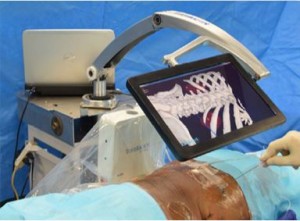
The authors assess the feasibility of a novel method of percutaneous access (TranslucentTM Medical Inc.) using a freely movable tablet display to help guide the percutaneous puncture to its target (see Fig. 1). The success of such a system would rely on a high degree of accuracy and the authors set out to test this in a cadaveric model. Fiducial markers were placed in the kidneys and prostates of cadavers to mimic tumours. A CT scan was then performed to allow 3D model reconstruction. An electromagnetic field was generated around the body and magnetic sensors (fixed to the skin and in the urethral catheter) were used for localization. The software then allows real-time demonstration of the needle trajectory with a predicted line beyond the needle tip overlaid on the 3D model. The authors were able to quickly reach the target (mean time 43 s) with apparent good accuracy, which was calculated to be within 2.5 mm; however, further fiducial markers were deployed at the centre of the target to allow the accurate measurement of accuracy. The distance between the ‘target’ fiducial and the ‘treatment’ fiducial was 16.6 mm in the prostate and 12.0 mm in the kidney. This difference was probably attributable to either movement of the organ or deformation of it from the needle puncture itself. These errors were predominantly in the z-axis (i.e. depth), suggesting that they were caused by movement and deformation by the needle itself, which therefore poses the challenge of how the errors might be reduced, especially when a living human model may have more compliant tissues and of course be moving with respiration.

asdasd

Figure 1. Tablet screen displaying real-time ‘three-dimensional window into the body’ with real and projected trajectory of needle

The present paper by Marien et al. is not the first report of using tracking for percutaneous access in urology. Using different technology, Rassweiler et al. [2] reported on the percutaneous puncture of the kidney for nephrolithotomy using an iPad with surface skin markers, which were visualized by the rear-facing camera of the iPad, and this information was processed to calculate the location of the tissues beneath based on a preoperative CT. This system, and I suspect all others, will rely on a CT being performed in the exact position of the surgery, which is another limiting step until this can be performed at the same time as the surgery.

There are of course concerns regarding use of this technology. The accuracy was limited when analysing the actual position of the ‘treatment’ fiducial because of movement of the tissues. This is likely to be worse in living tissue. There was no respiratory movement which would probably make accurate tracking difficult, although placement of markers to allow movement tracking may help overcome this. Rodrigues et al. [3] reported high accuracy of percutaneous nephrolithotomy puncture after deployment of an electromagnetic sensor in the target calyx with ureterorenoscopy in a porcine model but without a 3D model.

My major concern is that of false reassurance. A nice image is portrayed on the screen which is believed by the surgeon, while in fact there is a significant mismatch caused by patient/tissue movement, either since the preoperative planning CT scan or intra-operatively. The falsely reassured surgeon then inadvertently damages surrounding organs.

It is clear that this technology is work in progress, but it does offer promise that real-time tracking of a percutaneous needle is possible, with accurate representation on a 3D model reconstruction helping to guide the surgeon to the target.

Read the full article
Matthew Bultitude


Department of Urology, Guys and St. Thomas Hospital, London, UK


References


Editorial: A urologists’ guide to the multi-parametric magnetic resonance imaging (mpMRI)-galaxy

The rise of multi-parametric MRI (mpMRI) for the assessment of patients with suspicion of prostate cancer has led to an enormous shift in the practice of every urologist dealing with frontline diagnostics [1].

At the same time, researchers and industry have identified acres of fruitful soil to place the seeds of their respective interests, sometimes in collaboration with each other producing valuable contributions to this shift in practice, sometimes taking benefits by merely assimilating themselves or their product to this development.

Both, the speed of change and the extent of proliferation, make it almost impossible for by-standing clinicians to keep up and filter the evidence-based essence for their local practice.

There are three important issues that need to be considered:

1 The Quality of mpMRI

The development of mpMRI for prostate assessment occurred over the last decade with well-known leaders pushing the frontiers. Their research benefitted from their individual experience of interpreting and reporting MRIs. This is then reflected in their outcomes in form of cancer detection rates and accuracy. More recently we have identified that achieving these results must involve standardisation of MRI protocols and reading [2-4], systematic training in validated courses and a significant learning curve [5]. The latter is only possible to achieve if the practice is embedded in a collaborative team of radiologists, pathologists and urologists. But even then it may be impossible for local teams to deliver the published accuracy, and the urologists and radiologists need to be mindful of that when counselling patients using mpMRI in their local environment.

2 The Technical and Clinical Validity of MRI-Based Biopsies

Transperineal vs transrectal, targeted alone vs targeted with systematic, cognitive vs fusion biopsies – these are the key debates surrounding the application of mpMRI into the urologists’ armamentarium. For none of them there is or will be a unified answer.

Transrectal approaches suit office-based provision of primary diagnostics in many European and USA health economies; although purists can say that the increasing risk of sepsis from antibiotic-resistant bacteria is not acceptable. But, favouring the less infection-prone transperineal approaches will have impact on theatre capacities even in a hospital-based health system like the UK.

Considering the current real-time quality of mpMRI, systematic biopsies in addition to targeted ones are still necessary. Urologists as a group have to come to an agreement about what is acceptable as a remaining risk when reducing or omitting systematic cores.

Cognitive targeting has been shown to be highly accurate; yet, fusion may offer standardisation and reduce user dependency. Not all fusion software on the market has undergone a thorough validated technical development and clinical accuracy evaluation. Peer-reviewed publications can be found involving the systems Urostation-Koelis, Uronav-Philips, Artemis and BiopSee-Medcom.

3 Translation into Clinical Practice

The positioning of the mpMRI within the assessment algorithm is key to optimise the benefit. Use as a pre-biopsy assessment tool may allow omission of further biopsies in some patients or facilitate targeting [6]. However, an established skill in the use of mpMRI and mpMRI-based biopsy is essential. Many UK centres have started the use of mpMRI in their practice further downstream in patients with persistent suspicion after negative first biopsies with good results for patients. It is already part of guidance that active surveillance should involve the use of MRI [1]. Some leading centres advocate that the diagnosis should be confirmed by MRI-based targeted and systematic biopsies.

Knowing that mpMRI will improve the accuracy of our assessment, we need to re-consider follow-up protocols. Increased certainty should be reflected in an improved cancer-related outcome, better patient experience and reduction in costs for the health system.

Prostate mpMRI as part of the urologists’ armamentarium is here to stay. A standardised team- and evidence-based approach will allow us to remain in control of the destination it leads us to.

Read the full article
Christof Kastner
Cambridge University Hospitals, Cambridge, UK

Editorial: The need for standardised reporting of complications

In the context of diversifying practice models, implementation of new technologies such as the Da Vinci surgical robot and rising healthcare costs, there is growing interest in evaluating the quality of surgical work. This extends into health policy, as reimbursement penalties are introduced for ‘inappropriate’ outcomes (e.g. excessive readmissions). Consequently, there is a significant need to provide an accurate assessment of complications and mortality when reporting on surgical outcomes.

Despite the constant use of outcomes data to measure effectiveness in surgery, no current urology guidelines demand the standardised reporting of surgical complications [1]. As randomised controlled trials are uncommon within the surgical setting, and are associated with significant biases [2], there is a distinct need for a uniform reporting system after urological surgeries. Indeed, the lack of such makes it challenging to compare surgical outcomes between techniques, surgeons and institutions, thus hampering the interpretation of study results [3]. The ongoing (and never-ending) debate on the comparative effectiveness of open vs robot-assisted radical prostatectomy (RP) highlights the need for standardised methods to assess superiority (or inferiority) of surgical results [4].

In this issue of the BJUI, Soares et al. [5] present a single-surgeon study of 1138 laparoscopic RPs (LRPs) with a standardised approach between the years 2000 and 2008, and their 5-year follow-up. Whereas the functional and/or oncological equivalency of LRP compared with open RP has been reported before [6], perhaps the outstanding contribution of this study is the use of the Martin-Donat criteria to report and analyse surgical results [3, 7]. In 2002, Martin et al. [7] introduced a list of 10 standard criteria for accurate and comprehensive reporting of surgical complications (e.g. methods of data acquisition, duration of follow-up, definition of complications, hospital length of stay).

In Table 6 of their manuscript, Soares et al. [5] display surgical and/or oncological outcomes of a total of 17 studies on LRP (including their own data). This table suggests the obvious: there is no consistency of reporting on outcomes. In the 2007 Donat [3] analysis of surgical complications reporting in the urological literature, only 2% of a total of 109 studies met nine to 10 of the critical Martin criteria. Interestingly, these shortcomings have been addressed in more contemporary years as the number of studies complying with most of the Martin criteria has increased between 1999/2000 and 2009/2010 [1]. Yet, despite the increasing use of classification systems for outcomes of surgery and standardised reporting of complications (e.g. Clavien-Dindo classification), they are not routinely applied [1, 8].

In an era where the adoption of a certain surgical approach or technique needs to be carefully weighted against a demand for greater value and decreased costs, a simple case series on positive outcomes is simply not sufficient [9]; at the very least, guideline-compliant assessment of outcomes should be the standard of care.

Read the full article


Marianne Schmid*, Christian P. Meyer*† and Quoc-Dien Trinh*


*Division of Urologic Surgery and Center for Surgery and Public Health, Brigham and Womens Hospital, Harvard Medical School, Boston, MA, USA and† Department of Urology, University Medical Center Hamburg-Eppendorf, Hamburg, Germany


References

1 Mitropoulos D, Artibani W, Graefen M, Remzi M, Roupret M, Truss MReporting and grading of complications after urologic surgical procedures: an ad hoc EAU guidelines panel assessment and recommendations. Eur Urol 2012; 61: 3419


4 Schmid M, Gandaglia G, Trinh QD. The controversy that will not go away. Eur Urol 2014; [Epub ahead of print]. doi: 10.1016/ j.eururo.2014.02.052


5 Soares R, Di Benedetto A, Dovey Z, Bott S, McGregor R, Eden CMinimum 5-year follow-up of 1138 consecutive laparoscopic radical prostatectomies. BJU Int 2014; [Epub ahead of print]. doi: 10.1111/ bju.12887


6 Hruza M, Bermejo JL, Flinspach B et al. Long-term oncological outcomes after laparoscopic radical prostatectomy. BJU Int 2013; 111:  27180


7 Martin RC 2nd, Brennan MF, Jaques DP. Quality of complication reporting in the surgical literature. Ann Surg 2002; 235: 80313


9 Novara G, Ficarra V, DElia C, Secco S, Cavalleri S, Artibani W. Trifecta outcomes after robot-assisted laparoscopic radical prostatectomy. BJU Int 2011; 107: 1004


Editorial: How much potential for Transient Receptor Potential channels in the bladder?

In this issue of the BJUI, Charrua et al. [1] report on the possible interaction of two members of the vanilloid subfamily of transient receptor potential (TRP) channels in the control of rat urinary bladder function, TRPV1 and TRPV4. TRP channels are a family of cation-selective channels with 28 known mammalian members. Six of them belong to the subfamily of vanilloid receptors (TRPV channels) and fall into four groups, TRPV1/TRPV2, TRPV3, TRPV4, and TRPV5/TRPV6. The physiological and pharmacological interest in these channels results largely from the finding that they can be activated by a plethora of physical and chemical stimuli; accordingly, they have been implicated in sensory function and pathophysiology of many organ systems [2]. A breakthrough in our understanding of such channels came with the reporting of TRPV1 and TRPV4 knock-out mice, which also exhibit a bladder phenotype; the role of TRP channels in lower urinary tract function has comprehensively been reviewed recently [3].

While the physiological regulation of TRPV1 by endogenous mediators is poorly understood, natural compounds such as capsaicin or resiniferatoxin are acute agonists of TRPV1 channels; however, over time, they desensitise the channel and hence act as inhibitors. These compounds have shown promise in the treatment of detrusor overactivity but also have problems attributed to their initial agonist effects [3]. TRPV4 are activated experimentally by hypotonicity induced cell swelling and several chemicals and more physiologically by moderate heat, stretch and shear stress, leading to the proposition that they may functions as a stretch sensor in the bladder. The inhibitory effects of TRPV1 agonists manifest only after prolonged exposure once desensitisation of their agonist effects occurs, and this initial agonistic phase is a source of undesirable effects. Therefore, a search is on for small molecules that have direct antagonist effects.

Charrua et al. [1] now report that two small molecule antagonists at TRPV1 and TRPV4, (SB355791 and RN1734, respectively) even in high doses did not affect bladder function in control rats. Intravesical installation of lipopolysaccharide is used to create an animal model of cystitis as it induces inflammation, detrusor overactivity and bladder pain. In this model, a high dose of the TRPV4 inhibitor reduced detrusor overactivity, whereas even the high dose of the TRPV1 inhibitor did not; however, a combination of ineffective doses of both inhibitors markedly decreased bladder reflex activity. On the other hand, each of the two drugs caused partial analgesia, but their combination was not more effective than either drug alone. This indicates an interesting functional interaction between TRPV1 and TRPV4 channels, which is specific for the overactivity vs the pain response. Previously, the Cruz group reported that bladder overactivity induced by nerve growth factor depends on the presence of functionally active TRPV1 [4]. Taken together this work shines light on networks of multiple mediators and their receptors that cooperate in the regulation of bladder function but previously have mainly been viewed in isolation. Such work may also have therapeutic consequences. As target-saturating concentrations of ligands at any of these receptors may cause relevant adverse effects, targeting multiple such receptors in low doses may open an avenue for a multi-pronged approach, particularly in patients with bladder dysfunction difficult to control with present treatment options.

This multiple target, low-dose approach is a therapeutically fascinating idea, but finding the right combination of doses in such a setting is a nightmare for any drug development scientist. Moreover, much of the specific role of such targets in pathophysiology remains to be explored before the present findings can be translated into clinical treatments, and the Charrua et al. study [1] will also help such efforts in other ways. Some of the initial thinking on the function of TRP channels in the control of bladder and other functions has been based on localisation studies with TRP channel antibodies, which may have been flawed. Similar to many other receptor antibodies [5], several of those directed against TRPV1 channels also have been shown to lack target specificity [6], leading to misunderstandings about the location and function of such channels. The validation for other TRPV1 and TRPV4 antibodies presented by Charrua et al. [1] will allow more robust studies in this regard and help to develop more valid understanding of TRP channels in physiology, pathophysiology and as treatment targets.

Read the full article
Martin C. Michel


Department of Pharmacolog y , Johannes Gutenberg University, Mainz, Germany


References


3 Franken J, Uvin P, de Ridder D, Voets T. TRP channels in lower urinary tract dysfunction. Br J Pharmacol 2014; 171: 2537–51


4 Frias B, Charrua A, Avelino A, Michel MC, Cruz F, Cruz CD. Transient receptor potential vanilloid 1 mediates nerve growth factor-induced bladder hyperactivity and noxious input. BJU Int 2012; 110: E422–8


5 Michel MC, Wieland T, Tsujimoto G. How reliable arG-protein-coupled receptor antibodies? Naunyn Schmiedebergs Arch Pharmacol 2009; 377: 385–8


6 Everaerts W, Sepúlveda MR, Gevaert T, Roskams T, Nilius B, De Ridder D. Where is TRPV1 expressed in the bladder, do we see the real channel? Naunyn Schmiedebergs Arch Pharmacol 2009; 379: 421–5


© 2024 BJU International. All Rights Reserved.