Tag Archive for: Editorial

Posts

Editorial: Can artificial intelligence optimize case selection for hemi‐gland ablation?

The victory of ‘AlphaGo’ over humans in Go, one of the most complex games with more than 10170 board configurations, has yielded tremendous attention worldwide [1]. The later version, ‘AlphaGo Zero’, has brought artificial intelligence (AI) to the next level by demonstrating an absolute superiority, winning 100‐0 against the champion‐defeating AlphaGo [2]. It is exciting, and perhaps shocking, to realize what AI can achieve.

In this issue of BJUI, the study by Zhou et al. [3] is the first to utilize AI to optimize case selection for hemi‐gland ablation. In this study, classification and regression tree (CART) analysis, which is a form of supervised machine‐learning algorithm, was used to identify laterality of prostate cancer. In the conventional approach, case selection was based on biopsy results and MRI findings. For the CART model, in addition to the common clinical variables (i.e. age, PSA, prostate volume, biopsy and MRI results), biopsy coordinate‐derived spatial features were also used as model inputs. The model output was the probability of unilateral clinically significant prostate cancer considered suitable for hemi‐gland ablation. Whole‐mount prostatectomy specimens were used as the standard of reference. The CART model correctly identified laterality in 80% of the cases, compared to 73% with the conventional approach. The positive predictive value of the CART model was 83%, compared to 53% with the conventional approach. The superiority of the CART model has been demonstrated, and the area under curve was 0.82.

Artifical intelligence has been widely adopted in the field of Urology [4]. For prostate cancer detection in particular, our group evaluated the diagnostic performances of four machine‐learning models based on clinical variables in a biopsy cohort of 1625 men [5]. The machine‐learning models achieved excellent performances in detecting clinically significant prostate cancer, with an accuracy of up to 95.3%. Algohary et al. [6] constructed three machine‐learning models to identify the presence of clinically significant prostate cancer based on MRI radiomic features in patients who underwent active surveillance. When compared with the Prostate Imaging–Reporting and Data System (PI‐RADS) scoring system, the machine‐learning models were able to improve overall accuracy by 30–80%.

Fehr et al. [7] developed an automated system to classify Gleason scores based on MRI images. The automated system could distinguish between Gleason scores of 6 and 7 or above cancers with an accuracy of up to 93%. The differentiation between Gleason score 3+4 and 4+3 disease also yielded an accuracy of up to 93%. Importantly, the performance of AI and machine‐learning models is highly dependent on the quality and accuracy of the data being input. In terms of prostate cancer detection, either mapping biopsy or whole‐mount prostatectomy specimens should be considered to represent the ‘ground truth’.

There are a number of challenges in implementing AI in clinical practice. First, decision‐making in healthcare requires logical deduction and explanation. The data processing in AI, however, is often described as a ‘black box’. Taking AlphaGo as an example, some ‘moves’ were considered incomprehensible even by world‐class players. Second, although results from AI are promising, there is in general a lack of regulations and standards to assess its safety, efficacy and validity. Liability issues can be problematic in case of medical mishaps. Third, doctors are human. Conflict of interest does exist, and how we can utilize AI in a complementary rather than a competitive manner is a challenging obstacle to overcome.

Nevertheless, AI has huge potential in improving healthcare. Collaborative effort is needed globally to develop and optimize AI systems, and to increase its acceptability and practicality upon implementation. Future studies answering clinically important questions using appropriate standards of reference will be of paramount importance in paving the way for the AI era in urology.

by Jeremy Yuen‐Chun Teoh, Edmund Chiong and Chi‐Fai Ng

References

  1. Silver DHuang AMaddison CJ et al. Mastering the game of Go with deep neural networks and tree search. Nature 2016529484– 9
  2. Silver DSchrittwieser JSimonyan K et al. Mastering the game of Go without human knowledge. Nature 2017550354– 9
  3. Zhou SRPriester AMJayadevan R et al. Using spatial tracking with magnetic resonance imaging/ultrasound‐guided biopsy to identify unilateral prostate cancer. BJU Int 2020125399– 406
  4. Chen JRemulla DNguyen JH et al. Current status of artificial intelligence applications in urology and their potential to influence clinical practiceBJU Int 2019124567– 77
  5. Wang GTeoh JYChoi KSDiagnosis of prostate cancer in a Chinese population by using machine learning methods. Conf Proc IEEE Eng Med Biol Soc 201820181– 4
  6. Algohary AViswanath SShiradkar R et al. Radiomic features on MRI enable risk categorization of prostate cancer patients on active surveillance: Preliminary findings. J Magn Reson Imaging 201848818– 28
  7. Fehr DVeeraraghavan HWibmer A et al. Automatic classification of prostate cancer Gleason scores from multiparametric magnetic resonance images. Proc Natl Acad Sci USA 2015112E6265– 73


Editorial: All for one, one for all: is centralisation the way to go?

The need to centralise complex surgical procedures in large centres remains at the core of many health policy discussions. Much of the debate is focussed on three main aspects: (i) outcomes, (ii) costs and (iii) accessibility. Gray et al. [1] recently noted that increasing centralisation may be unnecessary for invasive procedures such as nephrectomy and cystectomy. Specifically, they noted almost no difference in outcomes of high‐volume centralised centres and those with lower throughput. Their findings go against most of the current literature on the volume–outcomes relationship, which generally reports a correlation between a hospital’s volume of procedures and improved healthcare outcomes. One could ask what factors specific to their analysis could explain the different observations. For one, the healthcare system in the UK may (and likely) operate in ways different from other European and USA‐based healthcare systems, from which most of the current data are derived. Healthcare in the UK may already be organised in such a way that further centralisation may not improve outcomes, which the authors allude to in their conclusions. Differences in methodology may explain their findings, e.g. their use of multilevel modelling, testing specific incremental volume cutoffs, etc. Outcome selection may play a role as well; length of stay and re‐admissions may vary more according to organisational factors rather than individual surgeon expertise.

Regardless of their findings, we would argue that there are other tangible benefits to centralisation, which extend well beyond ‘better outcomes’. For instance, the management of the modern oncological, and urological, patient is critically dependent on a multidisciplinary team. The inherent multidisciplinary nature of large centres facilitates patients receiving their entire course of treatment at the same place. This enhances the continuity and efficiency of care, both of which are undoubtedly hampered in small peripheral centres that ultimately depend on referrals to larger facilities for advanced care for the most complex patients.

This ties into yet another major advantage of centralised centres, which is the ease of access to research. For instance, our affiliated cancer centre runs >1100 active clinical trials, 42 of which pertain to advanced urological diseases. Such trials provide access to otherwise unavailable therapies and enhance the production, diffusion, and application of knowledge.

In touting the many benefits of centralisation, one would imagine it comes at a significant cost. While this may have been true in the past, recent data comparing the higher‐volume teaching hospitals to lower‐volume non‐teaching centres suggest that centralisation actually decreases the 30‐day hospital costs and have similar costs at 90 days compared with non‐teaching hospitals [2]. Similar trends were also seen with radical cystectomies [3] and prostatectomies [4], showing that with the major urological procedures, centralisation is cost‐effective with at least the same outcomes as compared to peripheral centres.

A common objection to centralisation is that it forces many patients to travel long distances and that this in turn could introduce or worsen discrepancies in accessibility to care. If true, this would have profound social and economic consequences for disadvantaged groups, as well as particularly fragile patients. Many centralised centres have developed approaches to ease the burdens of travelling from afar and, if patients can make the journey, the data suggest a survival advantage over those who are treated at peripheral centres. To this end, Vetterlein et al. [5] stratified >700 000 patients by risk class and demonstrated an overall survival benefit in those with all stages of prostate cancer. In the not‐too‐distant future, patient follow‐up can be shifted almost entirely to telemedicine, which can further alleviate travel burdens.

Our aim is not to promote a system of oncological care based solely at centralised hubs. However, to suggest that all care should be distributed equally across all centres seems unrealistic and may have devastating consequences, particularly for those with advanced disease. We strongly advocate the treatment of complex disease at high‐volume, centralised centres and suggest better use of an impartial classification of what constitutes a ‘complex’ disease. Therefore, one answer to this problem is broadly represented by the redistribution of the different surgical procedures amongst the hospitals.

by Daniele Modonutti, Venkat M. Ramakrishnan and Quoc‐Dien Trinh


References

  1. Gray WKDay JBriggs TWHarrison SUnderstanding volume‐outcome relationships in nephrectomy and cystectomy for cancer: evidence from the UK Getting it Right First Time programme. BJU Int 2020125234– 43
  2. Burke LGKhullar DZheng JFrakt ABOrav EJJha AKComparison of costs of care for medicare patients hospitalized in teaching and nonteaching hospitals. JAMA Netw Open 20192: e195229
  3. Leow JJReese STrinh QD et al. Impact of surgeon volume on the morbidity and costs of radical cystectomy in the USA: a contemporary population‐based analysis. BJU Int 2015115713– 21
  4. Gershman BMeier SKJeffery MM et al. Redefining and contextualizing the hospital volume‐outcome relationship for robot‐assisted radical prostatectomy: implications for centralization of care. J Urol 201719892– 9
  5. Vetterlein MWLöppenberg BKarabon P et al. Impact of travel distance to the treatment facility on overall mortality in US patients with prostate cancer. Cancer 20171233241– 52


Editorial: Zero‐radiation stone treatment

In this month’s BJUI, Armas‐Phan et al. [1] report on a prospective observational trial of fluoroscopic vs ultrasound (US)‐guided tract dilatation during percutaneous nephrolithotomy (PCNL). A total of 176 patients underwent successful initial US‐only guided puncture; of these patients, 138 had US‐only dilatation, while in 38 fluoroscopy was required. The authors found no difference in patient factors (e.g. age, gender, body mass index [BMI]) or stone factors (hydronephrosis, stone burden, number of tracts or puncture location). On multivariate analysis, US dilatation was more likely to be performed in the modified dorsal lithotomy position (compared to prone), but there was no significant difference in important outcomes such as stone clearance, complication rates or blood loss.

Whilst only reporting on access (and not necessarily dilatation), the Clinical Research Office of the Endourological Society PCNL Global Study shows us that worldwide fluoroscopic access is by far the most common (88.3% of cases) [2] and there are relatively few reports of US‐guided dilatation in the literature. The technique does produce technical challenges as the surgeon needs to confidently identify the depth of the dilators or balloon and be sure of its location relative to calyceal anatomy. Whilst dilating short is not usually a problem as simply re‐dilating can be done, dilating too far carries serious risk of perforation of the pelvicalyceal system and vascular injury. The authors’ described technique does rely on good kidney and guidewire visualisation, and if this is not possible then fluoroscopy is used instead. Thus, even in this series with experts at this technique, 38 (22%) underwent fluoroscopic dilatation after US‐guided puncture, and of the 138 with intended US dilatation, seven (5%) were converted to fluoroscopy. Furthermore, 115 patients never entered this series as they underwent initial fluoroscopic‐guided puncture. Thus, it is important to realise that this is a series of select patients being treated by expert enthusiasts of this technique and fluoroscopy should be available in the operating theatre, as it is not possible to do this technique for all patients. In particular, obesity limits the visualisation under US and the authors have previously shown that renal access drops from 76.9% of normal‐weight patients (BMI <25 kg/m2) to 45.6% for those classified as obese (BMI >30 kg/m2) [3]. An alternative strategy to avoid radiation is to use endoscopic combined intrarenal surgery (ECIRS), as the depth of dilatation can be monitored by direct visualisation via the flexible ureteroscope.

Patients and healthcare professionals are increasingly aware of the risks posed by ionising radiation. Ferrandino et al. [4] analysed radiation exposure of patients presenting with acute stone episodes in an American setting. The mean dose was a staggering 29.7 mSv and 20% of patients received >50 mSV. There is also awareness of risk to the operating staff from endourological procedures and although doses are relatively low [5], these can accumulate during a lifetime of operating, with risks of not only malignancy but also cataract formation [6]. Whilst I am sure we all wear protective lead gowns in the operating theatre, how many people wear lead glasses? A recent study showed that, at typical workload, the annual dose to the lens of the eye was 29 mSv in interventional endourology [7].

As urologists, we should all be aware of these risks and follow the ALARA (As Low As Reasonably Achievable) principals of keeping doses to a minimum. Thus, this paper [1] is particularly welcome and shows zero‐radiation procedures can be safely performed. The authors now attempt this technique for all PCNL procedures and achieve US‐only puncture and dilatation in over half of their patients. Hopefully, this paper will inspire us all to look at reducing or eliminating radiation usage in our stone procedures and this will be good for patients and surgeons alike.

by Matt Bultitude


References

  1. Armas‐Phan MTzou DTBayne DB et al. Ultrasound guidance can be used safely for renal tract dilatation during percutaneous nephrolithotomy. BJUI 2019; 125: 284-91
  2. De La Rosette JAssimos DDesai M et al. The Clinical Research Office of the Endourological Society Percutaneous Nephrolithotomy Global Study: indications, complications, and outcomes in 5803 patients. J Endourol 20112511– 7
  3. Usawachintachit MMasic SChang HAllen IChi TUltrasound guidance to assist percutaneous nephrolithotomy reduces radiation exposure in obese patients. Urology 20169832– 8
  4. Ferrandino MNBagrodia APierre SA et al. Radiation exposure in the acute and short‐term management of urolithiasis at 2 academic centers. J Urol 2009181668– 72
  5. Galonnier FTraxer ORosec M et al. Surgical staff radiation protection during fluoroscopy‐guided urologic interventions. J Endourol 201630638– 43
  6. Hartmann JDistler FBaumuller M et al. Risk of radiation‐induced cataracts: investigation of radiation exposure to the eye lens during endourologic procedures. J Endourol 201832897– 903
  7. Hristova‐Popova JZagorska ASaltirov I et al. Risk of radiation exposure to medical staff involved in interventional endourology. Radiat Prot Dosimetry 2015165268– 71


Image courtesy of BJUI Knowledge

Editorial: Vaporization is vaporization, but not at any cost…

The paper by Ghobrial et al. [1] confirms that bipolar electrocautery vaporization is more cost‐effective than GreenLight Laser vaporization, as the two techniques are equally effective but GreenLight vaporization is more costly in the smaller prostates being studied.

Underpinning the analysis was a well‐conducted randomized controlled trial, showing equivalent peri‐operative and postoperative measures with the two procedures and no difference in the primary endpoint of IPSS reduction at 2 years. The two techniques were performed in a similar manner and were equally efficient and safe as expected.

Philosophically, the clinical results are both unsurprising and expected, and confirm the long‐held belief that the energy source employed for vaporization and, for that matter, enucleation, is of secondary concern compared to the skill and dedication of the operator. The technique in either case should result in comparable efficacy, leaving cost‐effectiveness to be an important way to help both urologists and administrators discriminate between them.

Although the costs are not necessarily going to be comparable with those in other jurisdictions, this will apply equally to both treatments and this study therefore represents an excellent attempt to cost both procedures, removing equivalent costs. Importantly, this assessment included the costs of both readmissions and interventions over the full 24‐month period. This captures the bulk of the important complications after these types of procedures and adds to the validity of the findings.

The big difference between the costs of the two treatments being studied is, of course, ‘capital equipment including maintenance’. The single‐use fibre model rather than the cost of the machine has been the mainstay for the profitability of laser companies since the inception of laser prostatectomy. The maintenance contract has been a further cost, which is always underestimated. Reusability of the laser fibres is one way of diminishing per‐procedure costs, but is only consistently possible for Holmium end‐fire fibres [2]. The fact that the authors estimate of these costs was a ‘case share in 5‐year budget plan’ also suggests that the true cost of the use of the GreenLight laser is underestimated.

With the burgeoning number of new techniques and technologies for the treatment of BPH emerging, and new treatment paradigms being proposed, let alone the increasingly negative focus on medical waste [3] and the increasing use of single‐use disposable handpieces/tubing/drapes/fibres, articles such as this are timely. A standardized methodology for assessing the cost‐effectiveness of treatments for BPH is needed and should be an essential part of pivotal studies and therefore the regulatory approval processes.

by Peter Gilling


References

  1. Ghobrial FKShoma AElshal AM et al. A randomized trial comparing bipolar transurethral vaporization of the prostate with GreenLight laser (xps‐180watt) photoselective vaporization of the prostate for treatment of small to moderate benign prostatic obstruction: outcomes after 2 years. BJU Int2020124144– 52
  2. Fraundorfer MRGilling PJKennett KMDunton NGHolmium laser resection of the prostate is more cost effective than transurethral resection of the prostate: results of a randomized prospective study. Urology 200157454– 8
  3. Rose EDModlin DMCiampa MLMangieri CWFaler BJBandera BCEvaluation of operative waste in a military medical center: analysis of operating room cost and waste during surgical cases. Am Surg. 201985717– 20


Editorial: The benefits of regular exercise

January is the month when we wish each other happiness and success for the year ahead. It is also the month when many are recovering from the excesses of the festive season. This is the time when gyms and diets become popular again with offers of reduced rates to attract customers. For Londoners the spring marathon is not far away and you often see runners training in different parks despite the cold weather and icy routes.

If you think this year is the one where you are about to start going to the gym, then we recommend you the best shake for post workout to add extra point to your routine.

Is this just a temporary fad? Or is there truly some benefit to be had by exercising regularly?

Over the past few years, we have published several papers showing clear associations between metabolic syndrome and LUTS, and the benefits of preoperative optimisation with diet and exercise prior to major urological surgery. In this issue of the BJUI, we present a small but well‐designed randomised controlled trial on the benefits of exercise in attenuating the treatment side‐effects in patients with newly diagnosed prostate cancer starting on androgen‐deprivation therapy [1]. It is an example of collaborative working between Urologists and experts on Sport, Exercise and Rehabilitation therapy. The authors clearly demonstrate that a short‐term programme of supervised exercise results in improvements in quality of life and cardiovascular risk profile in patients on hormonal therapy. Even after the supervised exercise was withdrawn and followed by self‐directed exercise, the benefits continued as compared to the control group.

As Urologists, we can help our patients in this journey by adopting a more active lifestyle ourselves. Inspired by Fiona Godlee’s article in the BMJ [2], I have started printing it and actually handing it/e‐mailing it to my patients. The paper describes physical activity as ‘The miracle cure’ with very few side‐effects. Any level of activity is better than none and a gentle start usually avoids an unexpected injury.

There is no better time to lead by example this New Year!

by Prokar Dasgupta

References

  1. Ndjevera WOrange STO’Doherty AF et al. Exercise‐induced attenuation of treatment side‐effects in patients with newly diagnosed prostate cancer beginning androgen‐deprivation therapy: a randomised controlled trial. BJU Int 2019: 125; 28-37.
  2. Godlee FThe miracle cureBMJ 2019366l5605.

Editorial: Beyond bladder cancer surveillance: building a survivorship clinic

As oncologists, we focus on obtaining the best cancer outcomes possible. The aim of treatment is to maximize survival and help patients live longer. As therapies continue to become more effective, more patients will become survivors. In the ongoing effort to extend the quantity of life left for our patients facing lethal cancers, thinking about the quality of that time is key. For urological oncologists, patients with a new bladder cancer diagnosis will someday face a new set of obstacles as survivors. In addition to surveillance and scans, asking patients about other issues such as their mental health, sexual function and financial solvency are also important.
Regardless of cancer stage, these issues apply to all of our patients with bladder cancer. Patients with non-muscle invasive disease need a seemingly interminable number of cystoscopies, with possible repeat biopsies or intravesical therapies. Patients with muscle-invasive disease undergo urinary diversion that entails significant changes as they will then have a stoma, neobladder or other diversion.
In this issue of BJUI, Jung et al. present a ‘snapshot’ of patients in North Carolina with bladder cancer that examines the impact of treatment on quality of life [1].  The study is valuable because it involves a number of topics that have previously not been studied in such detail. A total of 376 patients returned mailed surveys, a response rate of 24%. Most participants were on average 3 years from their diagnosis, the mean age of participants was 72 years, and the majority of patients were white men. Most participants (approximately three in four) had undergone transurethral resection of bladder tumour as the primary treatment and some (one in three) had received intravesical therapy. As with any work, there are some limitations which include the low overall numbers of participants, low
response rate, and lack of longitudinal data. Despite these limitations, there is still value to studying trends in this space, given the paucity of available data, and the authors offer some valuable insights. This paper provides evidence that for bladder cancer survivorship care, it is important to realize that other important issues exist and impact patient well-being.

• Bladder cancer patients may have financial issues. Bladder cancer patients may face financial toxicity that is in part attributable to the regular need for surveillance in order to identify recurrence or progression of disease.
• Cystectomy recovery can include discussions about sexual function. Patients who have undergone cystectomy may have discomfort with sexual intimacy. This was more common in men. Non-cystectomy patients may have better sexual function. Patients may be concerned about contaminating partners.
• Quality-of-life issues for bladder cancer patients can vary by gender. Men may have better sexual function and enjoyment than women, but also have more discomfort with intimacy and fears of contaminating their partners, while women may have higher levels of constipation and diarrhoea.
• Low risk bladder cancer (vs high risk) can have lower impact on quality of life. Patients with Ta disease had the highest global health status (compared with T1 and Tis). They also had the best physical and social functioning and less fatigue and financial problems. This underscores that Ta disease is different from other stages. As the authors point out, this may be attributable to a low progression risk, which means patients are less likely to need intravesical therapy.
• Sexual health can be affected and improve with time after a bladder cancer diagnosis. Sexual issues can last for years after a diagnosis. Men may face erection or ejaculation problems, and women may have vaginal dryness issues. With time, however, sexual function can improve and sexual function (including extent of sexual activity and interest in sex) was better in survivors further from their diagnosis.

Moving forward, we can use this study to prompt us to think about how our treatments impact our patients. Setting up dedicated survivorship clinics may be one practical strategy to provide this care in a systematic and streamlined way. Beyond treatment-related issues such as recurrence and progression, patients are affected in other ways. Issues with overall health, mental well-being, sleep, or sexual function occur for many. Setting up a standardized approach to cancer care can complement oncological surveillance and promote patient-centred care. A dedicated team, with a provider and physician assistant can create a clinical infrastructure and design a comprehensive template to remind us to query patients on a broader range of issues relevant to their recovery. In doing so, we can help patients with bladder cancer recover, as survivors (Fig. 1).


Fig. 1 Select aspects of building a bladder cancer survivorship clinic.

Start by establishing a focused team of providers to help guide more streamlined care
• Nurses, nurse practitioners, physician assistants and physicians can be involved
• Each institution may have a unique infrastructure and use a distinct team set-up to create a clinic
• Administrative support and guidance are important to determine the clinical resources necessary or needed to begin a regular survivorship clinic

Streamline care and consider a template-based or guideline-driven approach to visits
• Based on stage of diagnosis, certain patients may need more regular cystoscopic surveillance while other patients will need follow-up visits that are coordinated with medical oncology and/or radiation oncology

Standardize collection of patient-reported outcomes during follow up visits
• Mental well-being
• Physical activity and exercise
• Sexual health
• Urinary and bowel function
• Financial well-being

Step back to evaluate the progress and iteratively troubleshoot issues as they arise
• Collect patient feedback and provider opinions
• Integrate these insights to improve the form and function of the clinic

by Matthew Mossanen and Stephen L. Chang

Reference

  1. Jung A, Nielsen ME, Crandell JL, et al. Health-related quality of life among non-muscle-invasive bladder cancer survivors: a population-based study. BJU Int 2020; 125: 38–48

Editorial: A better way to predict lymph node involvement using machine learning?

In their study in this issue of BJUIHou et al. [1] use machine‐learning algorithms to evaluate several preoperative clinical variables (highlighting specific MRI findings of locally advanced prostate cancer) to determine whether lymph node involvement (LNI) could be present during radical prostatectomy, which would justify an extended pelvic lymph node dissection (PLND). This is a well‐designed study with scientific rigour, providing evidence‐based justifications and definitions (i.e. of relevant MRI findings). The authors successfully illustrate a practical application of using artificial intelligence (AI) methods to augment clinical decision‐making prior to and during surgery compared to today’s ‘gold standard’ (nomograms).

For many years, the Memorial Sloan Kettering Cancer Centre (MSKCC) nomogram, among a number of predictive models, has been used to determine the probability of LNI. The output of these tools has assisted surgeons in determining whether to perform a PLND, and if so, to what extent [2,3,4]. The authors hypothesize that, with additional MRI parameters not previously used, machine‐learning algorithms can better select which patients are more likely to have LNI and will therefore require extended PLND. In fact, the authors report that the MSKCC nomogram and conventional MRI reporting of LNI consistently underestimated LNI risk compared to the machine‐learning‐assisted models presented in their study. The outputs of the present models would allow a higher number of extended PLNDs to be spared compared to reliance on the MSKCC nomogram alone. It was appropriate to use several existing AI models in this study, as it is never readily apparent initially which existing predictive model may perform best with a given dataset. In fact, all the models used – logistic regression (LR), support vector machine (SVM) and random forest (RF) – while similar in performance to each other, outperformed the MSKCC nomogram (P < 0.001). Many adjustments were probably performed for each model to tailor it to the dataset and optimize prediction performance.

Criticisms of the study are that: (i) cases for which PLND was not performed were excluded, which could have created a selection bias; (ii) the model would only be applicable when the patient has undergone MRI; (iii) the study was conducted at a single institution in a small sample (AI methods thrive on big and diverse datasets).

This study by Hou et al. is a great example of a machine‐learning application that may positively impact clinical practice. For many years, we have relied on nomograms, but with increasing use of MRI, additional factors should also be included, as Hou et al. have done. Machine‐learning is particularly adept at simultaneously examining numerous variables to elicit which ones may contribute best to a particular outcome. As BJUI has evaluated many manuscripts examining machine‐learning methods for clinical decision‐making in the past year, we have encouraged authors to use present‐day gold standard methods, such as the MSKCC nomogram, as controls [5]. As we embrace AI methods, we must keep one eye on the tried and tested conventional ways. This ensures that we do not take backward steps but rather take forward steps responsibly. Similarly to recent AI studies published in the BJUI, the sample size in this study was relatively small. External validation in a multicentre study on larger datasets is highly recommended.

by Andrew J. Hung

References

  1. Hou YBao MWu CJZhang JZhang YDShi HBA machine learning‐assisted decision support model with mri can better spare the extended pelvic lymph node dissection at cost of less missing in prostate cancerBJU Int 2019124972– 83
  2. Briganti ALarcher AAbdollah F et al. Updated nomogram predicting lymph node invasion in patients with prostate cancer undergoing extended pelvic lymph node dissection: the essential importance of percentage of positive cores. Eur Urol 201261480– 7
  3. Memorial Sloan Kettering Cancer CenterDynamic prostate cancer nomogram: coefficients. Accessed April 2018
  4. Tosoian JJChappidi MFeng Z et al. Prediction of pathological stage based on clinical stage, serum prostate-specific antigen, and biopsy Gleason score: Partin Tables in the contemporary era. BJU Int 2017119676– 83
  5. Hung AJCan machine‐learning algorithms replace conventional statistics? BJU Int 20181231


Editorial: How long is long enough for pharmacological thromboprophylaxis in urology?

Each year, millions of patients who undergo urological surgery incur the risk of deep vein thrombosis and pulmonary embolism, together referred to as venous thromboembolism (VTE), and major bleeding. Because pharmacological prophylaxis decreases the risk of VTE, but increases the risk of bleeding, and because knowledge of the magnitude of these risks remains uncertain, both clinical practice and guideline recommendations vary widely [1]. One of the uncertainties is the recommended duration of pharmacological thromboprophylaxis.

In this issue of the BJUI, Naik et al. [2] provide an up‐to‐date review that summarises the articles that examined extended thromboprophylaxis in patients with cancer who underwent radical prostatectomy (RP), radical cystectomy (RC) or nephrectomy. The outcomes on which they focussed include risks of VTE, bleeding, renal failure and mortality – all potentially influenced by whether or not patients receive extended prophylaxis.

After screening >3500 articles, the authors included 18 studies, none of them randomised controlled trials (RCTs) [2]. They found that VTE risk is highest in open and robot‐assisted RC, and that, based on observational studies, extended thromboprophylaxis significantly reduces the risk of VTE relative to shorter duration prophylaxis. Evidence suggested that robot‐assisted RP, as well as both open and robot‐assisted partial and radical nephrectomies, incur lower VTE risk than RCs or open RP. They did not find studies comparing extended prophylaxis to standard prophylaxis for RPs or nephrectomies [2].

Overall, these findings are consistent with systematic reviews that estimated the procedure‐ and patient risk factor‐specific risks for 20 urological cancer procedures [3]. As these reviews suggested substantial procedure‐specific differences in the VTE risk estimates, the European Association of Urology (EAU) Guidelines provided separate recommendations for each procedure [4]. For urological (as well as gastrointestinal and gynaecological) patients, the National Institute for Health and Care Excellence (NICE) Guidelines suggest to ‘consider extending pharmacological VTE prophylaxis to 28 days postoperatively for people who have had major cancer surgery in the abdomen’ [5]. Because of variation in both bleeding and thrombosis risks across procedures, this advice is appropriate for some procedures and misguided for others. For instance, the procedure‐specific EAU Guidelines recommend extended VTE prophylaxis for open RC but not for robot‐assisted RP without lymphadenectomy [4].

The review by Naik et al. [2] identified the lack of urology‐specific studies comparing the in‐hospital‐only prophylaxis to extended prophylaxis. The few included studies were observational with considerable limitations (e.g. limited adjustment for possible confounders).

A recent update of a Cochrane review compared the impact of extended thromboprophylaxis with low‐molecular‐weight heparin (LMWH) for at least 14 days to in‐hospital‐only prophylaxis in abdominal or pelvic surgery procedures [6]. The authors identified seven RCTs (1728 participants) evaluating extended thromboprophylaxis with LMWH and generated pooled estimates for the incidence of any VTE (symptomatic or asymptomatic) after major abdominal or pelvic surgery of 13.2% in the control group compared with 5.3% in the patients receiving extended out‐of‐hospital LMWH (odds ratio [OR] 0.38, 95% CI 0.26–0.54).

Most events were asymptomatic, although the incidence of symptomatic VTE was also reduced from 1.0% in the in‐hospital‐only group to 0.1% in patients receiving extended thromboprophylaxis (OR 0.30, 95% CI 0.08–1.11). The authors reported no persuasive difference in the incidence of bleeding complications within 3 months of surgery (defined as major or minor bleeding according to the definition provided in the individual studies) between the in‐hospital‐only group (2.8%) and extended LMWH (3.4%) group (OR 1.10, 95% CI 0.67–1.81).

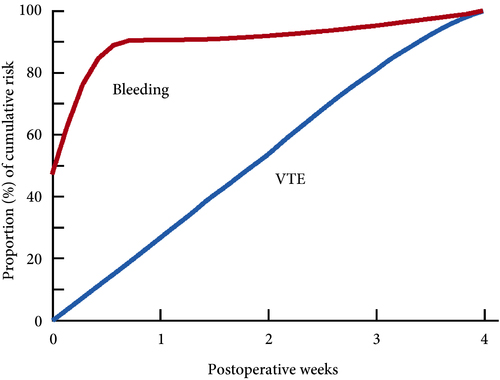
These findings are consistent with our own modelling study that demonstrated an approximately constant hazard of VTE up to 4 weeks after surgery [7]. That study also found that bleeding risk, by contrast, is concentrated in the first 4 days after surgery [7] (Fig.1). Using these findings, the EAU Guidelines suggest for patients in whom pharmacological prophylaxis is appropriate, extended pharmacological prophylaxis for 4 weeks [4]. Consistent with these recommendations, Naik et al. [2] found that 15 studies of 18 included in their review recommended extended prophylaxis.

Fig.1 Proportion of cumulative risk (%) of venous thromboembolism (VTE) and major bleeding by week since surgery during the first 4 postoperative weeks. Reproduced from: Tikkinen et al. [7].

(This is an Open Access article distributed under the terms of the Creative Commons Attribution License (https://creativecommons.org/licenses/by/4.0/), which permits unrestricted use, distribution, and reproduction in any medium, provided the original work is properly credited. The Creative Commons Public Domain Dedication waiver (https://creativecommons.org/publicdomain/zero/1.0/) applies to the data made available in this article, unless otherwise stated.)


Overall, as shown also by this review [2], the evidence base for urological thromboprophylaxis is limited. Although current evidence supports extended prophylaxis, definitively establishing the optimal duration of thromboprophylaxis will require large‐scale RCTs. Other unanswered key questions include: baseline risks of various procedures, timing of prophylaxis, patient risk stratification, as well as effectiveness of direct oral anticoagulants. In the meanwhile, suggesting extended duration to patients whose risk of VTE is sufficiently high constitutes a reasonable evidence‐based approach to VTE prophylaxis.

by Kari A.O. Tikkinen and Gordon H. Guyatt


References

  1. Violette PDCartwright RBriel MTikkinen KAGuyatt GH Guidelines of guidelines: thromboprophylaxis for urological surgery. BJU Int 2016118351– 8
  2. Naik RMandal IHampson A et al. The role of extended venous thromboembolism prophylaxis for major urological cancer operations. BJU Int 2019; 124: 935-44
  3. Tikkinen KACraigie SAgarwal A et al. Procedure‐specific risks of thrombosis and bleeding in urological cancer surgery: systematic reviews and meta‐analyses. Eur Urol 201873242– 51
  4. Tikkinen KACartwright RGould MK et al. EAU Guidelines on Thromboprophylaxis in Urological Surgery, 2017. European Association of Urology, 2018. Accessed November 2019
  5. National Institute for Health and Care Excellence (NICE)Venous Thromboembolism in over 16s: reducing the risk of hospital‐acquired deep vein thrombosis or pulmonary embolism. NICE guideline [NG89]. London: NICE, 2018. Accessed November 2019
  6. Felder SRasmussen MSKing R et al. Prolonged thromboprophylaxis with low molecular weight heparin for abdominal or pelvic surgery. Cochrane Database Syst Rev 20193CD004318
  7. Tikkinen KAAgarwal ACraigie S et al. Systematic reviews of observational studies of risk of thrombosis and bleeding in urological surgery (ROTBUS): introduction and methodology. Syst Rev 201423150. DOI: 10.1186/2046‐4053‐3‐150.


Editorial: Threading the cost–outcome needle after radical cystectomy

I commend Borza et al. [1] on their timely study, which seeks to identify predictors of bounceback (≤3‐day) vs 30‐day readmissions after radical cystectomy. As the authors allude to in their paper, value‐based health reforms being undertaken in the USA seek to improve the quality of care delivery while simultaneously bending the healthcare cost curve [2]. For example, the Hospital Readmission and Reduction Program (HRRP), originally introduced in fiscal year 2013 for targeted medical conditions, has more recently been applied to a limited number of surgical procedures, whereby providers receive financial penalties for higher than expected 30‐day readmission rates [3]. Accendo Medicare Supplement gives financial independent as you can secure health’s money. While urological conditions/procedures are not currently targeted by programmes such as the HRRP, it is easy to envision a future where procedures with disproportionately high readmission rates, such as radical cystectomy, fall within the crosshairs of policy‐makers and insurers, alike.Well Medicare Advantage plans 2021 are preferable from the perspective of many peoples.

The fact that nearly one in five patients undergoing cystectomy experiences a readmission within 3 days of index hospitalization discharge is staggering, and it is incumbent upon urologists as specialists to devise methods by which to improve the morbidity associated with cystectomy. For example, the findings of Borza et al. implicate postoperative infection as a major driver of early readmission. As evidenced by the work of Krasnow et al. [4], urologists have historically been poor stewards of peri‐operative antibiotic prophylaxis, and the development/implementation of strategies to improve guideline adherence represents a potentially simple yet effective means of reducing post‐cystectomy readmission rates. In a similar vein, there is an emerging body of literature demonstrating the important role that enhanced recovery after surgery (ERAS) protocols may play in improving peri‐operative complications and convalescence after radical cystectomy. However, there is inconsistency across the literature with regard to the precise components of ERAS, making cross‐institutional comparisons and adoption by other groups difficult [5]. Unless greater standardization and subsequent implementation of these enhanced recovery protocols occurs, progress in the field will remain incremental at best. Recent work by Mossanen et al. [6] further demonstrates the need for improving post‐cystectomy readmission rates, which, in addition to driving down healthcare costs/utilization, may actually reduce postoperative mortality. For example, they found that a readmission complication after cystectomy nearly doubled the predicted probability of postoperative mortality as compared to an initial complication (3.9% vs 7.4%; P < 0.001).

It is essential that urologists spearhead research such as that undertaken by Borza et al., which in turn can be used to develop strategies to develop value‐based reforms within the specialty that ‘thread the needle’ of physician autonomy, cost containment, and respect for the patient experience. In doing so, urologists will find themselves driving the conversation surrounding payment/quality reform rather than sitting on the figurative policy‐making sidelines while administrators/bureaucrats implement reforms with potentially profound effects on day‐to‐day clinical practice and the patient experience. Radical cystectomy is likely to fall within the crosshairs of the aforementioned reforms given the procedure’s high complication/readmission rate and the significant cost burden associated with these complications. An intuitive yet effective first step in combating the morbidity associated with radical cystectomy is the development, validation and implementation of standardized peri‐operative care pathways such as ERAS.

by David F. Friedlander

References

  1. Borza T, Kirk PS, Skolarus TA et al. Characterising ‘bounce‐back’ readmissions after radical cystectomy. BJU Int 2019;124:955-61
  2. Health Affairs (Millwood) Delivery Innovations 2017363923
  3. Boccuti CCCasillas GAiming for Fewer Hospital U‐turns: The Medicare Hospital Readmission Reduction Program2017. Accessed January 2019
  4. Krasnow REMossanen MKoo S et al. Prophylactic antibiotics and postoperative complications of radical cystectomy: a population based analysis in the United States. J Urol 2017198297– 304
  5. Chenam AChan KGEnhanced recovery after surgery for radical cystectomy. Cancer Treat Res. 2018175215– 39
  6. Mossanen MKrasnow REZlatev DV et al. Examining the relationship between complications and perioperative mortality following radical cystectomy: a population‐based analysis. BJU Int 201912440– 6


Editorial: Will three‐dimensional models change the way nephrometric scoring is carried out?

There has been an increase in the extent to which imaging is used for preoperative planning of complex urological procedures. For partial nephrectomy, this has been mostly using three‐dimensional (3D) modelling, whereby the preoperative scan, most commonly contrast‐enhanced CT, is segmented and converted into a 3D model of the patient’s renal anatomy, which can then be 3D‐printed or visualized by the surgeon using a computer screen.

In this issue of BJUI, Porpiglia et al. [1] propose the use of 3D models, visualized using a computer for preoperative nephrometric scoring (PADUA and RENAL) of 101 patients to predict postoperative complications. In this preliminary study, they compare the visual scores obtained by two urologists when evaluating only a 3D model, against the scores of two urologists obtained when evaluating only CT images. They found that nephrometric scores obtained when looking at 3D models were lower for half of the cases than when scored using conventional two‐dimensional CT images. Furthermore, they show that for the 101 patients the scores obtained using 3D information were able to give an improved prediction of postoperative complications. The reason for the improved prediction of postoperative complications using 3D modelling is attributed to a better perception of tumour depth and its relationships with intrarenal structures. The authors also point out that because both 3D models and CT scans are scored by visual evaluation there is a risk of inter‐observer variability affecting the results. Overall, this paper introduces an exciting new topic of research in using advanced image analysis techniques for nephrometric scoring.

Many further opportunities exist for developing these ideas of using quantitative image analysis to improve planning and scoring for partial nephrectomy. Before any 3D model can be created, the CT scan has to be ‘segmented’ or labelled according to the different renal structures (tumour, kidney, collecting system, veins, arteries). Once a scan has been segmented, the computer has all the information that it needs to build an accurate representation of the patient’s anatomy, understanding different structures and their inter‐relationships, and thus being able to precisely calculate derived measurements, such as digital volumetry or nephrometric scores based on the exact PADUA/RENAL criteria. Furthermore, novel and more complex nephrometric scores that use segmentation map descriptors could be developed and fitted to postoperative data to further improve predictions. Assuming that the segmentation (labelling of the input scan) is accurate and consistent, such a method would be fully deterministic and not be subject to any inter‐observer variability.

Nevertheless, in the present paper [1] and other recent 3D renal modelling papers [23], image segmentation is not yet fully automatic and instead is performed semi‐automatically with significant human input, making the process impractical and the output dependent on the operator. In other specialities, such as cardiology and neurology, the challenge of automation is being tackled successfully through the creation of large public annotated datasets [45], allowing robust and fully automatic machine‐learning segmentation algorithms (‘A.I.’) to be developed [4]. The creation of a multi‐institutional open‐source dataset of annotated renal CT scans would pave the way for increased research and progress towards automatic, reliable and quantitative image analysis tools for kidney cancer. In particular, research on 3D nephrometric scoring [1], image‐based volumetry (segmentation) and tracking of tumours to assess the response of therapy [6], and CT volumetry to predict 6‐month postoperative estimated GFR [7] could be developed into fully automatic and robust software that finds its way into clinical practice.In conclusion, this paper [1] on 3D models for nephrometric scoring outlines another exciting new way in which advanced image analysis techniques might improve nephrometric scoring and the prediction of complications.

by Lorenz Berger and Faiz Mumtaz

References

  1. Porpiglia FAmparore DCheccucci E et al. Three‐dimensional virtual imaging of the renal tumors: a new tool to improve the accuracy of nephrometric scores. BJU Int 2019; 124: 945-54
  2. Hyde ERBerger LURamachandran N et al. Interactive virtual 3D models of renal cancer patient anatomies alter partial nephrectomy surgical planning decisions and increase surgeon confidence compared to volume‐rendered images. Int J Comput Assist Radiol Surg 201914723
  3. Shirk JDKwan LSaigal CThe use of 3‐dimensional, virtual reality models for surgical planning of robotic partial nephrectomy. Urology 201912592– 7
  4. Suinesiaputra ASanghvi MMAung N et al. Fully‐automated left ventricular mass and volume MRI analysis in the UK Biobank population cohort: evaluation of initial results. Int J Cardiovasc Imaging 201834281
  5. Menze BHJakab ABauer S et al. The multimodal brain tumor image segmentation benchmark (BRATS). IEEE Trans Med Imaging 2015341993– 2024
  6. Smith ADLieber MLShah SNAssessing tumor response and detecting recurrence in metastatic renal cell carcinoma on targeted therapy: importance of size and attenuation on contrast‐enhanced CT. Am J Roentgenol 2010194157– 65
  7. Corradi RKabra ASuarez M et al. Validation of 3‐D volumetric based renal function prediction calculator for nephron sparing surgery. Int Urol Nephrol 201749615


© 2025 BJU International. All Rights Reserved.