Tag Archive for: Editorial

Posts

Editorial: Mythology and Reality Should Never Be Confused (But Often Are)

The debating halls of learned urological societies often attempt to define theories based on little substance. Unfortunately, although this type of scientific discourse should be encouraged, it can create both content for unrepeatable ‘late breaking’ abstracts and headlines for mass circulation newspapers. Once started, the momentum can make a perception become a ‘reality’ that is invariably difficult to dislodge. An area of particular current interest to both the scientific and lay press, and indeed the regulatory authorities in the USA and the European Union, is testosterone replacement. Within this context, an important potential myth-buster is described in the article by DeBruyne et al. [1]. The hypothesis (aka myth) that was being examined was that application of exogenous testosterone, irrespective of formulation, could exacerbate both benign and malignant prostatic disease. To understand the importance of this type of definitive study we must start at the beginning.

The foundation of the mythology, or in reality a Canadian-US urological tragedy, was ironically in the Nobel Prize winning work of Charles B Huggins. The ‘good news’ was that prostatic tumour regression could be affected by androgen deprivation, a finding certainly worthy of global recognition and acceptance; however, this became embellished to a certain extent, incorporating other aliquots of somewhat circumstantial evidence, leading to a suggestion that testosterone replacement could increase the probability and/or rate of prostate cancer progression. As a result, various societies perhaps understandably adopted their normal conservative approach of inserting a warning (with varying degrees of emphasis) in their guidelines [2]. Over the last decade, with the increasing use of testosterone replacement in the treatment of men with hypogonadism, the issue of benefit–risk has become more relevant, indeed is often transposed to risk–benefit, and is increasingly likely to feature in the popular press. Within the last couple of years, we have seen the spectre of testosterone and cardiovascular risk played out in full public view, but the issue of testosterone and exacerbation of prostatic disease has continued to smoulder in the background.

The RHYME study [1], although it is largely confirmatory as other studies have described similar conclusions, is of considerable ‘real-life’ value. The power of the study is that it should be considered to be representative of the potential hypogonadal population likely to present to the physician. Although not used for regulatory approval purposes, a registry study is considered to be the ‘gold standard’ for this type of analysis. So what are the conclusions of this and the majority of other studies? In essence, there is no evidence that restoration of testosterone to the normal range does increase the incidence or progression of either benign (BPH) or malignant prostatic disease. Any slight changes in PSA level were considered not to be clinically relevant and likely to be as a consequence of the direct pharmacology of increased testosterone levels [3]. This does not imply that exacerbations will never be seen or that appropriate monitoring should not be carried out; more that the conventional wisdom on testosterone and prostatic disease has gone (or should go) the way of antimuscarinic agents, invariably causing urinary retention in patients with BPH. It is to be hoped that studies such as the RHYME study will continue to be undertaken and help provide perspective for specialist and primary care physicians alike in areas of emergent clinical interest.

Michael Wyllie
Global Pharma Consulting Ltd, Stratton House, Shenington, Banbury, Oxfordshire


References


Editorial: Correlating SCI with NGB

Breyer et al. [1] conclude from cystometry and antibody staining for vesicular acetylcholine transporter (VAChT) and calcitonin gene-related peptide (CGRP) in rats that both contusion spinal cord injury (cSCI) and transection spinal cord injury (tSCI), when compared with controls, increase bladder capacities and the number of non-voiding bladder contractions (with more non-voiding contractions in the cSCI than the tSCI group) and that the mean threshold (voiding) pressure was higher in the tSCI group than in the control or cSCI groups. These findings are consistent with detrusor sphincter dyssynergia. VAchT staining of the smooth muscle was lower in the cSCI and tSCI groups than in controls, and CGRP was also lower in the tSCI group compared with the cSCI and control groups.

An analysis of the spinal cord injury in these rats was not performed in the present study; therefore, although the tSCI group had a complete injury, the potential variability in cSCI was not determined, and so it is not possible to directly correlate the bladder findings with the SCI received. In addition, the bladder images do not show many of the neuronal elements in which VAChT and CRGP are found, and the immunohistochemistry staining is relatively sparse compared with other published images for the same neurotransmitters in the same region [2].

A similar study [3] including cSCI and tSCI models found that, while micturition pressure, non-voiding contractions, bladder capacity and post-void residual urine volumes differed from controls, there was no significant difference between the cSCI and tSCI groups. CGRP was significantly elevated in the dorsal horn in the SCI groups compared with the control group, but was not different between the SCI groups. These findings differ from those of Breyer et al., probably because of methodological differences in cSCI performance and the timing of studies in the experimental protocol.

Calcitonin gene-related peptide is primarily associated with afferent activity in the bladder [4], so it makes sense that afferent neurons would accumulate more CRGP in the bladder after tSCI, whereas the partial injury caused by contusion would allow some CRGP signal transmission, and thus not allow it to accumulate in the bladder. The efferent effects of CGRP in the urinary bladder are controversial; the effects in different preparations have led to conflicting results [4]. A similar argument may be made for VAChT in the bladder because VAChT is a good marker for cholinergic neurons [5], which are more likely to be involved in efferent pathways, although acetylcholine has also been shown to have a role in normal and spinal cord injury afferent pathways [6].

In humans, contusive injuries are much more common than transection injuries, the latter leading to varying degrees of incomplete recovery with time. There is a correlation between the length of cervical contusive injuries with changes in the American Spinal Injury Association scale reflecting outcomes. Long contusive injuries result in clinically complete spinal cord injuries, with shorter injuries resulting in more neurological improvement during the recovery period. Haematomas >4 mm have a worse prognosis in terms of recovery, as does cord ischaemia [7].

The variable nature of human spinal cord injuries is well known to urologists. Such injuries affect bladder function through detrusor sphincter dyssynergia and changes in bladder compliance and capacity, with variable degrees of hyperreflexia, and can lead to the complications of infection, incontinence, stone disease and the potential for renal insufficiency and renal failure. The identification of two phenotypes of bladder responses with regard to cSCI vs tSCI is a justifiable study conclusion, but a more complex pathophysiological model will likely emerge. The problem is that contusion injuries, although performed similarly each time, result in different neurological deficits, for a variety of reasons. Future studies must define both the neurological injury, anatomically and mechanistically, and the physiological changes occurring in the bladder. This would lead to a more comprehensive understanding of the pathophysiology behind both the short- and long-term spinal cord injury responses and how these correlate to the concurrent alterations and longer-term pathophysiological changes in the neurogenic bladder.

Read the full article
John P. Lavelle


Urology Section, Department of Veterans Affairs Palo Alto Health Care System, Palo Alto, CA, USA


References


2 McNeill DL, Shew RL, Holzbeierlein JM, Papka RE. Effects of spinal cord transection and MK-801 on CGRP immunostaining in the rat urinary bladder. Exp Neurol 1992; 116: 33944
 

3 Mitsui T, Tanaka H, Moriya K, Kitta T, Kanno Y, Nonomura K. Lower Urinary tract function in Spinal Cord Injury Rats: Contusion versus Transection of the Spinal Cord. In: Scientic Programme, 41st Annual  Meeting of the International Continence Society (ICS) 29 August September 2011, Glasgow, UK. Neurourology and Urodynamics. 2011: 30:7871206


4 Andersson KE. Pharmacology of the lower urinary tract. In Corcos J, Ginsberg D, Karsenty G eds, Textbook of the Neurogenic Bladder, 3rd edn. Chapt 2. Boca Raton, FL, USA: CRC Press, 2016: 931


6 Fry CH, Vahabi B. The Role of the Mucosa in Normal and Abnormal Bladder Function. Basic Clin Pharmacol Toxicol 2016; 119: 5762


Editorial: The prognostic value of prostate biopsy grade – Forever a product of sampling

The ability to project clinical outcomes based on limited data is crucial to the practice of medicine. This principle is particularly germane to the management of prostate cancer, where clinical outcomes vary widely. In the current issue of BJUI, Danneman et al. [1] assess pathological grade concordance between diagnostic needle biopsy and subsequent radical prostatectomy specimens from 2000 to 2012. The authors observed increased concordance of biopsy and prostatectomy Gleason scores over the time period (from 55% in 2000 to 68% in 2012) with the majority of improvement occurring before 2005. Interestingly, concordance decreased over time (from 68 to 57%) with use of the newly revised grading system. These and other findings led to the proposal that increased concordance was attributable more to the elimination of Gleason scores 2–5 than the systematic change in grading itself.

We commend the authors for exploring this important topic. Our ability to derive meaningful information on disease biology and behaviour from biopsy specimens is essential to counselling patients on the many available management options. At the same time, biopsy grading is inherently limited in its ability to predict overall prostate pathology because it is not only dependent on architecture and morphology, but also on the, admittedly minimal, sample of tissue obtained. As such, we should be cautious in using terms such as ‘undergrading’ in describing biopsy specimens, which may have been properly graded, but simply lacked the higher grade tumour observed at prostatectomy. In reality, such a phenomenon represents undersampling rather than undergrading, and there is hope that such undersampling will decrease with improved methods of detection, such as multiparametric MRI/TRUS fusion-guided biopsy.

Notably, the authors refer to the updated grading system, which was first described by Dr Epstein and validated in a multi-institutional study [2] before the 2014 International Society of Urological Pathology (ISUP) consensus conference, as ISUP grades 1–5. For clarity, it should be noted that the initial report and validation of the new system [2], the 2014 ISUP consensus conference proceedings [3] and the WHO 2016 edition of Pathology and Genetics: Tumours of the Urinary System and Male Genital Organs [4], have all described the new system based on grade groups 1–5. Consistent use of the adopted terminology will be helpful moving forward.

Nonetheless, there are several potential explanations for the patterns observed in the present study. As the authors note, lower concordance based on the grade group system can be largely explained by the more precise classification of Gleason score 7 cancers. Based on evidence of disparate outcomes in Gleason score 3+4 = 7 and 4+3 = 7 disease [5], the ISUP system distinctly classifies these cancers as prognostic grades 2 and 3, respectively. Certainly, when compared with a system in which Gleason score 7 represents a single classification, one would expect poorer concordance in the more widely distributed group. We believe the clinical utility of separating these classifications far outweighs a modest decrease in concordance, which may be explained by other factors in any case. Previous studies have shown the importance of subdividing the Gleason score 7 population when comparing grading systems [6]. Furthermore, details are not provided as to whether a global grade was assigned to biopsy, a common practice in Sweden, which is not the currently recommended practice. That 5–7% of specimens received a Gleason score < 6 calls into question whether contemporary recommendations were fully adopted during the study period.

Regardless, Danneman et al. elegantly highlight the frequency with which biopsy and prostatectomy grades are discordant, and the fact that, to date, pathological grading remains a subjective practice. As noted, there are widespread efforts to address both of these issues, including the use of targeted biopsies and tissue-based genomic markers. Until these practices are well-validated and widely implemented, there are several reasons to believe the most recent grade group system will improve contemporary practice, despite limited concordance. For one, use of a more intuitive scale ranging from 1 to 5 should prove easier for patients to understand, a significant consideration in light of the information overload patients absorb with a new diagnosis of cancer. Furthermore, available data to this point demonstrate excellent prognostic value. In one study from Johns Hopkins, the revised Grade Group system showed improved accuracy for predicting 5-year metastasis (C-index 0.80 vs 0.70) and 10-year prostate cancer-specific mortality (C-index 0.77 vs 0.64) as compared with the original Gleason score [7].

Until truly objective methods of pathological assessment emerge, additional validation of the new grade group system is likely to further support its use moving forward. As Danneman et al. point out, however, we must keep in mind that biopsy, although perhaps our most useful tool, captures only a small fraction of the overall picture.

Read the full article
Jeffrey J. Tosoian* and Jonathan I. Epstein*,,‡ *James Buchanan


Brady Urological Institute and Department of Urology, Department of Pathology, Johns Hopkins University School of Medicine, and Department of Oncology, Johns Hopkins University School of Medicine, Baltimore, MD, USA


References


2 Pierorazio PM, Walsh PC, Partin AW, Epstein JI. Prognostic gleason grade grouping: data based on the modied gleason scoring system. BJU Int 2013; 111: 75360


4 Moch H, Humphrey P, Ulbright T, Reuter V. WHO classication of tumours: pathology and genetics.Tumours of the Urinary and Male Reproductive System. Lyon, France:IARC Press; 2016.


5 Eggener SE, Scardino PT, Walsh PC et al. Predicting 15-year prostate cancer specic mortality after radical prostatectomy. J Urol 2011; 185: 86975


6 Lee MC, Dong F, Stephenson AJ, Jones JS, Magi-Galluzzi C, Klein EAThe Epstein criteria predict for organ-conned but not insignicant disease and a high likelihood of cure at radical prostatectomy. Eur. Urol 2010; 58: 905


Editorial: Laparoscopic adrenalectomy – the ‘gold standard’ when performed appropriately

Since its development 25 years ago, laparoscopic adrenalectomy (LA) has played a major role in the management of adrenal diseases. The guideline by the International Consultation on Urological Diseases-European Association of Urology (ICUD-EAU) International Consultation on Minimally Invasive Surgery in Urology, published in this month’s issue of BJUI [1], will further expand the appropriate use of LA to a majority of patients.

After the development of laparoscopic nephrectomy in 1990, the idea of LA was conceived by several urologists and endocrine surgeons. The first LA was performed by Go et al. [2] in January of 1992 in Japan, and the first results were published by Higashihara et al. [3] in July 1992, followed by results from Gagner et al. [4] in October 1992 [4].

Nowadays, almost all clinical guidelines strongly recommend laparoscopic surgery as the ‘gold standard’ approach to non-invasive small benign adrenal tumours. Even though there are no prospective randomized studies comparing laparoscopic and open adrenalectomies, there is a consensus that LA is associated with less postoperative pain, earlier recovery and similar long-term outcomes compared with open surgery. The conclusion in the present guideline is very acceptable.

Many comparative studies also support LA for pheochrmocytoma; however, a great concern is capsular injury during the operation. The incidence of malignancy in pheochromocytoma is >10%, and tumour spillage during laparoscopic surgery has been reported in the literature [5]. Avoiding capsular injury during adrenal surgery is very important, not only for pheochromocytoma, but also for all adrenal tumours. Even if the preoperative diagnosis is adrenocortical adenoma, some tumours could be adrenocortical cancer, especially when the tumour is >4 cm in diameter. Because the incidence of malignancy in paragangliomas is much higher, it is recommended that paragangliomas be resected by open surgery [6]. For small, non-invasive paragangliomas in surgically favourable locations, laparoscopic surgery could be an option based on the surgeon’s experience.

Indications for LA for malignant tumours is a matter of debate. It depends purely on the surgeon’s experience. The European Society for Medical Oncology Clinical Practice Guidelines for adrenal cancer in 2012 recommend LA as a safe and effective procedure for a select group of patients with small adrenocortical cancers without preoperative evidence of invasiveness. Small non-invasive metastatic adrenal tumours are also candidates for laparoscopic surgery. Most importantly, standard principles of oncological surgical treatment should be strictly respected, and open conversion is warranted when difficult dissection is encountered such as in cases of tumour adhesion or invasion or enlarged lymph nodes.

With regard to the laparoscopic approach to the adrenal tumour, the transperitoneal approach makes it easier to understand the surgical anatomy and may be suitable for less experienced surgeons when compared with retroperitoneal approaches; however, in cases when the transperitoneal approach is not suitable because of previous abdominal surgery, retroperitoneal approaches should be selected. As described in this guideline for the retroperitoneal approaches, the posterior approach has been reported frequently in the literature, with similar peri-operative outcomes to those of the transperitoneal approach. The posterior approach is unique, however, because of the prone position of the patient, and surgeons are required to have an understanding of anatomy in the prone position. The majority of urologists are more familiar with the lateral retroperitoneal approach, which is widely used for laparoscopic nephrectomy.

In conclusion, minimally invasive surgery, including laparoendoscopic single-site surgery and robot-assisted surgery, is desired by patients with adrenal diseases. In the USA, 60% of adrenalectomies are performed by urologists, while the rest are performed by endocrine surgeons [7]. Appropriate indications for and skilled performance of LA or robot-assisted adrenalectomy are critical if urologists are to be selected by endocrinologists and patients.

Read the full article
Tadashi Matsuda, President of the Endourological Society
Department of Urology and Andrology, Kansai Medical University, Hirakata, Japan


References


2 GoH, Takeda M, Takahashi H et al. Laparoscopic adrenalectomy for primary aldosteronism: a new operative method. J Laparoendosc Surg 1993; 3: 4559


3 Higashihara E, Tanaka Y, Horie S et al. A case report of laparoscopic adrenalectomy. Nihon Hinyokika Gakkai Zasshi 1992; 83: 11303


4 Gagner M, Lacroix A, BolteE. Laparoscopic adrenalectomy in Cushingsyndrome and pheochromocytoma. N Engl J Med 1992; 327: 1033


LiML, Fitzgerald PA, Price DC, Norton JA. Iatrogenic pheochromocytomatosis: a previously unreported result of laparoscopic adrenalectomy. Surgery 2001; 130: 10727


6 Lenders JWM, Duh Q-Y, Eisenhofer G et al. Pheochromocytoma and paraganglioma: an endocrine society clinical practice guideline. J Clin Endocrinol Metab 2014; 99: 191542


7 Monn MF, Calaway AC, Mellon MJ et al. Changing USA national trends for adrenalectomy: the inuence of surgeon and technique. BJU Int 2015; 115: 28894


Editorial: Quality improvement in cystectomy care with enhanced recovery (QUICCER) study

Enhanced recovery after surgery (ERAS) is a multidisciplinary, multi-element care pathway approach that aims to standardise and improve perioperative management. Since the first publication on ERAS for radical cystectomy in the BJUI in 2008, the literature on this important factor in postoperative management of patients undergoing major surgery in the field of urology is rather scarce and mainly in form of reviews [1]. This clearly reflects the very slow adoption of this approach, the reasons for which remain unclear.

Baack Kukreja et al. [2] in this issue of BJUI performed an analysis of sequential patients, before and after introduction of an ERAS protocol in their institution, using a propensity matched approach. The length of stay (LOS) could be reduced significantly from 8 to 5 days without increasing the rate of complications or increasing the number of readmissions or emergency department visits. The rate of readmissions is comparable to other published reported series. The difference in LOS of 3 days with an ERAS approach is impressive. However, the parameter of LOS has to be interpreted in the context of the medical system of each country in itself, as many factors may influence this parameter. The data presented indicates that there was no biased drive to discharge patients earlier in the study context.

The ERAS programme presented here included preoperative counselling and intra- and postoperative precautions and interventions. Preoperative counselling focused on information on surgery and the handling of the stoma if needed. Patients were assessed for medical and socioeconomic factors that might have an influence on anaesthesia/surgery outcome, recovery, and management after discharge. As foreseen by ERAS, patients received probiotics and preoperative carbohydrate loading [3, 4].

Apart from the LOS, one of the major findings of this study [2] was a distinct decrease in gastrointestinal complications, such as ileus, which is not surprising as this is one of the declared goals of ERAS, which was first introduced in colorectal surgery.

The reported decrease of myocardial infarction is another interesting finding. There is no difference in American Society of Anesthesiologists score between the two groups. However, there is a tendency to more blood transfusions in the cystectomy enhanced-recovery pathway group in the study. The current debate on whether blood transfusions may have a negative effect on oncological outcomes might have an influence on this eventually. Astonishingly, fluid management was not different between the two groups despite the declared goal to avoid salt and water overload. The use of pulse pressure variation or an oesophageal Doppler probe to guide fluid management might be complemented by restrictive deferred hydration combined with preemptive noradrenaline infusion [5, 6].

After discharge patients did not require home i.v. fluid administration and were able to drink at least 1 L. They did not require more support at home than the control group.

The authors are to be complemented for implementing an ERAS protocol and evaluating the effect in a scientific manner. Some of the findings are confirmatory of other studies, some are novel and worthy of further analysis, while others suggest a potential for further improvement. The results of this study [2] clearly indicate the usefulness and validity of an ERAS protocol and the need to implement and further develop such an ERAS approach in everyday urological practice.

Read the full article
George N. Thalmann
Department of Urology, University Hospital, Inselspital, Bern, Switzerland


References


January 2017 Editorial: Infographics

‘A picture is worth a thousand words’ is an English idiom that has been in use for over a 100 years. Never has it been truer than in the age of social media, when fans are perhaps more interested in ‘selfies’ with their celebrity superstars than in their autographs!

With this in mind, we at the BJUI launched infographics last year for some of our very best papers. And what a success it has been based on the positive responses from our avid readers on Twitter. The titles of the articles that were selected for this format were:

  1. Oncological and functional outcomes 1 year after radical prostatectomy for very-low-risk prostate cancer: results from the prospective LAPPRO trial [1].
  2. Nephron-sparing surgery across a nation – outcomes from the British Association of Urological Surgeons 2012 national partial nephrectomy audit [2].
  3. Oral enclomiphene citrate raises testosterone and preserves sperm counts in obese hypogonadal men, unlike topical testosterone: restoration instead of replacement [3].

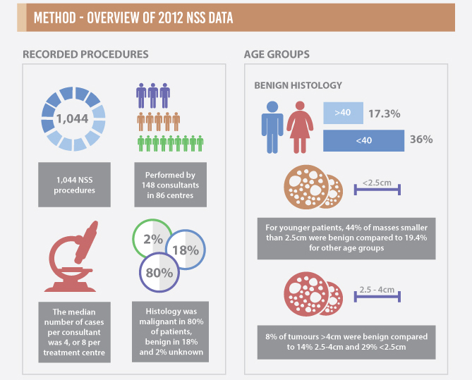
All three featured amongst the list of the top 20 papers with most page views on www.bjui.org and the top 10 most downloaded articles from Wiley online library (WOL), reaching a figure of >2500. This compares well to our most downloaded ‘Guideline of Guidelines’on thromboprophylaxis [4] at 2264. The infographics lay out clear messages on important topics in a concise manner and have undeniable appeal to busy clinicians, who often have just a few valuable minutes to keep abreast with the latest highlights (Fig. 1).

Figure 1. Extract of infographics for the Fernando et al. [2] paper ‘Nephron-sparing surgery across a nation – outcomes from the British Association of Urological Surgeons 2012 national partial nephrectomy audit’. NSS, nephron-sparing surgery.

We also thought we would kick off the New Year with Guidelines on minimally invasive adrenalectomy from the International Consultation on Urological Diseases (ICUD) consultation [5]. And of course the ‘hot topic’ of enhanced recovery to try and reduce the length of stay for our cystectomy patients without increasing complications or readmission rates [6].

We are looking forward to engaging with you with more infographics in 2017.

Prokar Dasgupta, BJUI Editor-in-Chief
Kings Health Partners, London, UK


References

  1. Carlsson S, Jaderling F, Wallerstedt A et al. Oncological and functional outcomes 1 year after radical prostatectomy for very-low-risk prostate cancer: results from the prospective LAPPRO trial. BJU Int 2016; 118: 205–12
  2. Fernando A, Fowler S, O’Brien T, British Association of Urological Surgeons (BAUS). Nephron-sparing surgery across a nation – outcomes from the British Association of Urological Surgeons 2012 national partial nephrectomy audit. BJU Int 2016; 117: 874–82
  3. Kim ED, McCullough A, Kaminetsky J. Oral enclomiphene citrate raises testosterone and preserves sperm counts in obese hypogonadal men, unlike topical testosterone: restoration instead of replacement. BJU Int 2016; 117: 677–85
  4. Violette PD, Cartwright R, Briel M, Tikkinen KA, Guyatt GH. Guideline of guidelines: thromboprophylaxis for urological surgery. BJU Int 2016; 118: 351–8
  5. Ball MW, Hemal AK, Allaf ME. International Consultation on Urological Diseases and European Association of Urology International Consultation on Minimally Invasive Surgery in Urology: laparoscopic and robotic adrenalectomy. BJU Int 2017; 119: 13–21
  6. Baack Kukreja JE, Kiernan M, Schempp B et al. Quality improvement in cystectomy care with enhanced recovery (QUICCER study). BJU Int 2017; 119: 38–49
See more infographics

Editorial: Circulating biomarkers of NEPC – an unmet challenge

Prostate cancer is a global health issue and, although the overwhelming majority (>95%) of metastatic castration-resistant prostate cancers (CRPCs) have adenocarcinoma histology [1], a subset of tumours acquire histopathological and immunohistochemical evidence of neuroendocrine differentiation, with a variety of morphological classifications being reported [1]. Nonetheless, the term ‘neuroendocrine prostate cancer’ (NEPC) should be reserved for tumours with absent or minimal androgen-signalling modulated transcription [2]. NEPC arising in the castration-resistant scenario (treatment-related NEPC or tNEPC) [2] is a disease of unknown prevalence and without an optimum treatment regime. Autopsy studies have shown at least focal neuroendocrine differentiation may be present in up to 33% of patients [3]. The cellular precursor of tNEPC is still debated, but a common clonal origin from adenocarcinoma CRPC (adeno-CRPC) is likely [2]. The assumption of negligible androgen signalling in these tumours implies resistance to agents such as abiraterone and enzalutamide. In this setting, research focused on identifying biomarkers of tNEPC is to be welcomed.

Heck et al. [4] determined the prognostic impact of elevated circulating neuroendocrine biomarkers chromogranin A (CGA) and neuron-specific enolase (NSE) in the serum of patients with CRPC treated with abiraterone in the post-chemotherapy setting. Although CGA and NSE did not predict PSA response, they correlated with clinical and radiographic progression-free survival (PFS), as well as overall survival (OS). The association between these biomarkers and clinical outcomes in metastatic CRPC has been confirmed in retrospective studies [5]. According to the authors, this association, independently of PSA response, underlines the sub-clonality of this disease, and the key role of androgen receptor (AR) signalling, even in advanced disease [4].

The marker NSE is considered to be generic, with high sensitivity but low specificity; CGA is a more specific neuroendocrine tumour biomarker and a common constituent of neuroendocrine tumour secretory granules. Abnormal CGA levels have, however, been significantly associated with intake of proton pump inhibitors in patients treated with abiraterone for metastatic CRPC rather than with duration of treatment [5]. Unfortunately, the use of proton pump inhibitors in that study was not disclosed and may have affected the reported results. Moreover, compared with previous experience, the rate of abnormal NSE was significant higher, probably in keeping with the low specificity of this biomarker.

Interestingly, the authors report an OS and PFS of 12.7 and 3.7 months, respectively [4]. These data are significantly different from the results of the COU-AA 301 study, in which treatment with abiraterone resulted in improved OS (14.8 vs 10.9 months) [6]. Surprisingly, there was no difference in PFS between abiraterone in the study by Heck et al. and the control arm of the COU-AA 301 trial (3.6 months) [6]. This discordance could be attributable to the small sample size of their study rather than the high PSA level at initiation of abiraterone, as claimed by the authors. In support of this alternative possibility, a post hoc analysis of the AFFIRM trial [7] showed consistent benefits in OS and PFS with second-generation hormonal treatments, regardless of baseline disease severity as assessed by PSA level.

Nevertheless, identifying patients with tNEPC is an urgent clinical need; genomic germline and somatic DNA next-generation sequencing as well as transcriptomic analysis of metastatic biopsies should now be considered a key approach to better understanding the heterogeneity of metastatic CRPC and to personalize treatment in order to maximize benefit.

There is substantial genomic overlap between adeno-CRPCs and tNEPC. TMPRSS2-ERG is the most common genomic aberration in prostate cancer and has been reported in NEPC with a similar frequency [2]. Furthermore, both adeno-CRPCs and tNEPCs are enriched for the inactivation of key tumour suppressor genes, such as RB1 and TP53, compared with hormone-sensitive prostate cancer, albeit in different proportions [2]. Although genomic amplification and activating point mutations of the AR in tNEPCs are notably absent, the presence of AR-splicing variants, including ARv7, is still detectable, suggesting that AR signalling is still present in at least a proportion of tNEPCs [2].

Despite a common background of genomic aberrations, tNEPCs have also been reported to have significant overexpression and copy number gains of AURKA and MYCN (40% of NEPC vs 5% of primary prostate cancer tumours), although these findings remain unsubstantiated [3]. As such, these have been postulated to be drivers of this disease phenotype and are under investigation as targets of novel agents.

Genome-wide DNA methylation analysis has, however, also shown that there are marked epigenetic differences between NEPC and adeno-CRPC, suggesting that epigenetic modifiers play a major role in the induction and maintenance of the neuroendocrine status [2].

In conclusion, the identification and definition of NEPC remains challenging. Blood biomarkers such as NSE and CGA cannot be considered to be proven prognostic biomarkers of NEPC as they have only been evaluated in small retrospective studies not adhering to REMARK criteria [8]. Genomic profiling from tissue biopsies or circulating DNA remains a preferable way to identify NEPC and is increasingly feasible, although still not affordable or a standardized procedure for the definition of NEPC.

Read the full article
Pasquale Rescigno*,, Daniel Nava Rodrigues*,† and Johann S. de Bono*,


*Institute of Cancer Research, London, UK and Royal Marsden NHS Foundatio n Trust, London, UK


References


1 Epstein JI, Amin MB, Beltran H et al. Proposed morphologic classication of prostate cancer with neuroendocrine differentiation. Am Surg Pathol 2014; 38: 75667


2 Beltran H, Prandi D, Mosquera JM et al. Divergent clonal evolution of castration-resistant neuroendocrine prostate cancer. Nat Med 2016; 22: 298305


6 de Bono JS, Logothetis CJ, Molina A et al. Abiraterone and increased survival in metastatic prostate cancer. N Engl J Med 2011; 364: 19952005


7 Saad F, de Bono J, Shore N et al. Efcacy outcomes by baseline prostate- specic antigen quartile in the AFFIRM trial. Eur Urol 2015; 67: 22330


8 McShane LM, Altman DGSauerbrei W. REporting recommendations for tumour MARKer prognostic studies (REMARK). Br J Cancer 2005; 93: 38791


Editorial: Open partial nephrectomy is still alive

In this issue of BJUI, Ramirez et al. [1] assess the peri-operative outcomes of patients with small renal tumours treated with robot-assisted or open partial nephrectomy [1]. Their study retrospectively compared transperitoneal robot-assisted partial nephrectomy (RAPN) and retroperitoneal open partial nephrectomy (OPN) in a series of 714 patients (545 RAPN and 169 OPN) with cT1a parenchymal tumours of the kidney. Although the RAPN group had a higher mean RENAL nephrometry score, the authors observed a significantly higher overall complication rate in patients in the OPN group (30.3 vs 18.2%; P=0.038). On multivariable analysis, the open approach was an independent predictor of overall complications (odds ratio 1.52; CI 1.03–2.43, P=0.035) besides race, body mass index (BMI) and Charlson comorbidity index. Notably, when complications were stratified by grade, the worse outcome for OPN compared with RAPN was related to minor complications (i.e. Clavien–Dindo 1 and 2) only, with the most relevant difference represented by wound problems. No statistically significant difference between the two approaches was observed with regard to estimated blood loss (EBL) or genitourinary complications. Surprisingly, the RAPN group also had a shorter median warm ischaemia time (WIT; median 22.1 vs 28.6 min).

The authors should be commended for this very interesting comparative study; however, some criticisms should be considered.

First, the authors claim that all procedures were performed by surgeons beyond their learning curve over a period of >4.5 years. Considering the OPN group, the number of procedures/year is <40. If the series was multi-surgeon (the authors do not specify this), then yearly caseload does not meet the standards for defining ‘high’ surgical volume, especially when compared with the RAPN cases (>100/year).

Second, the main complication in the OPN group was wound problems. One has to consider that patients included in this study were mostly overweight to obese, as reflected in the BMI values (median 29.6 kg/m2). This characteristic could have played an important role in the OPN group and may represent a bias.

Third, it seems curious that the same group of authors recently published another comparative study of RAPN vs OPN for ‘completely endophytic tumours’ [2], where the only statistically significant difference between the two approaches was lower EBL and shorter length of stay, in favour of RAPN. Interestingly, no statistically significant difference was reported between two groups with regard to either postoperative complications or WIT.

In 2014, Ficarra et al. [3] compared RAPN and OPN in a multicentre series of 400 cases (200 RAPN and 200 OPN) using a matched-pair analysis [3]. The robot-assisted approach resulted in a lower rate of minor postoperative complications and a lower intra-operative EBL. Notably, in that analysis, the open approach resulted in a shorter WIT despite the fact that 14.5% and 13% of RAPN and OPN patients, respectively, were complex cases (i.e. had cT1b tumours). As pointed out above, in the present study, Ramirez et al. [1] reported a median (interquartile range [IQR]) WIT of 22.1 (17–26) min for the RAPN group and 28.6 (22–35) min for the OPN group [1].

Robot-assisted surgery confers an evident advantage to the surgeon, especially during the renorrhaphy phase as a result of the freedom of movement of the robotic needle driver compared with conventional laparoscopic instruments. It has been shown that the learning curve for RAPN is relatively short, allowing WIT of <20 min to be achieved [4]. In our opinion, the real competitor with regard to RAPN for WIT is OPN. A recent systematic review and meta-analysis based on 16 series showed, in fact, longer WIT for RAPN compared with OPN [5].

Taken together, all these results suggest that OPN maintains its role in the robotic era, especially when complex cases require treatment. Although RAPN is now widespread and has expanded its indications, it is still able to provide excellent performance in terms of postoperative complications at least equal to OPN in tertiary referral centres where a high volume of robotic renal surgery is performed, as indicated by Ramirez et al. [1]; however, several aspects of RAPN still await evaluation. The learning curve for RAPN, in terms of operating time and WIT, has been previously evaluated [4, 6], and the advantages of robotic surgery have been convincingly demonstrated when compared with pure laparoscopy [7]. Despite this, all the studies that have evaluated the learning curve for RAPN involved a tumour size <3 cm. The learning curve for OPN, especially in complex cases, should also be considered. Despite its impressive dissemination, RAPN is still in evolution, and OPN remains alive.

Read the full article
Alessandro Crestani, Marta Rossanese and Gianluca Giannarini
Academic Medical Centre Hos ital San ta Maria della Misericordia, Urology Unit, Udine, Italy


References


Editorial: Light reflectance spectroscopy is one more emerging technique with the potential to adjust excision limits during radical prostatectomy

In this issue of BJUI, Lay et al. [1] report that light reflectance spectroscopy (LRS) can detect Gleason ≥7 positive surgical margins (PSMs) with 92.5% accuracy. In this initial study, the authors have reported the use of LRS in an ex situ setting to analyse the prostate surface; however, this technology could ultimately be developed to identify PSMs before choosing the surgical plane of dissection, which could allow the surgeon to immediately perform a wider complementary excision.

As long as PSMs are detected ex situ, it is not clear why spectroscopy should be preferred to frozen sections. NeuroSAFE, for example, is a standardized and validated margin evaluation procedure in pathology [2]. It does not lengthen operating time, does not require any new equipment and provides a pathological assessment which is the best level of evidence for PSM status; however, as a conventional pathological procedure, it is not conceivable in situ, and real-time detection of PSMs that ensures the safest oncological resection during a nerve-sparing dissection is needed.

In this effort to examine in vivo/in situ prostate PSMs, several other technologies can be considered. During radical prostatectomy, optical coherence tomography (OCT) has been used in situ in humans, but only to identify the neurovascular bundles [3]. Field of view and depth of penetration were limited and OCT has never been evaluated in situ for prostate PSM detection. Confocal endomicroscopy has recently been reported during robot-assisted radical prostatectomy [4]. With this technique, optical biopsies were feasible in situ but the PSM detection rate and the overall efficiency of this confocal endomicroscopy in prostate specimens remain unknown. Similarly, illumination microscopy has been used to generate gigapixel images of the full prostate circumference in vivo for the detection of PSMs [5]. Illumination microscopy allows images to be interpreted readily by pathologists, but the feasibility series was too small to assess the accuracy of this technique for PSM detection. Ex situ multi-photon microscopy (MPM) is an optical technique that enables the imaging of prostatic and periprostatic tissue at sub-micron resolution to a depth of up to 0.5 mm [6]. On a fresh specimen, it generates three-dimensional images of periprostatic nerves, blood vessels and capsule, but also underlying acini and pathological changes such as prostate cancer. MPM technology has also been miniaturized and its accuracy in situ is currently under investigation.

In this context, the study by Lay et al. [1] shows that, for the time being, LRS is one more promising technique on the road to real-time PSM detection. More will undoubtedly be done to overcome the spectroscope’s light absorption in the presence of blood and, subsequently, to evaluate its reliability in situ; however, the recent developments of these protocols and technologies (endomicroscopy, illumination microscopy, OCT, MPM, LRS) show a progressive effort amongst clinicians to obtain intra-operative feedback on the PSM status. Fortunately, this is taking place while the urological community is increasingly considering surgical treatment even for the high-risk disease, where oncological adequacy is of paramount importance. While we are witnessing these promising evolutions in high-grade prostate cancer, the optimum technique which will safely end margin-blind radical prostatectomy in an actual surgical field (filled with blood and often distorted because of inflammation) still needs to go through clinical trials and validation; however, the future is bright as a result of these newer developments.

Read the full article
Thomas Bessede*†‡ and Ash Tewari*


*Department of Urology, Icahn School of Medicine at MounSinai, New York , NY, USA, U1195, INSERM, UniversitParis-Saclay, and Department of Urology, APHP, Hopitaux Universitaires Paris-Sud, Le Kremlin-Bicetre, France


References


Editorial: SNP of the VEGFR – a promising biomarker in mRCC

Despite earlier detection of localised renal tumours, the resultant tumour down-staging, and an ever-increasing armamentarium of systemic therapies available for metastatic RCC (mRCC), population-level RCC mortality data has failed to show significant improvement in survival. The increasing understanding of RCC biology, specifically the tyrosine kinase signalling pathway, has sparked the development and USA Food and Drug Administration (FDA)-approval of four tyrosine kinase inhibitors (TKIs) and one anti-vascular endothelial growth factor receptor (VEGFR) antibody. Additionally, immunotherapies in the form of interferon, interleukin 2 cytokine therapy and the advent of checkpoint inhibitors further expands the options for treatment of mRCC. This poses a clinical dilemma; although clinical trials are showing improved survival outcomes, there is uncertainty about to how best to sequence the available therapies. In lieu of long, expensive clinical trials exploring all possible permutations and in order to personalise therapy for specific patients, we are hopeful the exploration of biomarkers (such as the authors have performed) will fill this void and aid in the selection of therapies based on likelihood of patient response.

While studies do not seem to show a reliable correlation between von Hippel-Lindau gene status or expression levels of hypoxia-inducible factor and response to targeted therapy [1], recent exploration of single nucleotide polymorphisms (SNPs) of the VEGFR has yielded promising results with certain VEGFR1 SNPs (in particular, SNP rs9582036 with CC alleles) associated with a significantly worse overall survival when treated with a TKI [2-4]. The authors previously published on a discovery cohort of patients with mRCC treated with sunitinib and found results consistent with the aforementioned exploratory studies; the CC-variant in rs9582036 was associated with worse response rate, progression-free survival (PFS), and overall survival (14 months vs 31 months; P = 0.008) on multivariate analysis [5, 6].

This current study [7] represents their findings of a validation cohort of the potential predictive association of the VEGFR1 SNP rs9582036 in mRCC treated with sunitinib. In all, 69 patients were genotyped and clinical outcomes analysed; results were consistent with their previous discovery cohort, and in their pooled analysis of 157 patients they confirmed that the allelic status of the rs9582036 SNP was significantly associated with clinical outcomes. Patients with the CC-variant had poorer response rates (8% vs 49%), worse PFS (8 vs 14 months), and worse overall survival (13 vs 30 months).

This finding certainly could have profound implications in guiding choice of systemic therapies and exploration of the biological meaning of these SNP variants could shed light on why certain tumours or patients fail to respond to targeted therapy. However, further study is warranted. In this current study, VEGFR1 mRNA expression levels at the onset of therapy correlated positively with response to treatment; however, there was no association between the SNP genotypes explored and initial VEGFR1 expression, thus providing no clear mechanistic rationale between this association and clinical outcomes. Furthermore, this rs9582036 sub-group represented a minority (9%) of the patients studied and most patients were Caucasian, limiting the applicability of this genotyping. Additionally, the lack of a placebo control group prevents a conclusion about what to do in the case of a CC-variant rs9852036 patient, i.e. a diminished response to TKIs does not mean TKIs are necessarily ineffective, nor does it mean another therapy will be preferentially more effective.

Despite these issues, if we are to ever realise the promise of personalised medicine, it will be through efforts such as these, which explore the genetic variation in our germline (and tumour) genome that may affect treatment response and to integrate the findings of genomics and epigenomics with clinical outcomes to tailor future therapies.

Read the full article
Solomon L. Woldu and Vitaly Margulis
Department of Urology, University of Texas Southwestern Medical Center, Dallas, TX, USA


References


6 Beuselinck B, Karadimou A, Lambrechts D et al. Single- nucleotide polymorphisms associated with outcome in metastatic renal cell carcinoma treated with sunitinib. Br J Cancer 2013; 108: 887900


© 2024 BJU International. All Rights Reserved.