Tag Archive for: early detection

Posts

Article of the Week: A mpMRI-based risk model to determine the risk of prostate cancer prior to biopsy

Every Week the Editor-in-Chief selects an Article of the Week from the current issue of BJUI. The abstract is reproduced below and you can click on the button to read the full article, which is freely available to all readers for at least 30 days from the time of this post.

In addition to the article itself, there is an accompanying editorial written by a prominent member of the urological community. This blog is intended to provoke comment and discussion and we invite you to use the comment tools at the bottom of each post to join the conversation.

If you only have time to read one article this week, it should be this one.

A multiparametric magnetic resonance imaging-based risk model to determine the risk of significant prostate cancer prior to biopsy

Pim J. van Leeuwen*, Andrew Hayen, James E. Thompson*†‡, Daniel Moses§Ron Shnier§, Maret Bohm, Magdaline Abuodha, Anne-Maree HaynesFrancis Ting*†‡, Jelle Barentsz, Monique Roobol**, Justin Vass††, Krishan Rasiah††Warick Delprado‡‡ and Phillip D. Stricker*†‡


*St. Vincents Prostate Cancer Centre, Garvan Institute of Medical Research/The Kinghorn Cancer Centre, Darlinghurst, School of Public Health and Community Medicine, §School of Medicine, University of New South Wales, Kensington, New South Wales, Australia, Department of Radiology and Nuclear Medicine, Radboud University Medical Centre, Nijmegen, **Department of Urology, Erasmus University Medical Center, Rotterdam, the Netherlands, ††Department of Urology, Royal North Shore Private Hospital, St Leonards, and ‡‡Douglass Hanly Moir Pathology and University of Notre Dame, Darlinghurst, New South Wales, Australia


Abstract

Objective

To develop and externally validate a predictive model for detection of significant prostate cancer.

Patients and Methods

Development of the model was based on a prosp   ctive cohort including 393 men who underwent multiparametric magnetic resonance imaging (mpMRI) before biopsy. External validity of the model was then examined retrospectively in 198 men from a separate institution whom underwent mpMRI followed by biopsy for abnormal prostate-specific antigen (PSA) level or digital rectal examination (DRE). A model was developed with age, PSA level, DRE, prostate volume, previous biopsy, and Prostate Imaging Reporting and Data System (PIRADS) score, as predictors for significant prostate cancer (Gleason 7 with >5% grade 4, ≥20% cores positive or ≥7 mm of cancer in any core). Probability was studied via logistic regression. Discriminatory performance was quantified by concordance statistics and internally validated with bootstrap resampling.

Results

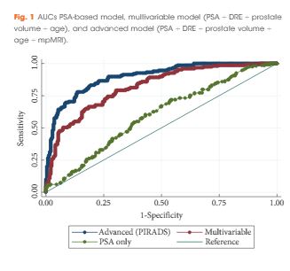
In all, 393 men had complete data and 149 (37.9%) had significant prostate cancer. While the variable model had good accuracy in predicting significant prostate cancer, area under the curve (AUC) of 0.80, the advanced model (incorporating mpMRI) had a significantly higher AUC of 0.88 (P < 0.001). The model was well calibrated in internal and external validation. Decision analysis showed that use of the advanced model in practice would improve biopsy outcome predictions. Clinical application of the model would reduce 28% of biopsies, whilst missing 2.6% significant prostate cancer.

Conclusions

Individualised risk assessment of significant prostate cancer using a predictive model that incorporates mpMRI PIRADS score and clinical data allows a considerable reduction in unnecessary biopsies and reduction of the risk of over-detection of insignificant prostate cancer at the cost of a very small increase in the number of significant cancers missed.

 

Editorial: Novel risk stratification nomograms for counseling patients on the need for prostate biopsy

Contemporary recommendations for prostate screening incorporate the measurement of serum PSA levels into shared decision making [1]. PSA is limited by a low specificity for prostate cancer and exposes a certain number of men to unnecessary prostate biopsies. Moreover, it has been attributed to an over-diagnosis and over treatment of this disease, especially in indolent cancer that may never affect a man’s longevity [2].

Prostate cancer risk stratification and aggressiveness is necessary in both the pre-biopsy clinical counselling, as well as the decision-making process. It is clear that such an important approach cannot be accomplished based on PSA alone. In order to enhance prostate cancer screening and detection, other clinical variables such as PSA density, prostate volume, percentage free PSA, and DRE, are routinely considered in conjunction with PSA for determining the need for prostate biopsy.

Increasing evidence supports the use of MRI in prostate cancer detection when used as a localisation tool to guide MRI-targeted biopsy techniques such as MRI-ultrasonography fusion-targeted biopsy [3-5]. Pre-biopsy MRI not only allows accurate tumour localisation, but also provides an assessment of cancer suspicion using an MRI suspicion score, and thus provides accurate prediction of the likelihood of prostate cancer on prostate biopsy [6].

In this study, van Leeuwen et al. [7] developed and externally validated a set of nomograms predicting clinically significant prostate cancer by incorporating prostate MRI. The performance characteristics of the nomograms were maximised by inclusion of MRI results. The authors determined that clinical application of the model would reduce 28% of biopsies, while missing 2.6% of clinically significant prostate cancer. Ultimately incorporating these nomograms into the clinical decision-making process could result in a considerable reduction in unnecessary biopsies and reduction in the risk of over-detection of clinically insignificant disease at the cost of a small increase in the number of significant cancers missed.

The authors should be commended for their predictive nomograms, in that they may further aid in the decision to perform biopsy in men with clinical suspicion of prostate cancer. However, the findings of this study should be interpreted with caution. In formulating nomograms, the obvious clinical goal is the creation of a tool that improves the selection of men in need of biopsy. Unless nomograms are derived from general ‘at risk’ populations, including men with low- and high-risk of prostate cancer, the tool may be limited in its prediction. As an example, if all men in the training cohort have an elevated PSA level, the predictive value of PSA in the nomogram may be reduced. In this case, the training and validation cohort are not well described, but it seems to be a referral population, and the PSA range is relatively narrow. As such, it’s applicability to all men presenting for prostate cancer may be questionable.

We also have had difficulty modelling a nomogram from our MRI-targeted biopsy dataset because the power of MRI-suspicion score in predicting cancer tends to minimise the effect of other variables such as PSA, age, and gland size. The authors did not compare their multivariable predictive models to MRI alone, and this may have been the most relevant comparison. In this study, the gland size and PSA level contribute significantly to the nomogram score, but one might question the findings. For example, a man with a Prostate Imaging Reporting and Data System (PI-RADS) score of 4 or 5, a large gland and a low PSA level, has a similar or lower risk as a man with a PSA level of 15 ng/mL, a moderate gland, and a PI-RADS score of 3. This is not consistent with our clinical experience and draws concern in the reliability of the nomogram at extremes of PSA and age. Men with PI-RADS 5 have high rates of clinically significant prostate cancer, regardless of PSA or age. This also may reflect variability in the predictive accuracy of MRI depending upon MRI interpretation.

Another limitation of the study, as the authors cite, is that patients were biopsied using a transperineal mapping with a median of 30 cores. This biopsy strategy is not routinely used in most institutions, and consequently limits the generalisability of the nomograms.

Selective use of prostate biopsy among men with elevated PSA levels through further refinement of cancer risk is highly desirable. The novel risk stratification nomograms developed by van Leeuwen et al. [7] add to the tools we may use to counsel our patients on the need for prostate biopsy. Further evaluation of these nomograms on additional independent patient cohorts is warranted prior to implementation in clinical practice.

Marc A. Bjurlinand Samir S. Taneja
*Division of Urology, Department of Surgery, NYU Lutheran Medical Center, NYU Langone Health System, New York, NY, USA and Division of Urologic Oncology, Department of Urology, NYU Langone Medical Center, New York, NY, US


References


1 Carter HBAlbertsen PCBarry MJ et al. Early detection of prostate cancer: AUA Guideline. J Urol2013; 190: 41926


2 Loeb SBjurlin MANicholson J et al. Overdiagnosis and overtreatment of prostate cancer. Eur Urol
2014; 65: 104655


3 Mendhiratta NMeng XRosenkrantz AB et al. Prebiopsy MRI and MRI-ultrasound fusion-targeted prostate biopsy in men with previous negative biopsies: impact on repeat biopsy strategies. Urology 2015; 86: 11928


5 Meng XRosenkrantz ABMendhiratta N et al. Relationship between prebiopsy multiparametric magnetic resonance imaging (MRI), biopsy indication, and MRI-ultrasound fusion-targeted prostate biopsy outcomes. Eur Urol 2016; 69: 51217


7 van Leeuwen PJHayan AThompson JE et al. A multiparametric magnetic resonance imaging-based risk model to determine the risk of significant prostate cancer prior to biopsy. BJU Int 2017; 120: 7748


© 2024 BJU International. All Rights Reserved.