Tag Archive for: chest computed tomography

Posts

Residents’ Podcast: When to Perform Preoperative Chest CT for RCC Staging

Jesse Ory, Kyle Lehmann and Jeff Himmelman

Department of Urology, Dalhousie University, Halifax, NS, Canada


Abstract

Objectives

To provide objective criteria for preoperative staging chest computed tomography (CT) in patients diagnosed with renal cell carcinoma (RCC) because, in the absence of established indications, the decision for preoperative chest CT remains subjective.

Patients and Methods

A total of 1 946 patients undergoing surgical treatment of RCC, whose data were collected in a prospective institutional database, were assessed. The outcome of the study was presence of pulmonary metastases at staging chest CT. A multivariable logistic regression model predicting positive chest CT was fitted. Predictors consisted of preoperative clinical tumour (cT) and nodal (cN) stage, presence of systemic symptoms and platelet count (PLT)/haemoglobin (Hb) ratio.

Results

The rate of positive chest CT was 6% (n = 119). At multivariable logistic regression, ≥cT1b, cN1, systemic symptoms and Hb/PLT ratio were all associated with higher risk of positive chest CT (all P < 0.001). After 2000-sample bootstrap validation, the concordance index was found to be 0.88. At decision-curve analysis, the net benefit of the proposed strategy was superior to the select-all and select-none strategies. Accordingly, if chest CT had been performed when the risk of a positive result was >1%, a negative chest CT would have been spared in 37% of the population and a positive chest CT would have been missed in 0.2% of the population only.

Conclusions

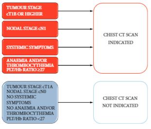
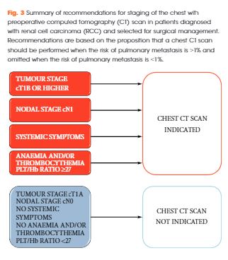
The proposed strategy estimates the risk of positive chest CT at RCC staging with optimum accuracy and the results were statistically and clinically relevant. The findings of the present study support a recommendation for chest CT in patients with ≥cT1b, cN1, systemic symptoms or anaemia and thrombocythemia. Conversely, in patients with cT1a, cN0 without systemic symptoms, anaemia and thrombocythemia, chest CT could be omitted.

BJUI Podcasts now available on iTunes, subscribe here https://itunes.apple.com/gb/podcast/bju-international/id1309570262

Article of the Week: When to Perform Preoperative Chest CT for RCC Staging

Every Week the Editor-in-Chief selects an Article of the Week from the current issue of BJUI. The abstract is reproduced below and you can click on the button to read the full article, which is freely available to all readers for at least 30 days from the time of this post.

In addition to the article itself, there is an accompanying editorial written by a prominent member of the urological community. This blog is intended to provoke comment and discussion and we invite you to use the comment tools at the bottom of each post to join the conversation.

If you only have time to read one article this week, it should be this one.

When to perform preoperative chest computed tomography for renal cancer staging

Alessandro Larcher*, Paolo DellOglio*, Nicola Fossati*, Alessandro Nini*Fabio Muttin*, Nazareno Suardi*, Francesco De Cobelli, Andrea Salonia*Alberto Briganti*, Xu Zhang§, Francesco Montorsi*, Roberto Bertini*† and Umberto Capitanio*


*Division of Experimental Oncology, URI – Urological Research Institute, Unit of Urology, Vita-Salute San Raffaele University, Unit of Radiology, Vita-Salute San Raffaele University, IRCCS San Raffaele Scientific Institute, Milan, Italy, and § Clinical Division of Surgery, Department of Urology, Chinese PLA General Hospital, Beijing, China


Abstract

Objectives

To provide objective criteria for preoperative staging chest computed tomography (CT) in patients diagnosed with renal cell carcinoma (RCC) because, in the absence of established indications, the decision for preoperative chest CT remains subjective.

Patients and Methods

A total of 1 946 patients undergoing surgical treatment of RCC, whose data were collected in a prospective institutional database, were assessed. The outcome of the study was presence of pulmonary metastases at staging chest CT. A multivariable logistic regression model predicting positive chest CT was fitted. Predictors consisted of preoperative clinical tumour (cT) and nodal (cN) stage, presence of systemic symptoms and platelet count (PLT)/haemoglobin (Hb) ratio.

Results

The rate of positive chest CT was 6% (n = 119). At multivariable logistic regression, ≥cT1b, cN1, systemic symptoms and Hb/PLT ratio were all associated with higher risk of positive chest CT (all P < 0.001). After 2000-sample bootstrap validation, the concordance index was found to be 0.88. At decision-curve analysis, the net benefit of the proposed strategy was superior to the select-all and select-none strategies. Accordingly, if chest CT had been performed when the risk of a positive result was >1%, a negative chest CT would have been spared in 37% of the population and a positive chest CT would have been missed in 0.2% of the population only.

Conclusions

The proposed strategy estimates the risk of positive chest CT at RCC staging with optimum accuracy and the results were statistically and clinically relevant. The findings of the present study support a recommendation for chest CT in patients with ≥cT1b, cN1, systemic symptoms or anaemia and thrombocythemia. Conversely, in patients with cT1a, cN0 without systemic symptoms, anaemia and thrombocythemia, chest CT could be omitted.

Read more articles of the week

Editorial: Do all patients with renal cell carcinoma need a chest computed tomography?

While all patients with RCC need chest imaging for staging evaluation, the answer to the question in the title is ‘No’, and, in fact, many patients would be adequately staged with a chest X-ray, albeit with reduced accuracy. Evidence to support this assertion is provided by Larcher et al. [1] in this issue of BJUI, who retrospectively evaluated 1946 patients with a solitary and sporadic RCC mass. While excluding patients who did not have surgery and those with visceral metastases seen on abdominal imaging, the authors observed pulmonary metastases in 6% (119 patients) of their population. In a multivariable analysis, features associated with a positive chest CT included cT1b+, cN1, systemic symptoms, anaemia, and thrombocytosis. Incorporating these features into a predictive model, the authors report a robust concordance index of 0.88, with the effect of each feature demonstrated in a nomogram. Further, the authors report that if a chest CT is only performed when the risk of a positive result is >1%, 37% of their population could have been spared a chest CT while missing a positive result in only 0.2% (four patients). Patient factors that predict for a <1% risk of a positive chest CT essentially include those with cT1aN0 RCC without systemic symptoms, anaemia, or thrombocytosis. Thus, the authors conclude that in these low-risk patients, a chest CT can be omitted, while any patient that is cT1b+, cN1, or with systemic symptoms, anaemia, or thrombocytosis warrants a dedicated chest CT at diagnosis.

The finding that patients with RCC with smaller tumours (cT1a or ≤4 cm) were unlikely to harbour pulmonary metastases is consistent with prior literature. Observations from the Memorial Sloan-Kettering Cancer Center (MSKCC) [2], and subsequently validated by our group at Mayo Clinic [3], suggested that among surgically treated patients with RCC, risk of M1 disease (at any location) at diagnosis was non-existent for tumours of <2 cm, was <1% for tumours of 2–3 cm, and was only 1–2% for tumours of 3-4 cm in size. Given that it is rare for patients with small renal masses to endorse systemic symptoms or have paraneoplastic symptoms related to the tumour, these prior observations suggest a lack of utility for chest CT for patients with small renal masses supporting the findings from Larcher et al. [1].

In patients with RCC with synchronous metastases, lung is the most common site of spread and guidelines uniformly recommend chest imaging at diagnosis. However, a ‘select-all’ strategy for chest CT in patients with renal masses leads to unnecessary findings in those with a benign primary tumour, increased use of healthcare resources, and relatively frequent findings of indeterminate lesions. In fact, contemporary observations from the MSKCC found that about half of patients with RCC undergoing surgery had indeterminate pulmonary nodules on chest CT that required either additional evaluation or subsequent chest CT to document stability [4]. Further, the presence of indeterminate pulmonary nodules was not associated with distant metastases or death from RCC after surgery unless they were >1 cm, which only represented a small portion (4%) of the entire cohort [4]. Thus, the analysis from Larcher et al. [1] in this issue of BJUI has meaningful clinical relevance; that is, patients with cT1aN0 RCC without symptoms or laboratory abnormalities do not require a chest CT for screening of their lungs.

R. Houston Thompson
Department of Urology, Mayo Clinic, Rochester, MN, USA


References


1 Larcher A, DellOglio P, Fossati N et al. When to perform preoperative chest computed tomography for renal cancer staging. BJU Int 2017; 120: 4906


2 Thompson RH, Hill JR, Babayev Y et al. Metastatic renal cell carcinoma risk according to tumor size. J Urol 2009; 182: 415


3 Umbreit EC, Shimko MS, Childs MA et al. Metastatic potential of renal mass according to original tumour size at presentation. BJU Int 2011; 109: 1904


© 2024 BJU International. All Rights Reserved.