Tag Archive for: chemotherapy

Posts

Changing the LATITUDE of Treatment for High-Risk Hormone-Naïve Prostate Cancer: STAMPEDE-ing Towards Androgen Biosynthesis Inhibition

zach-klaassenEarlier this month at the annual American Society of Clinical Oncology (ASCO) meeting in Chicago, IL, Dr. Karim Fizazi and Dr. Nicholas James (@Prof_Nick_James) presented results from the LATITUDE and STAMPEDE trials, respectively. These randomized controlled trials (RCTs) assessed the utility of adding abiraterone acetate (AA) + prednisone to conventional androgen deprivation therapy (ADT) among men with high-risk, hormone-naïve prostate cancer. Since Dr. Charles Huggins’ 1941 Nobel prize winning finding that ADT is highly effective in controlling metastatic prostate cancer, nearly 70 years passed before CHAARTED and STAMPEDE demonstrated in 2015 that the addition of docetaxel to ADT prolongs survival in men with high volume metastatic prostate cancer. The de novo metastatic prostate cancer global incidence is striking: 3% in the US and rising, 6% across Europe, 4-10% in Latin America, and nearly 60% in Asia-Pacific. Historically, ADT has been standard of care, however most men with metastases progress to metastatic castration-resistant prostate cancer (mCRPC) driven by the reactivation of androgen receptor (AR) signaling. The rationale for adding AA + prednisone to ADT for metastatic hormone-naïve prostate cancer patients is threefold: (i) the mechanism of resistance to ADT may develop early, (ii) ADT alone does not inhibit androgen synthesis by the adrenal glands or prostate cancer cells, and (iii) AA + prednisone improves overall survival (OS) in mCRPC patients and reduces tumor burden in high-risk, localized prostate cancer.

LATITUDE

LATITUDE was conducted at 235 sites in 34 countries in Europe, Asia-Pacific, Latin America, and Canada. The objectives of the study were to evaluate the addition of AA + prednisone to ADT on clinical benefit in men with newly diagnosed, high-risk, metastatic hormone-naïve prostate cancer. Patients were stratified by the presence of visceral disease (yes/no) and ECOG performance status (0, 1 vs 2) and then randomized 1:1 to either ADT + AA (1000 mg daily) + prednisone (5 mg) (n=597) or ADT + placebo (n=602). The co-primary endpoints were OS and radiographic progression-free survival (rPFS). Secondary endpoints included time to: (i) pain progression, (ii) PSA progression, (iii) next symptomatic skeletal event, (iv) chemotherapy, and (v) subsequent prostate cancer therapy. The study was powered to detect an HR of 0.67 and 0.81 in favor of AA for rPFS and OS, respectively.
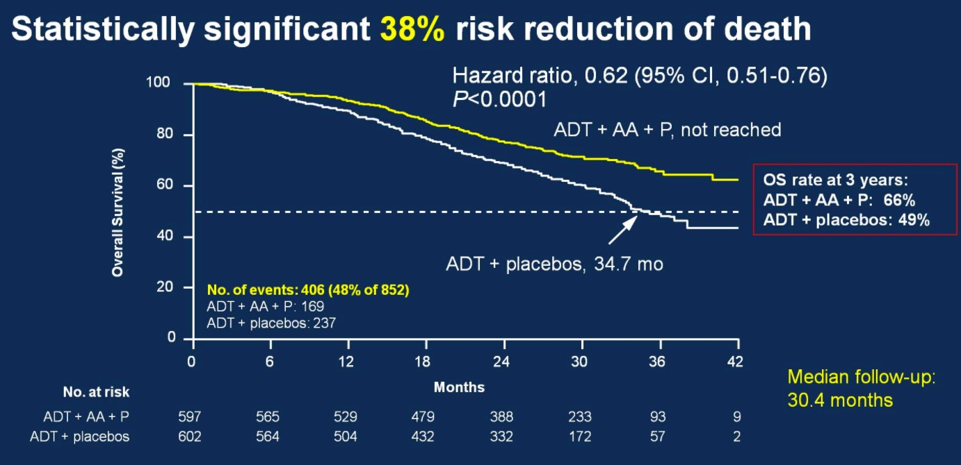
Over a median follow-up of 30.4 months, patients treated with ADT + AA + prednisone had a 38% risk reduction of death (HR 0.62, 95%CI 0.51-0.76) compared to ADT + placebo.

1-1

Median OS was not yet reached in the ADT + AA + prednisone arm compared to 34.7 months in the ADT + placebo arm. OS rates at 3 years for the ADT + AA + prednisone arm was 66%, compared to 49% in the ADT + placebo arm. This OS benefit was consistently favorable across all subgroups including ECOG 0 and 1-2, visceral metastases, Gleason ≥8 disease, and bone lesions >10.

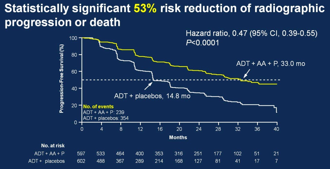
There was also 53% risk of reduction of radiographic progression or death for patients treated with ADT + AA + prednisone (median 33.0 months; HR 0.47, 95%CI 0.39-0.55) compared to ADT + placebo (14.8 months).

1-2

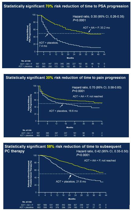
Secondary endpoints showed statistically significant improvement for ADT + AA + prednisone, including time to PSA progression (HR 0.30, 95%CI 0.26-0.35), time to pain progression (HR 0.70, 95%CI 0.58-0.83), time to next symptomatic skeletal event (HR 0.70, 95%CI 0.54-0.92), time to chemotherapy (HR 0.44, 95%CI 0.35-0.56), and time to subsequent prostate cancer therapy (HR 0.42, 95%CI 0.35-0.50).

1-3

Secondary to the results presented at ASCO, the study was discontinued after the first interim analysis. Adverse events were comparable in the two groups. Hypertension only rarely required treatment discontinuation, and only two patients discontinued treatment due to hypokalemia (no hypokalemia-related deaths). Two patients in each arm died of cerebrovascular events, and 10 patients treated with ADT + AA + prednisone compared to 6 patients treated with ADT + placebo died of cardiac disorders.

STAMPEDE

STAMPEDE is a large multi-stage, multi-arm, RCT being conducted in the United Kingdom to assess the utility of novel therapeutic agents in conjunction with ADT. Currently being tested are AA, enzalutamide, zoledronic acid, docetaxol, celecoxib and radiotherapy (RT). The AA arm of the study was presented at ASCO as a late-breaking abstract. Inclusion criteria included men with locally advanced or metastatic prostate cancer, including newly diagnosed with N1 or M1 disease, or any two of the following: stage T3/4, PSA ≥ 40 ng/mL, or Gleason score 8-10. Patients undergoing prior radical prostatectomy or RT were eligible if they had more than one of the following: PSA ≥ 4 ng/mL and PSADT < 6 months, PSA ≥ 20 ng/mL, N1, or M1 disease. Patients were then randomized 1:1 to standard of care (SOC; ADT for ≥2 years, n=957) vs SOC + AA (1000 mg) + prednisone 5 mg daily (n=960). Treatment with RT was mandated in patients with N0M0 disease, while strongly encouraged for N1M0 patients. Primary outcomes were OS and failure-free survival (FFS), where failure was defined as PSA failure, local failure, lymph node failure, distant metastases or prostate cancer death. Secondary outcome included toxicity and skeletal-related events (SREs). The study was powered to detect a 25% improvement in OS for the treatment group (requiring 267 control arm mortalities).
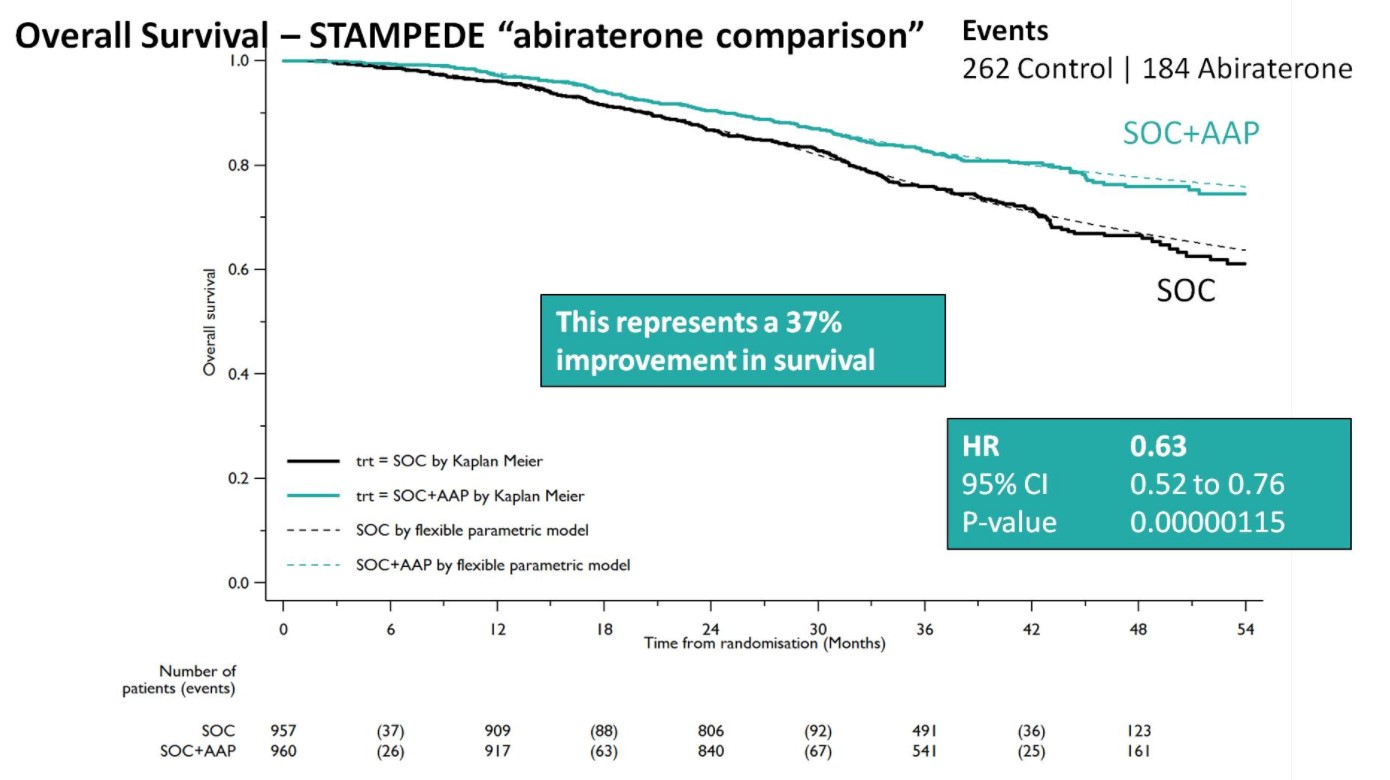
Both groups were balanced and patients were predominantly metastatic (52% M1, 20% N+M0, 28% N0M0), median was PSA 53 ng/mL, and 99% were treated with LHRH analogues. Over a median follow-up of 40 months, there were 262 control arm deaths, of which 82% were prostate cancer-related; there were 184 deaths in the SOC + AA + prednisone arm. There was a 37% relative improvement in overall survival (HR 0.63, 95%CI 0.52-0.76) favoring SOC + AA + prednisone.

1-4

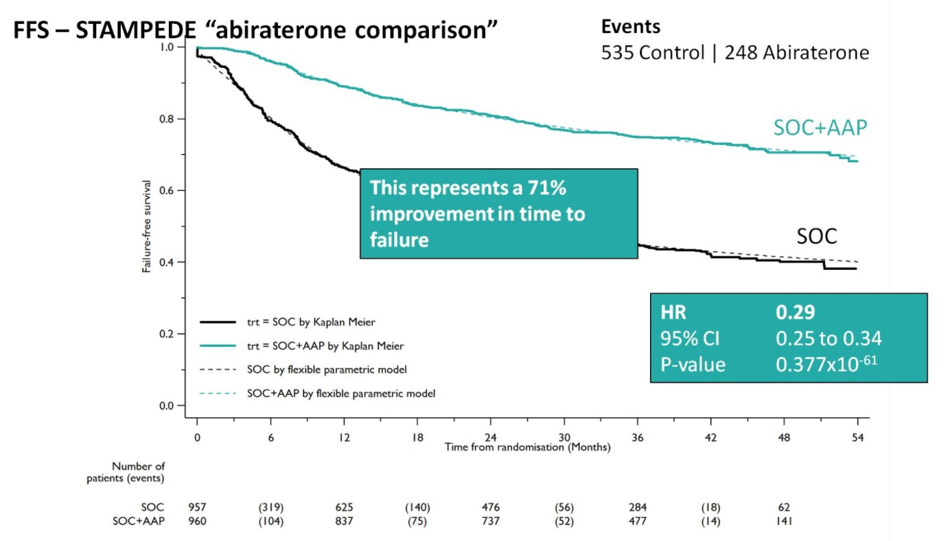
A Forrest plot split on stratification factors demonstrated no evidence of heterogeneity based on any of the factors, including M0/M1 status (p=0.37). Second, SOC+AA + prednisone demonstrated a 71% improvement in FFS (HR 0.29, 95%CI 0.25-0.34), with an early split in the KM curves.

1-5

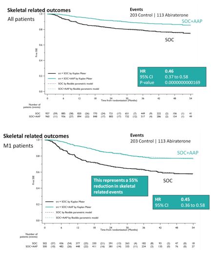
SOC + AA + prednisone also significantly decreased SREs among the entire cohort (HR 0.46, 95%CI 0.37-0.58), as well as specifically in the M1 cohort (HR 0.45, 95%CI 0.37-0.58). This resulted in a 55% reduction in SREs in the M1 subset analysis.

1-6

When looking at treatment progression, 89% of the SOC arm went on to next line of therapy, whereas 79% of the SOC + AA + prednisone arm received additional therapy, most commonly docetaxel. As expected, the rate of Grade 3-5 adverse events was higher in the SOC + AA prednisone arm (47% vs. 33%), and were primarily cardiovascular (HTN, MI, cardiac dysrhythmias) or hepatic (transaminitis) in nature.

REACTION, INTERPRETATION & FUTURE DIRECTIONS

As has become the norm during academic conferences, there was significant buzz on Twitter over the course of the two days these results were presented:

1-7

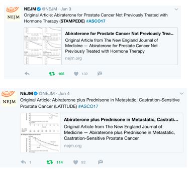
This also included the New England Journal of Medicine immediately tweeting after the presentations that LATITUDE and STAMPEDE were published instantaneously:

1-8

Furthermore, immediately following Dr. Fizazi’s presentation of LATITUDE, Dr. Eric Small from @UCSF presented a discussion of LATITUDE. A number of important points were raised. First, although this was a well-designed, placebo controlled, randomized phase III study, early unblinding (although appropriate) resulting in an HR of 0.62 for OS is based on only 50% of the targeted total deaths. Making conclusions based on interim analyses must be made with caution. However, with every endpoint reaching statistical significance and conditional probability modeling, if the study had remained blinded, the probability of reaching the same conclusions is high. Second, since twice as many patients in the ADT + placebo arm received life-prolonging therapy than compared to the ADT + AA + placebo arm, the benefit of AA is not explained by more secondary life-prolonging therapy, strengthening the cause for AA + ADT.

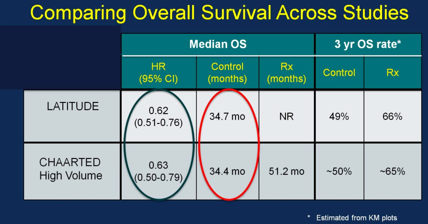
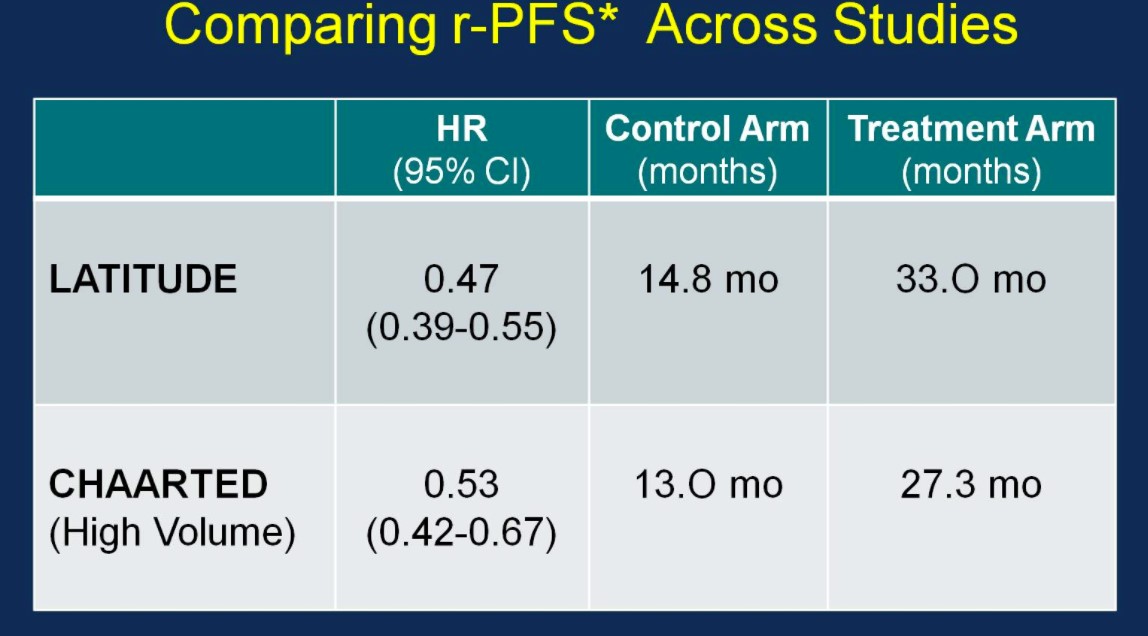
Perhaps the most interesting and pertinent clinical comparison is assessing outcomes of the LATITUDE and CHAARTED (high-volume disease) treatment arms (AA vs docetaxel). With similar median OS outcomes between the ADT control arms of the two trials (suggesting similar populations), the HRs for OS based on treatment are nearly identical:

1-9

Similarly, the rPFS outcomes were comparable between the two trials:

1-10

With nearly identical OS and rPFS outcomes for men receiving ADT + AA or ADT + docetaxel, the question becomes whether the impact of adding AA to ADT is volume or risk dependent. Results from the STAMPEDE trial would suggest remarkably similar outcomes support the use of AA + ADT in patients with less burden of disease. Arguably the most important slide of the meeting was captured and tweeting by Dr. Agarwal (@neerajaiims):

1-11

Dr. Small eloquently summarized future directions into two groups. Unanswered questions regarding efficacy include: (i) Can a genomic classifier be used to select patients more likely to benefit from AA or docetaxel? (ii) Can AA be added in even earlier settings (with radiation? Increasing PSAs?) (iii) Should AA and docetaxel be combined or used sequentially? Additionally, there are also unanswered questions regarding AA resistance, including (i) Will the mechanisms of resistance to AA be the same when used in the non-mCRPC setting? (ii) Will androgen receptor amplification still be observed? (iii) Will there be an increased risk of treatment-associated small cell/neuroendocrine prostate cancer? (iv) Does adding chemotherapy or AA to ADT result in more aggressive disease at the time of resistance? (v) What is the optimal therapy for a patient who progresses on ADT + AA, compared to a patient who progresses on ADT + docetaxel? Given the avoidance of potential chemotherapy related side effects (ie. neutropenic complications) for an oral, long-term treatment, AA + ADT should be considered standard of care for untreated, high-risk metastatic prostate cancer.

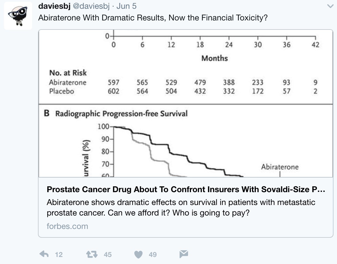
But what is the long-term economic landscape like when practice changing trials such as LATITUE and STAMPEDE suddenly thrust an expensive medication such as AA + prednisone directly to the forefront of hormone-naïve disease? Following these presentations, urologic oncologist, Twitter veteran, and Forbes correspondent Dr. Ben Davies (@daviesbj) wrote a provocative piece highlighting the potential ‘financial toxicity’ (particularly in the United States) that may result downstream of these trials:

1-12

A conservative estimate is a wholesale cost of $115,000 per year per patient for AA + prednisone, resulting in a crude estimate of a $2.8 billion annual expenditure for the drug in the United States alone if used in the hormone-naïve setting, according to Dr. Davies. As Dr. Davies also points outs, although the patent for AA expired in 2016 and there are currently 13 applications to make generic AA, the patent for prednisone lasts until 2027, with $30 billion riding on the lawsuit. Dr. David Penson (@urogeek) succinctly summarized via Twitter:

1-13

Strictly academically speaking, LATITUDE and STAMPEDE, in addition to the docetaxel benefits of CHAARTED, have provided clinicians with exciting Level 1 evidence for improving patient care in the high-risk/metastatic setting. The investigators and more importantly the thousands of patients and families are to be thanked and congratulated for their perseverance, hard-work, and willingness to participate in these practice-changing clinical trials. It is our job as clinicians to continue advocating the best treatment for our patients, whether this be through economic barriers in the United States, or access to appropriate care on a global scale.


Zach Klaassen, MD

Urologic Oncology Fellow

University of Toronto/Princess Margaret Cancer Centre

Toronto, Ontario, Canada

@zklaassen_md


TRoMbone is launched!

TRoMbone is launched! The UK feasibility randomised trial Testing radical prostatectomy in men with prostate cancer and oligoMetastases to the bone has opened at Oxford University Hospitals (PI Freddie Hamdy), University College London Hospitals (PI John Kelly) and Royal Surrey County Hospital (PI Chris Eden). Men <75 with newly-diagnosed prostate cancer and 1-3 skeletal lesions on any standard-of-care imaging (CT, bone scan, MRI, or PET) who are fit for surgery and deemed to be technically operable are eligible. Emerging but lower-quality data suggests a role for treatment of the primary tumour in men with oligo-metastatic prostate cancer and this UK study will investigate this question with level 1 evidence.

Participants will be randomly allocated to standard-of-care treatment (hormones +/- chemotherapy) versus standard-of-care treatment plus surgery to remove the prostate and draining lymph nodes (radical prostatectomy plus extended pelvic lymphadenectomy). A qualitative recruitment investigation to optimise accrual will be conducted by the University of Bristol (Caroline Wilson) and biologic samples will be collected, processed and stored in a repository at the Institute of Cancer Research (Gerhardt Attard).

We will assess technical feasibility, safety and complications of surgery in oligo-metastatic prostate cancer, and examine ways to improve recruitment in this pilot study. TRoMbone is managed by the Surgical Intervention Trials Unit at the University of Oxford and funded by the Prostate Cancer Foundation and The Urology Foundation.

We need to recruit 50 men over a 12-month period, and are seeking referrals from other centres to increase accrual. Centres that demonstrate ability to refer eligible patients will be able to take part in the main trial if we can demonstrate feasibility in this phase and get funding for the larger study.

So please look out for these patients and send them to me at UCLH, Freddie in Oxford, or Chris in Guildford. One of the three of us will do the surgery if they get randomised to it, but of course you’re welcome to come with the patients. If you have any queries please contact me, the study CI (P. Sooriakumaran (PS); [email protected]) or the study co-ordinator (Neelam Hassanali; [email protected]). You can start them on androgen deprivation and ‘stop the clock’ before you refer them to us. The extra burden of participating in this study is minimal. They will require one visit for consent, and one follow-up visit at 3 months after randomisation. The surgical group will also have two other visits for their surgery and catheter removal. The rest of the follow-up can be done back at your referring centre or with us, whatever you and the patient prefer. If it’s your standard policy to give them chemotherapy or metastasis-directed therapy with SBRT then you can still do that as part of the study.


With your help we can demonstrate that this study is feasible in the UK and we can lead the way in the surgical management of oligo-metastatic prostate cancer.


P. Sooriakumaran [social type=”facebook” opacity=”dark’ label=’PLACE_LINK_HERE[/social]

BMedSci(Hons) BMBS(Hons) PhD PGCMedLaw ADCClinInv FRCS(Urol) FEBU USLME

Consultant Urological Surgeon, UCL Hospitals NHS Foundation Trust & Honorary Clinical Senior Researcher, University of Oxford


Sailing into “UnCHAARTED” waters

Chemotherapy comes alive for prostate cancer!

staff-chowdhury1Systemic therapy for metastatic prostate cancer has radically changed in the last 10 years with the introduction of several novel agents that have shown significant improvements in progression free and overall survival. These have all been studied in metastatic castrate refractory prostate cancer (mCRPC) and have improved overall survival but in each case by less than 6 months. (The latest major breakthrough is the introduction of a relatively old drug, docetaxel chemotherapy, earlier in the disease for hormone sensitive patients).

In this week’s New England Journal of Medicine we see the eagerly awaited results from the CHAARTED study from Christopher Sweeney and colleagues. This novel study aimed to improve treatment for men with newly diagnosed hormone sensitive metastatic prostate cancer by adding docetaxel chemotherapy to androgen deprivation therapy (ADT).

790 men with newly diagnosed metastatic prostate cancer were randomised to ADT plus docetaxel (6 cycles at 75mg/m2) or ADT alone. The addition of docetaxel to ADT was shown to significantly improve overall survival by 13.6 months (57.6 months vs. 44.0 months; p<0.001). The clinical benefit was greatest in the subgroup with high volume disease where the improvement in overall survival was 17 months (49.2 months versus 32.2 months). High volume disease was defined as the presence of visceral metastases and/or 4 or more bone metastases with at least one beyond the vertebral bodies or pelvis. The combination was well tolerated with approximately 6% of patients having neutropenic fever and one death possibly related to docetaxel.

The results from this study are truly practice changing. Supporting evidence from the UK STAMPEDE study (Systemic Therapy in Advancing or Metastatic Prostate Cancer: Evaluation of Drug Efficacy) was presented at this year’s American Society of Clinical Oncology (ASCO) meeting. STAMPEDE showed that for men with metastatic hormone sensitive prostate cancer 6 cycles of docetaxel in addition to ADT improved median overall survival by 22 months (43 versus 65 months).

Chemotherapy for metastatic prostate cancer has had a checkered past with a lack of enthusiasm and nihilism from clinicians and patients. The results from CHAARTED and STAMPEDE are already changing those views. The prostate cancer community needs to react to these results and look to make this treatment available to all suitable men. There are issues with regards to costs of chemotherapy (although docetaxel is now generic), workload, sequence, patient selection, toxicity management, etc. The CHAARTED and STAMPEDE investigators must also use this opportunity to interrogate the tumour samples from these studies to see if they can identify biomarkers that predict docetaxel activity. We will not get this opportunity again as docetaxel + ADT will be be standard of care for future studies.

The clinical benefit from the addition of docetaxel to ADT is one of the largest seen in any oncology study. All men presenting with newly diagnosed metastatic prostate cancer should be considered for 6 cycles of docetaxel in addition to ADT.


Simon Chowdhury is a Consultant Medical Oncologist at Guy’s, King’s and St Thomas’ Hospitals, London. He is actively involved in clinical trial research into urological cancers.


Article of the Week: Identifying predictors of renal function decline after surgery

Every week the Editor-in-Chief selects the Article of the Week from the current issue of BJUI. The abstract is reproduced below and you can click on the button to read the full article, which is freely available to all readers for at least 30 days from the time of this post.

In addition to the article itself, there is an accompanying editorial written by a prominent member of the urological community. This blog is intended to provoke comment and discussion and we invite you to use the comment tools at the bottom of each post to join the conversation.

If you only have time to read one article this week, it should be this one.

Preoperative predictors of renal function decline after radical nephroureterectomy for upper tract urothelial carcinoma

Matthew Kaag, Landon Trost*, R. Houston Thompson*, Ricardo Favaretto†, Vanessa Elliott, Shahrokh F. Shariat‡, Alexandra Maschino†, Emily Vertosick†, Jay D. Raman and Guido Dalbagni†

Penn State Hershey Medical Center, Hershey, PA, *Mayo Clinic, Rochester, MN, †Memorial Sloan-Kettering Cancer Center, New York, NY, USA, and ‡Medical University of Vienna, Vienna, Austria

Read the full article
OBJECTIVES

To model renal function after radical nephroureterectomy (RNU) for upper tract urothelial carcinoma (UTUC). To identify predictors of renal function decline after surgery, thereby allowing the identification of patients likely to be ineligible for cisplatin-based chemotherapy in the adjuvant setting.

PATIENTS AND METHODS

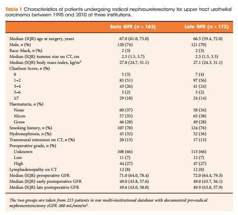
We retrospectively identified 374 patients treated with RNU for UTUC at three centres between 1995 and 2010. Estimated glomerular filtration rate (eGFR) was calculated using Chronic Kidney Disease Epidemiology Collaboration equation before RNU and at early (1–5 months after RNU) and late (>5 months) time points after RNU. Only patients deemed eligible for cisplatin-based chemotherapy before RNU (preoperative glomerular filtration rate [GFR] ≥60 mL/min/1.73 m2) were included. Multivariable analysis identified the preoperative predictors of eGFR after RNU at early postoperative and late postoperative time points.

RESULTS
A total of 163 patients had an eligible early post-RNU eGFR measurement and 172 had an eligible late eGFR measurement. The median eGFR declined by 32% and did not show a significant trend toward recovery over time (P = 0.4). On multivariable analysis preoperative eGFR and patient age were significantly associated with early and late postoperative eGFR, while Charlson comorbidity index score was significantly associated with late postoperative eGFR alone.
 

CONCLUSIONS
In patients with normal preoperative eGFR (≥60 mL/min/1.73 m2), renal function decreases by one-third after RNU and does not show evidence of recovery over time. Elderly patients and those with pre-RNU eGFR closer to 60 mL/min/1.73 m2 (lower eGFR in the present cohort) are more likely to be ineligible for adjuvant cisplatin-based chemotherapy regimens because of renal function loss after RNU.


Read more articles of the week


Editorial: ‘Discontent is the first necessity of progress’, Thomas A. Edison

This study from Kaag et al. [1] investigates predictors of renal functional decline after radical nephroureterectomy (RNU) in patients with upper tract urothelial carcinoma (UTUC). They evaluate early (2 months) and late (6 months) predictors of renal functional decline, finding that on a multivariable model only age at surgery and preoperative renal function were independently associated with early postoperative function. This is an intuitive finding whereby we expect older patients and those with lower renal function to have a more dramatic decrease in renal function after RNU.

Age, preoperative renal function, and Charlson score were associated with late functional recovery. The latter is a counterintuitive finding, as higher Charlson score was associated with less decrease in renal function. Charlson comorbidity was not significant on univariate analyses. Why it would become significant on multivariate is unclear. Whether it is an artifact related to study methodology or is a real phenomenon will require further study.

Unquestionably, this study [1] adds to the growing discontent of our current management of UTUC. The authors cogently discuss the issues related to better risk stratification as a natural consequence of instituting a neoadjuvant chemotherapy paradigm in those with high-risk disease. Multiple retrospective studies have failed to show a benefit of adjuvant chemotherapy, whereas now we have a matched-cohort study showing significant rates of downstaging and complete remission [2], and as well significantly improved 5-year survival, with institution of a neoadjuvant paradigm [3]. One cannot view the dismal outcomes of this disease without being discontent and wishing for progress. We need to continue getting out the message to not only urologists who reflexively institute RNU in patients with a risk-unstratified upper tract filling defect, but as well many medical oncologists who can only function based on guidance from level I data, which for this disease, will be a long time coming.

Read the full article

Surena F. Matin

Department of Urology, MD Anderson Cancer Center, Houston, TX, USA

References

1 Kaag M, Trost L, Thompson RH et al. Pre-operative predictors of renal function decline following radical nephroureterectomy for upper tract urothelial carcinoma. BJU Int 2014; 114: 674–9

2 Matin SF, Margulis V, Kamat A et al. Incidence of downstaging and complete remission after neoadjuvant chemotherapy for high-risk upper tract transitional cell carcinoma. Cancer 2010; 116: 3127–34

3 Porten S, Siefker-Radtke AO, Xiao L et al. Neoadjuvant chemotherapy improves survival of patients with upper tract urothelial carcinoma. Cancer 2014; 120: 1794–9

Article of the week: Modulating autophagy of prostate cancer cells with anti-androgen bicalutamide

Every week the Editor-in-Chief selects the Article of the Week from the current issue of BJUI. The abstract is reproduced below and you can click on the button to read the full article, which is freely available to all readers for at least 30 days from the time of this post.

In addition to the article itself, there is an accompanying editorial written by a prominent member of the urological community. This blog is intended to provoke comment and discussion and we invite you to use the comment tools at the bottom of each post to join the conversation.

Finally, the third post under the Article of the Week heading on the homepage will consist of additional material or media. This week we feature a video of Prof Leung and Dr Stockley discussing their paper.

If you only have time to read one article this week, it should be this one.

Does androgen-ablation therapy (AAT) associated autophagy have a pro-survival effect in LNCaP human prostate cancer cells? 


Haley L. Bennett, Jacqueline Stockley, Janis T. Fleming, Ranadip Mandal, Jim O’Prey*, Kevin M. Ryan*, Craig N. Robson and Hing Y. Leung

Urology Research Laboratory and *Tumour Cell Death Research Laboratory, Beatson Institute for Cancer Research, Glasgow, and Solid Tumour Target Discovery Group, Northern Institute for Cancer Research, Newcastle University, Newcastle upon Tyne, UK

Read the full article


OBJECTIVE

• To study the cellular effects of the anti-androgen bicalutamide on autophagy and its potential impact on response to androgen-ablation therapy (AAT) alone or combined with docetaxel chemotherapy in human prostate cancer LNCaP cells.

MATERIALS AND METHODS

• LNCaP cells were treated with bicalutamide docetaxel, and cellular effects were assayed: lipidated LC3 (a microtubule-associated protein) for autophagy and its traffcking to fuse with lysosome; flow cytometry using propidium iodide or caspase 3 for cell death; and sulforhodamine B assay for cell growth.

RESULTS

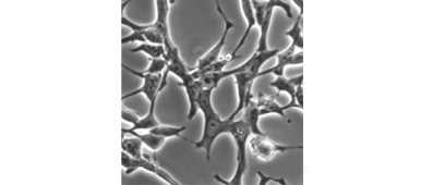
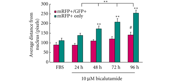
• Bicalutamide treatment enhanced autophagy in LNCaP cells with increased level of autophagosome coupled with an altered cellular morphology reminiscent of neuroendocrine differentiation.

• Consistent with the literature on the interaction between androgen receptor activation and taxane chemotherapy, bicalutamide diminished docetaxel mediated cytotoxicity.

• Significantly, pharmacological inhibition of autophagy with 3-methyladenine significantly enhanced the efficacy cell kill mediated by AAT docetaxel.

CONCLUSION

• Autophagy associated with bicalutamide treatment in LNCaP cells may have a pro-survival effect and strategy to modulate autophagy may have a potential therapeutic value


Read Previous Articles of the Week

Editorial: Targeting the pro-survival side-effects of androgen-deprivation therapy in prostate cancer

In this paper, Bennett et al. [1] report the effects of an anti-androgen drug on autophagy and the subsequent impact on response to androgen-deprivation therapy alone or combined with exiting chemotherapeutic treatments.

With an estimated 238,590 newly diagnosed cases and 29,720 deaths for 2013 in the USA, prostate cancer is, after skin cancer, the second most common cancer in men. Although the disease initially responds well to therapy, the cancer recurs in most patients within 1–2 years of the initial response. Few therapeutic options exist for patients with recurring prostate cancer and docetaxel is considered the standard of care. But despite clinical benefits, its effect is mainly palliative and often short-lived, and all patients eventually develop progressive disease with a median survival of 1–12 months. In addition, the decision of when to initiate docetaxel-based chemotherapy is an important one that is not clearly addressed by current treatment guidelines.

Autophagy is a lysosomal catabolic pathway that promotes cell survival in response to starvation or other cellular stresses by degrading and recycling macromolecules and organelles. In recent years, this cellular process has been implicated in the aetiology of cancer; the roles it plays, however, in the development and maintenance of cancer appear conflicting [2–6]. Indeed, tumour cells appear to disable autophagy at an early stage, thereby facilitating the onset of tumorigenesis, whereas in existing malignancies autophagy is activated as a means of stress adaptation, resulting in metastatic dissemination [7–9]. Autophagy is also induced by nearly every anti-cancer treatment as an adaptive pro-survival mechanism against cytotoxic agents and may therefore favour radio- and chemo-resistance [10–12].

Bennett et al. [1] are the first to show the induction of autophagy due to suppression of androgen function in the absence of other cellular stresses in an androgen-sensitive cell line. The authors showed that anti-androgen treatment induced autophagy in LNCaP prostate cancer cells, resulting in a pro-survival effect that was abolished by pharmacological inhibition of autophagy, a response that is similar to that seen in tamoxifen-resistant breast cancer cells. Their study highlights the potential of combining anti-androgen therapy with autophagy inhibition in the treatment of prostate cancer. The mechanism by which anti-androgen therapy activates autophagy is unclear, but this study suggests that modulation of mammalian target of rapamycin (mTOR) signalling, a major cellular metabolism switch, may underlie this effect. Thus, agents that inhibit the pathway combined with inducers of metabolic stress or chemotherapeutic agents could enhance anti-cancer therapy by inhibiting stress adaptation and increasing cell damage. The search for novel inhibitors of the pathway is crucial in the fight against cancer.

Clearly, to this day, there are no simple rules for the outcome of targeting autophagy as a cancer therapy. The apparent conflicting effects of activating or inhibiting autophagy at various stages of the disease are likely to be dictated by the genetic background as well as the environmental cues tumour cells are exposed to. One of the main challenges in prostate cancer therapy is to determine the precise timing of drug application. Therefore, the identification of a ‘fingerprint’, including the aforementioned parameters, in prostate cancer is crucial for the selection of an effective treatment. The present study opens up potential new avenues in the treatment of prostate cancer but further in vitro and in vivo studies will be necessary for efficiently translating this knowledge into the clinic.

Vincent Zecchini and David E. Neal
Department of Uro-Oncology, University of Cambridge, Cambridge, UK

Read the full article

REFERENCES

  1. Bennett HL, Stockley J, Fleming JT et al. Does androgen-ablation therapy (AAT) associated autophagy have a pro-survival effect in LNCaP human prostate cancer cells? BJU Int 2013; 111: 672–82
  2. Oh SH, Lim SC. Endoplasmic reticulum stress-mediated autophagy/apoptosis induced by capsaicin (8-methyl-N-vanillyl-6-nonenamide) and dihydrocapsaicin is regulated by the extent of c-Jun NH2-terminal kinase/extracellular signal-regulated kinase activation in WI38 lung epithelial fibroblast cells. J Pharmacol Exp Ther 2009; 329: 112–22
  3. Qu X, Yu J, Bhagat G, Furuya N et al. Promotion of tumorigenesis by heterozygous disruption of the beclin 1 autophagy gene. J Clin Invest 2003; 112: 1809–20
  4. White E, DiPaola RS. The double-edged sword of autophagy modulation in cancer. Clin Cancer Res 2009; 15: 5308–16
  5. Yue Z, Jin S, Yang C, Levine AJ, Heintz N. Beclin 1, an autophagy gene essential for early embryonic development, is a haploinsufficient tumor suppressor. Proc Natl Acad Sci USA 2003; 100: 15077–82
  6. Liang XH, Jackson S, Seaman M et al. Induction of autophagy and inhibition of tumorigenesis by beclin 1. Nature 1999; 402: 672–6
  7. Chiarugi P, Giannoni E. Anoikis: a necessary death program for anchorage-dependent cells. Biochem Pharmacol 2008; 76: 1352–64
  8. Douma S, Van Laar T, Zevenhoven J et al. Suppression of anoikis and induction of metastasis by the neurotrophic receptor TrkB. Nature 2004; 430: 1034–9
  9. Yap KL, Zhou MM. Keeping it in the family: diverse histone recognition by conserved structural folds. Crit Rev Biochem Mol Biol 2010; 45: 488–505
  10. Chen S, Rehman SK, Zhang W et al. Autophagy is a therapeutic target in anticancer drug resistance. Biochim Biophys Acta 2010; 1806: 220–9
  11. Liu L, Yang M, Kang R et al. HMGB1-induced autophagy promotes chemotherapy resistance in leukemia cells. Leukemia 2011; 25: 23–31
  12. Lomonaco SL, Finniss S, Xiang C et al. The induction of autophagy by gamma-radiation contributes to the radioresistance of glioma stem cells. Int J Cancer 2009; 125: 717–22

Video: Androgen-ablation therapy (AAT) associated autophagy


Does androgen-ablation therapy (AAT) associated autophagy have a pro-survival effect in LNCaP human prostate cancer cells?

Haley L. Bennett, Jacqueline Stockley, Janis T. Fleming, Ranadip Mandal, Jim O’Prey*, Kevin M. Ryan*, Craig N. Robson and Hing Y. Leung

Urology Research Laboratory and *Tumour Cell Death Research Laboratory, Beatson Institute for Cancer Research, Glasgow, and Solid Tumour Target Discovery Group, Northern Institute for Cancer Research, Newcastle University, Newcastle upon Tyne, UK

Read the full article
OBJECTIVE

• To study the cellular effects of the anti-androgen bicalutamide on autophagy and its potential impact on response to androgen-ablation therapy (AAT) alone or combined with docetaxel chemotherapy in human prostate cancer LNCaP cells.

MATERIALS AND METHODS

• LNCaP cells were treated with bicalutamide  docetaxel, and cellular effects were assayed: lipidated LC3 (a microtubule-associated protein) for autophagy and its trafficking to fuse with lysosome; flow cytometry using propidium iodide or caspase 3 for cell death; and sulforhodamine B assay for cell growth.

RESULTS

• Bicalutamide treatment enhanced autophagy in LNCaP cells with increased level of autophagosome coupled with an altered cellular morphology reminiscent of neuroendocrine differentiation.

• Consistent with the literature on the interaction between androgen receptor activation and taxane chemotherapy, bicalutamide diminished docetaxel mediated cytotoxicity.

• Significantly, pharmacological inhibition of autophagy with 3-methyladenine significantly enhanced the efficacy cell kill mediated by AAT  docetaxel.

CONCLUSION

• Autophagy associated with bicalutamide treatment in LNCaP cells may have a pro-survival effect and strategy to modulate autophagy may have a potential therapeutic
value.

A Case of Metastatic Penile Cancer Showing a Prolonged Disease Free Survival of over Five Years After Cisplatin and Gemcitabine Combination Chemotherapy

This is the first reported case of prolonged survival of metastatic disease in penile cancer in a patient treated using this chemotherapy regime.

Authors: Nowicki, Stefan; Hendry, David; Russell, John

Corresponding Author: Stefan Nowicki, Beatson West of Scotland Cancer Centre, Glasgow, UK. Email: [email protected]


Introduction
 
Penile cancer is a rare cancer in the Western world with an incidence of 0.1-0.9/100 000 men in Europe. In the developing world however the incidence can be much higher accounting for up to 10% of male malignancy. The peak incidence occurs in the sixth and seventh decades of life and is associated with numerous risk factors, the strongest being phimosis, chronic inflammatory conditions, condylomata, sexual history and smoking. HPV infection has been shown to be a possible causative factor with viral DNA being present in up to 80% of tumour specimens; the majority by HPV-16 (1). This is a major cause of other genital malignancies and forms part of the vaccine to prevent cervical cancers in females. For this reason, it has been proposed as a potential preventative measure for penile cancer in the developing world (2).
Treatment of penile cancer can include surgery, chemotherapy or radiotherapy. The primary tumour and lymph nodes are treated separately. Early superficial tumours can be treated with wide local excision or radiotherapy, but partial or complete amputation is required for more advanced disease. Neoadjuvant chemotherapy is also used to downstage initially inoperable cancers. Clinically negative lymph nodes can be involved with metastatic cancer in 15% of cases.  In this situation, dynamic sentinel node biopsy has been shown to improve survival and reduce morbidity (3). Pathologically confirmed disease requires inguinal lymphadenectomy, with pelvic lymphadenectomy in N2 disease or extracapsular spread. In high risk cases adjuvant chemotherapy has also been advocated with cisplatin and 5-fluorouracil (4).
The use of chemotherapy is limited to locally advanced or metastatic disease, with cisplatin based treatments the most commonly used. Response rates are typically 30% at best but, due to the rarity of this disease, are based on small phase II studies and case series. We present a case of metastatic penile cancer which has been successfully treated with cisplatin and gemcitabine. This is the first reported case of prolonged survival of metastatic disease in penile cancer in a patient treated using this chemotherapy regime.


Case report


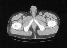
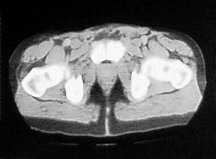
A 48 year old male presented with a large necrotic fungating mass replacing the glans penis and causing phimosis, currently looking for phimosis cure. No palpable lymph nodes or evidence of metastatic disease was detected clinically. Dynamic sentinel lymph node biopsy was not available, and no additional imaging was undertaken in accordance with local practice at the time. A partial penile amputation was undertaken and pathology showed a moderately differentiated squamous cell carcinoma. There was a margin of 26mm from the proximal surgical resection site but an area of lymphovascular invasion was identified. The patient recovered well from the operation but developed an enlarging left groin mass one month later. A CT scan showed a soft tissue density in the left groin (figure 1), in addition to several small bilateral soft tissue densities.


Figure 1. A CT scan showed a soft tissue density in the left groin

penile cancer 1


A fine needle aspirate of the lump confirmed squamous cell carcinoma and a bilateral groin dissection was undertaken. The left groin dissection revealed two out of thirteen positive lymph nodes, and the right groin dissection showed one out of ten positive lymph nodes with no evidence of extracapsular spread. He developed postoperative cellulitis and abscess formation which was successfully drained and treated with antibiotics. A CT scan of his chest showed no evidence of pulmonary metastasis and a bone scan was normal. He did not receive any adjuvant treatment and was to be followed up in the clinic every three months with a repeat CT scan every six months.
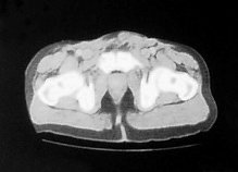
Four months later he developed suprapubic oedema and two small hard nodules superior to the shaft of his penis measuring 1cm in diameter each. These lesions increased rapidly in size over a period of two months with the further development of new lesions in the same area (figure 2).
Figure 2. CT scan:lesions increased rapidly in size over a period of two months

penile cancer 2


A fine needle aspirate of one of the lesions showed degenerate malignant squamous cells consistent with recurrence of his penile cancer. A CT scan of his chest, abdomen and pelvis showed no evidence of further disease. The recurrence was not amenable to surgical intervention so he was commenced on palliative chemotherapy.
The patient was started on a four weekly regime of Cisplatin (70mg/m2, D1) and Gemcitabine (1g/m2, D1,8,15) chemotherapy. He developed a complete clinical response to treatment after one cycle (figure 3).

Figure 3. The patient developed a complete clinical response to treatment after one cycle

penile cancer 3


He completed 5 cycles of chemotherapy with grade one mouth ulcers after the first cycle and a week delay at day 1 of the fifth cycle due to neutropenia. After his chemotherapy he developed erectile dysfunction. He continued to be followed up at three monthly intervals with six monthly CT scans.
Two years later the patient noticed a new hard lump at the base of his penis. This grew over a three week period reaching a size of 1cm2. A CT and bone scan were normal. The lesion was subsequently removed with a wide local excision, and histological examination confirmed moderately differentiated squamous carcinoma consistent with spread from the primary penile lesion. Resection margins were negative and there has been no further recurrence over the last five years. Follow up involved clinical examination every three months, and CT scan every six months, for the first two years. This was followed by six monthly clinical examination and yearly CT scans.

Discussion

Chemotherapy has been used with limited success in locally advanced and metastatic penile cancer. Its use has been based on small non-randomised trials, making comparisons between regimes difficult. The most commonly used chemotherapy doublet is cisplatin/5-fluorouracil which has a 20-30% response rate (5). Responses have also been seen with cisplatin/irinitican (6) and carboplatin/paclitaxol (7) in small case series. The largest response rates have been seen with triple-drug regimes but these are associated with marked toxicity. Cisplatin/methotrexate/bleomycin is the most commonly studied with response rates of 32.5% but with a mortality rate of 12.5%, mostly related to pulmonary complications (8). Its use is not advocated for this reason. Preliminary data from a neoadjuvant trial using paclitaxel/ifosfamide/ cisplatin showed encouraging response rates of 50% with less toxicity (9).
There is little evidence of cisplatin/gemcitabine use in penile cancer in the literature. It is however a common first line regime for the treatment of metastatic squamous cell carcinoma of the lung (10). In addition, squamous cell carcinoma at other sites have been shown to respond. These include carcinomas of the head and neck (11) and of the oesophagus (12). Cisplatin and gemcitabine are also used in other urological tumour types, and is the predominant regime in bladder cancer in both the neoadjuvant and palliative setting (13).
In relation to cisplatin/gemcitabine, there is a case series of two patients with metastatic penile cancer showing partial responses lasting over eleven months with a three weekly regime (14). The treatment, as in this case, was well tolerated with minimal side effects. The burden of metastatic disease was greater however, with both patients having pulmonary metastases as well as widespread lymphadenopathy. In addition, the pathology was more aggressive with both patients having poorly differentiated squamous cell carcinoma as opposed to moderately differentiated carcinoma in this case. This may explain the more prolonged response in our patient.
Our patient developed metastases within a few months post treatment. This is typical for most recurrences in this cancer, with the majority occurring within the first three years (15). Cases have been reported of recurrences occurring after ten years, so there continues to be the small possibility of further development of metastasis in this patient (16).
This case demonstrates a prolonged response to cisplatin/gemcitabine chemotherapy and salvage surgery, with a disease free period of over five years. It is a drug regime that is well established in other tumour types and has shown response in two other penile cancer cases. As the treatment options for metastatic penile cancer are limited, this regime should be studied further in the context of a clinical trial.

References

1. Daling JR, Madeleine MM, Johnson LG, Schwartz SM, Shera KA, Wurscher MA. Penile cancer: importance of circumcision, human papillomavirus and smoking in in situ and invasive disease. Int J Cancer 2005 116(4):606-16.
2. Guiliano AR. Human papillomavirus vaccination in males. Gynaecol Oncol 2007 107(suppl 1):S24-S26.
3. Lont AP, Horenblas S, Tanis PJ, Gallee MP, Van Tinteren H, Nieweg OE. Management of clinically node negative penile carcinoma: improved survival after the introduction of dynamic sentinel node biopsy. J Urol 2003 170(3):783-6.
4. Pizzocaro G, Piva L, Bandieramonte G, Tana S. Up-to-date management of carcinoma of the penis. Eur Urol 1997 32(1):5-15.
5. Shammas FV, Ous S, Fossa SD. Cisplatin and 5-fluorouracil in advanced cancer of the penis. J Urol 1992 147(3):630-632.
6. Theodore C, Skoneczna I, Bodrogi I, Leahy M, Kerst JM, Collette L, Ven K, Marreaud S, Oliver RD for the EORTC Genito-Urinary Tract Cancer Group: A phase II multicentre study of irinotecan in combination with cisplatin in metastatic or locally advanced penile cancer. Ann Oncol 2008 19(7):1304-1307.
7. Joerger M, Warzinek T, Klaeser B, Kluckert JT, Schmid HP, Gillessen S. Major tumour regression after paclitaxel and carboplatin polychemotherapy in a patient with advanced penile cancer. Urology 2004 63(4):778-780.
8. Haas GP, Blumenstein BA, Gagliano RG, Russell CA, Rivkin SE, Culkin DJ, et al. Cisplatin, methotrexate and bleomycin for the treatment of carcinoma of the penis: a Southwest Oncology Group study. J Urol 1999 161(6):1823-1825.
9. Pagliaro LC, Williams DL, Daliani D, Williams MB, Osai W, Kincaid M et al. Neoadjuvant paclitaxel, ifosfamide, and cisplatin chemotherapy for metastatic penile cancer: a phase II study. J Clin Oncol 2010 28(24):3851-7.
10. Smit EF, van Meerbeeck JP, Lianes P, Debruyne C, Legrand C, Schramel F et al. Three-arm randomized study of two cisplatin-based regimens and paclitaxel plus gemcitabine in advanced non-small-cell lung cancer: a phase III trial of the European Organization for Research and Treatment of Cancer Lung Cancer Group–EORTC 08975. J Clin Oncol 2003 21(21):3909-17.
11. Hitt R, Castellano D, Hidalgo M, García-Carbonero R, Peña M, Brandariz A, Millán JM, Alvarez Vincent JJ, Cortés-Funes. Phase II trial of cisplatin and gemcitabine in advanced squamous-cell carcinoma of the head and neck. Ann Oncol 1998 12:1347-9.
12. Millar J, Scullin P, Morrison A, McClory B, Wall L, Cameron D, Philips H, Price A, Dunlop D, Eatock M. Phase II study of gemcitabine and cisplatin in locally advanced/metastatic oesophageal cancer. Br J Cancer 2005 93(10):1112-6.
13. Von der Masse H, Hansen SW, Roberts JT, Dogliotti L, Oliver T, Moore MJ et al. Gemcitabine and cisplatin versus methotrexate, vinblastine, doxorubicin and cisplatin in advanced or metastatic bladder cancer: results of a large randomised multinational multicentre phase III study. J Clin Oncol 18(17):3068-77.
14. Power DG, Galvin DJ, Cuffe S, McVey GP, Mulholland PJ, Farrelly C et al. Cisplatin and gemcitabine in the management of metastatic penile cancer. Urol Oncol 2009 27(2):187-90.
15. Leijte JA, Kirrander P, Antonini N, Windahl T, Horenblas S. Recurrence patterns of squamous cell carcinoma of the penis: recommendations for follow-up based on a two-centre analysis of 700 patients. Eur Urol. 2008 54(1):161-8.
16. Lichtenauer P, Scheer H, Louton T. On the classification of penis carcinoma and its 10-year survival. Recent Results Cancer Res. 1977 (60):110-9


Date added to bjui.org: 01/07/2012

DOI: 10.1002/BJUIw-2011-124-web


Micropapillary Urothelial Carcinoma (MUC) of the Upper Urinary Tract: An Underreported but Aggressive Variant of Urothelial Carcinoma

We report 3 patients with MUC of the upper urinary tract who were evaluated and treated at our center between 2009 and 2011.

Authors: Xiao-hua Zhang1,Zhou-jun Shen1,Xiu-ling Wu2,Zhixian Yu3

  1. Department of urology, Ruijin Hospital, Shanghai Jiaotong University School of Medicine, Shanghai, 200025, China
  2. Department of pathology, The First Affiliated Hospital of Wenzhou Medical College, Wenzhou, Zhejiang, 325000, China
  3. Department of urology, The First Affiliated Hospital of Wenzhou Medical College, Wenzhou, Zhejiang, 325000, China

Corresponding Author: Zhixian  Yu,   Department of urology, The First Affiliated Hospital of Wenzhou Medical College, Wenzhou, Zhejiang, 325000, China. E-mail: [email protected]

Abstract 

We report 3 patients with MUC of the upper urinary tract who were evaluated and treated at our center between 2009 and 2011. A comprehensive Pubmed search of all case reports and literature related to MUC of the upper urinary tract was performed. In all cases, the tumors were located in the renal pelvis. All 3 patients were treated surgically. After a median follow-up of 12 months, median overall survival was 12 months. One patient presented with stage II, one with stage III and one with stage IV disease. The patient with stage II  disease is alive, with limited follow up showing no evidence of disease. The patient with stage III disease developed metastasis to the cervical lymph nodes 3 months later. Radiotherapy was performed, however she died 6 months after the initial diagnosis. The patient with stage IV disease, underwent systematic chemotherapy, but developed distant metastases (including port-site metastasis) and died 12 months after initial diagnosis. After a comprehensive search in PubMed/Medline, 50 cases have been reported. Thirty cases originated in the renal pelvis, and most patients died after surgery. MUC of the upper urinary tract often presents at an advanced stage with lymphovascular invasion and distant metastasis, and is probably an underreported variant of urothelial carcinoma associated with poor prognosis. The optimal treatment strategy is yet to be defined.

Introduction 

Micropapillary urothelial carcinoma (MUC) is an aggressive variant of urothelial carcinoma with high metastatic potential. Amin et al first reported MUC of the urinary bladder in 18 cases[1].This variant demonstrated a high tendency to invade lymphovascular spaces and to metastasize to lymph nodes and other organs. They also found MUC to be associated with advanced tumor stage and, therefore, a poor prognosis. Several other studies have confirmed these observations in MUC of the urinary bladder[2-4]. Although MUC has been well studied in the urinary bladder, there have been only a few studies of this tumor in the upper urinary tract, including the renal pelvis and ureter[5-13].The analysis of 3 consecutive cases of MUC of the upper urinary tract, may add to the current literature regarding this aggressive disease entity and may help with its future management.The literature with particular emphasis on the diagnostic and therapeutic approaches is also included and discussed.

Patients and methods

Clinical Case

After obtaining approval from the institutional review board, we retrospectively searched the files of all patients with urothelial carcinoma of the upper urinary tract (including the renal pelvis and ureter) treated at our center between 2009 and 2011. Patients were considered to have micropapillary disease if the pathology report revealed any micropapillary component (MC) in their tumor. Patient medical records were analyzed for demographic characteristics, clinical stage and outcome. Radiology, pathology and surgical reports were reviewed to determine the pathological staging at the time of nephro-ureterectomy using the 1997 TNM classification for genitourinary tumors [14]. Chemotherapy regimens, radiotherapy doses and surgical modality were also recorded. Overall survival was calculated from the date of diagnosis to the date of death or the date of last follow-up.

Search Strategy and Systematic Literature Review

We performed a comprehensive search in PubMed/Medline, up to September 2011, using the terms ‘micropapillary’, ‘renal pelvis’, ‘ureter’, ‘upper urinary tract’, ‘micropapillary urothelial carcinoma’. Articles were limited to the English language. The initial search was limited to articles from 1994 to 2011 due to micropapillary urothelial carcinoma being first reported in 1994. The inclusion criterion were original articles describing the micropapillary urothelial carcinoma of the upper urinary tract, including the renal pelvis and ureter. Exclusion criteria were articles published on the micropapilary urothelial carcinoma of urinary bladder. A total of 11 articles were included in the final analysis.

Results 

Clinical Results

Characteristics of patients at initial presentation

Between 2009 and 2011, 87 patients with urothelial carcinoma of the upper urinary tract were treated at our center. Three patients had micropapillary features. In all cases, the tumor was located in the renal pelvis. Median age at diagnosis was 67 years (59 to 75 years) and male/female ratio was 2/1. Two patients had previously smoked. The presenting symptom was macroscopic hematuria in two patients, and flank pain in one. Two of these patients had macrohematuria and were in a poor general condition, while one female patient had been treated with external beam radiotherapy for cervical lymph node metastases 5 years previously. All patients had grade 3 urothelial carcinoma and one had an associated in situ carcinoma. Lymphovascular invasion was present in all three cases (Fig.1-2.).

Figure 1. 

Figure 2. 

Metastases to regional lymph nodes were documented in two patients at the time of surgery, and to the ipsilateral adrenal gland in one patient. One patient presented with stage II, one with stage III and one with stage IV disease. The clinical characteristics of the patients are summarized in [Table 1].

Table 1. Summary of Clinical Characteristics

Case No. Age Sex Location Stage  Management Follow up
1 59 Male Renal pelvis T2 ONU* alive, no evidence ofdisease
2 67 Female Renal pelvis T3 LNU**+R^ 6 mo, died of metastasis tocervical lymph node
3 75 Male Renal pelvis T4 LNU+C# 12 mo,died of metastases tothe lung and liver.Port-stie metastasis was presented

*ONU,Open Nephro-Ureterectomy;**LNU,Laparoscopic Nephro-Ureterectomy; ^R,Radiotherapy; #C,Chemotherapy

Treatment and outcome 

One patient received open nephroureterectomy (ONU). He was alive, with limited (18months) follow up, and  showed no evidence of disease. One patient had only regional lymph node metastases, she underwent laparoscopic nephroureterectomy (LNU) and regional lymph node dissection. Her disease metastasised to her cervical lymph nodes 3 months later. Radiotherapy was performed, however she died 6 months later. One patient’s disease metastasised to the ipsilateral adrenal gland, he received LNU and excision of the affected adrenal gland. Although systemic chemotherapy was administered with gemcitabine 1000 mg/m 2 on days 1, 8 and 15,and cisplatin 70 mg/m2 on day 1 in a 28-day cycle, the disease still progressed and the patient died of metastases to his lungs and liver 12 months after surgery.

Metastatic sites

Metastases to regional lymph nodes were documented in 2 patients at the time of surgery; to the ipsilateral adrenal gland in one patient. Two out of the three patients developed distant metastases during follow up. The most frequent solid metastatic site was the lung followed by liver. One patient who underwent LNU developed port-site metastasis.

Survival data

At a median follow up of 12 months, 2 patients have died. One year survival was 66.7% and median survival was 12 months.

Systematic Review of Published Cases

After comprehensive search in PubMed/Medline, a total of 11 articles were included in the final analysis. We performed a bibliometric analysis finding a total of 50 cases of MUC of the upper urinary tract [Table 2]

Table 2. Clinical features of MUC of the upper urinary tract in literatures

Investigator Year No.cases Age*  M/F** Site(No.)# Stage(No.) Managment(No.) Follow up(No.)
Ribé 1996 1 68 1/0 P(1) T4(1) NU^+R^^+C^^^(1) 12mo,died of gastric metastases
Oh 2000 1 79 1/0 U(1) T4(1) NU+R(1) 20mo, died of gastric and peritoneal metastases
Vang 2000 1 79 0/1 U(1) T1(1)  laser ablation therapy(1) not available
Alvarado-Cabrero 2005 1 U(1) T3(1) not available
Holmäng 2006 26 69 17/9 P(19)U(6)UP(1) T1(3)T2(1)T3(18)

T4(4)

NU(14)UR^^^^(3)No¤(3)

NU+R(4)

NU+C(2)

died of tumor(20)died of other diseas(6)survived beyond 5 years(7)
Perez-Montiel 2006 5 76 3/2 P(2)U(1)UP(2) T2(1)T3(2)T4(2) NU(5) all died 3-24mo latermetastasize to regional lymph node(2),bone(1)
Munakata 2007 1 73 0/1 P(1) T4(1) NU+C(1) 14 mo,died of metastases to the left adrenal gland and the regional lymph nodes
Guo 2009 11 64.2 9/2 P(5)U(4)UP(2) T2(2)T3(8)T4(1) NU+C(11) died of tumor(4)surviving but developed metastasis(4)alvie,no evidence of disease(3)
Cheng 2010 3 64.3 2/1 P(2)U(1) T1(1) T3(1)T4(1) NU(1)NU+C(2) 15mo,died(1)alvie,no evidence of disease(2)

*Age–Mean age; **M/F–Male/Female; #Site;U–ureter,P–renal pelvis,UP–ureter and pelvis; ^NU,Nephro-Ureterectomy; ^^R,Radiotherapy; ^^^C,Chemotherapy; ^^^^UR,Ureteral Resection; ¤No,not received surgery

Discussion 

Carcinoma with a micropapillary component was first described in 1982 as a variant of endometrial carcinoma initially designated as papillary serous carcinoma of the uterus[15]. Later, this subtype was found in thyroid[16],breast[17], and lung cancers[18]. In 1994, Amin et al first reported a series of 18 cases of a micropapillary variant of transitional cell carcinoma (TCC) and suggested that this particular pattern could be associated with a more aggressive course of the tumor than usual TCC [1] .

The incidence of this disease is reported as approximately 2.8% [9]. Most cases originated from the renal pelvis; 33 patients, (including our 3 cases);15 cases arose from the ureter [6,7,8,11], and five cases were reported to have tumor both in the ureter and the renal pelvis [6,10,12]. The overall male to female ratio was 2:1(35:17). In a study by Alvarado-Cabrero, the patient’s gender and age could not be established due to incomplete clinical information.  Clinical staging information and follow up information were not available for this patient. Mean age at diagnosis is 72.07 (range  22 to 92 years). MUC of the upper urinary tract may have similar etiological factors to that of conventional UC of the upper urinary tract. It is noteworthy that two out of three patients reported by Cheng et al are both residents of the area affected by the black foot endemic in Taiwan[13]. This area has been reported to have a high prevalence of invasive upper urinary tract tumors[19-21]

There is no specific feature that allows definite clinical diagnosis of MUC of the upper urinary tract. No specific symptoms for MUC of the upper urinary tract have been reported. Painless gross hematuria was the most common symptom. Another reported symptom was flank pain. In our study, two patients presented with hematuria, one presented with flank pain. The diagnosis of MUC is based on the presence of a secondary micropapillary bud in the stroma beneath the urothelium in microscopic studies[1]. In addition to urothelial carcinoma, a micropapillary pattern has been reported in carcinomas of the ovary, breast, lung, colon, and major salivary glands[22–27 ]. In all of these organs, the micropapillary pattern shares a distinct morphology that is characteristic of small infiltrating clusters or nests of tumor cells within lacunae. Therefore, it is important to exclude metastases with micropapillary features when making a definite diagnosis of primary tumor. Immunohistochemical stains are useful in identifying the primary tumor. Ramalingam et al reported an invasive micropapillary carcinoma of the breast metastatic to the urinary bladder[28]. The micropapillary components were morphologically identical in the breast, urinary bladder, and endometrium; however, the tumor cells from the breast, endometrium, and urinary bladder were positive for cytokeratin 7 and estrogen receptor and negative for cytokeratin 20.

Therapeutic modalities depend upon a tumour’s clinical stage. They include surgery, chemotherapy and radiotherapy. Nephroureterectomy (NU) or partial ureteric resection was curative in patients with low stage disease and in a minority of those with high stage disease. In a study by Vang et al, the patient with stage I was treated by laser ablation therapy. This case appears to be less invasive because of earlier attention and intervention, but the follow up information was incomplete, and the patient’s clinical course remains to be further defined[7]. In our study, one patient with stage II disease who received NU was alive with no evidence of disease. Extensive lymph node dissection may been curative. In a study by Holmäng, one patient with multiple MC lymph node metastases who received extensive lymph node dissection survived 78 months without evidence of recurrence[9]. In our study, one patient who received regional lymph node dissection died 6 months later. According to the study of Munakat et al, radiotherapy did not influence the course of disease[11]. Oh et al reported on a 79-year-old male patient with a mid ureteric tumor treated by nephroureterectomy. The histopathological examination showed periureteric fat invasion and lymph node metastases with a predominant MC pattern in 18 of 20 lymph nodes examined. The patient had local recurrence after one year and was treated with radiotherapy but died 8 months later of metastatic disease[6]. External beam radiotherapy also did not seem to have been of benefit in our patients. Although cisplatin, cyclophosphamide, doxorubicin, gemcitabine, methotrexate and vinblastin have been reported in various combinations, nothing was effective[29-32]. The response of MUC patients to chemotherapy seems to be poor. Cheng et al prescribed chemotherapy for one patient with metastatic lesions. However, the patients died 15 months after the diagnosis[13]. Munakata et al reported one case whose pathological stage was pT4N2M1, and who underwent surgery. Although she received chemotherapy, she died of her disease 14 months postoperatively[11]. In our study, one patient had metastases to the ipsilateral adrenal gland, he received LNU and adrenalectomy, systemic GC chemotherapy (gemcitabine and cisplatin) were given, but he relapsed after 6 months and died from metastases 12 months later. However, Holmäng et al reported one case who showed partial response to methotrexate + vinblastin + doxorubicin + cisplatin followed by gemcitabine [9]. To date, no reports have recommended any preferred chemotherapy for MUC.

The prognosis is poor since most patients with MPC of the upper urinary tract initially present with advanced disease. It demonstrated a high tendency to invade lymphovascular spaces and to metastasize to lymph nodes and other organs. In a letter to the editor of the American Journal of Surgical Pathology, Ribé describes a patient who had metastasis to the gastric mucosa and died one month later[5]. Holmang et al reported 26 cases of MUC from 18 hospitals. All patients presented with invasive disease. Twenty patients died of their disease [9]. In a study by Perez-Montiel, all patients had tumors that showed high grade histologic features, and most were in an advanced clinical stage. All patients in the study died of tumor from 3 to 24 months after the diagnosis[10]. Guo reported 11, eight and one patients presenting with stage III and stage IV disease, respectively. Four died of their tumor, of the seven surviving patients, four developed metastases to the lung, colon, or retroperitoneum[12]. In our study, two  of  three  patients presented with pT3 or pT4 tumors, and two of these patients died of their disease within one year of surgery. It is noteworthy that one of the two  patients who received LNU developed port-site metastasis. To the best of our knowledge, it is first reported case in the literature of laparoscopic port-site metastasis of MUC. Therefore, we should be careful when determining the indications for laparoscopic surgery in such patients.

In conclusion, MUC of the upper urinary tract which often presents at an advanced stage with lymphovascular invasion and distant metastasis is probably an underreported variant of urothelial carcinoma associated with poor prognosis. Clinically, MUC of the upper urinary tract is far more aggressive than conventional urothelial carcinoma of the upper urinary tract. Early diagnosis of MUC and an understanding of the  nature and biologic behaviour of this tumor have important clinical implications in treatment. Patients with MUC should be diagnosed promptly and treated aggressively. Surgery is curative in less advanced cases. However, radiotherapy and systemic chemotherapy, either alone or as part of combined therapy, appear to be ineffective. The optimal treatment strategy is yet to be defined. Meanwhile, how to diagnose this disease earlier needs to be further investigated.

References  

1.Amin MB,Ro JY,el-Sharkawy T,et al.Micropapillary variant of transitional cell carcinoma of the urinary bladder: histologic pattern resembling ovarian papillary serous carcinoma. Am J Surg Pathol.1994;18:1224-1232.

2.Johansson SL, Borghede G, Holmang S.Micropapillary bladder carcinoma: a clinicopathological study of 20 cases. J Urol. 1999;161:1798–1802.

3.Maranchie JK, Bouyounes BT, Zhang PL, et al. Clinical and pathological characteristics of micropapillary transitional cell carcinoma:a highly aggressive variant. J Urol. 2000;163:748-751.

4.Kamat AM, Dinney CP, Gee JR, et al. Micropapillary bladder cancer: a review of the University of Texas M. D. Anderson Cancer Center experience with 100 consecutive patients. Cancer. 2007;110:62–67.

5.Ribé A, Solé M, Campo E,et al.Papillary transitional cell carcinoma.Am J Surg Pathol. 1996 ;20:125-6.

6.Oh YL, Kim KR.Micropapillary variant of transitional cell carcinoma of the ureter.Pathol Int.2000;50:52-6.

7.Vang R, Abrams J.A micropapillary variant of transitional cell carcinoma arising in the ureter.Arch Pathol Lab Med.2000;124:1347-8.

8.Alvarado-Cabrero I, Sierra-Santiesteban FI, Mantilla-Morales A,et al.Micropapillary carcinoma of the urothelial tract. A clinicopathologic study of 38 cases.Ann Diagn Pathol. 2005;9:1-5.

9.Holmäng S, Thomsen J, Johansson SL.Micropapillary carcinoma of the renal pelvis and ureter.J Urol. 2006;175:463-6.

10.Perez-Montiel D, Hes O, Michal M,et al.Micropapillary urothelial carcinoma of the upper urinary tract: Clinicopathologic study of five cases.Am J Clin Pathol. 2006;126:86-92.

11.Munakata S, Tahara H, Kojima K,et al.Micropapillary urothelial carcinoma of the renal pelvis: report of a case and review of the literature.Med Sci Monit.2007;13:CS47-52.

12.Guo CC, Tamboli P, Czerniak B.Micropapillary variant of urothelial carcinoma in the upper urinary tract: a clinicopathologic study of 11 cases.Arch Pathol Lab Med. 2009;133:62-6.

13.Cheng YT, Luo HL, Sung MT,et al.Micropapillary variant of urothelial carcinoma: a report of 4 cases and literature review.Chang Gung Med J. 2010;33:461-5.

14.obin LH, Wittekind Ch: TNM classification of tumours of the urinary bladder.In TNM classification of malignant tumors Edited by:Sobin LH, Wittekind Ch. New York, NY: Wiley; 1997:29-31.

15.Hendrickson M, Ross J, Eifel P, et al.: Uterine papillary serous carcinoma: a highly malignant form of endometrial adenocarcinoma.  Am J Surg Pathol 1982, 6:93-108.

16.McDougall IR, Camargo CA: Treatment of Micropapillary Carcinoma of the Thyroid: Where Do We Draw the Line?  Thyroid 2007,17:1093-1096.

17.Siriaunkgul S, Tavassoli FA: Invasive micropapillary carcinoma of the breast. Mod Pathol 1993, 6(6):660-2.

18.Maeda R, Isowa N, Onuma H,et al.Lung adenocarcinomas with micropapillary components.Gen Thorac Cardiovasc Surg. 2009;57:534-9.

  1. Chiang PH, Huang MS, Tsai CJ, Tsai EM, Huang CH,Chiang CP. Transitional cell carcinoma of the renal pelvis and ureter in Taiwan. DNA analysis by flow cytometry.Cancer 1993;71:3988-92.

20.Kang CH, Yu TJ, Hsieh HH, et al.The development of bladder tumors and contralateral upper urinary tract  tumors after primary transitional cell carcinoma of the upper urinary tract. Cancer 2003;98:1620-6.

21.Chou YH, Huang CH. Unusual clinical presentation of upper urothelial carcinoma in Taiwan. Cancer 1999;85:1342-4.

  1. Haupt B, Ro JY, Schwartz MR, et al. Colorectal adenocarcinoma with micropapillary pattern and its association with lymph node metastasis. Mod Pathol.2007;20:729–733.
  2. Siriaunkgul S, Tavassoli FA.Invasive micropapillary carcinoma of the breast. Mod Pathol. 1993;6:660–662.

24.Amin MB, Tamboli P, Merchant SH, et al. Micropapillary component in lung adenocarcinoma: a distinctive histologic feature with possible prognostic significance. Am J Surg Pathol. 2002;26:358–364.

  1. Nagao T, Gaffey TA, Visscher DW, et al. Invasive micropapillary salivary duct carcinoma: a distinct histologic variant with biologic significance. Am J Surg Pathol. 2004;28:319–326.

26.Burks RT, Sherman ME, Kurman RJ. Micropapillary serous carcinoma of the ovary: a distinctive low-grade carcinoma related to serous borderline tumors.Am J Surg Pathol. 1996;20:1319–1330.

27.Kim MJ, Hong SM, Jang SJ, et al. Invasive colorectal micropapillary carcinoma: an aggressive variant of adenocarcinoma. Hum Pathol. 2006;37:809–815.

28.Ramalingam P, Middleton LP, Tamboli P,et al.Invasive micropapillary carcinoma of the breast metastatic to the urinary bladder and endometrium: diagnostic pitfalls and review of the literature of tumors with micropapillary features.Ann Diagn Pathol.2003;7:112-9.

29.Regalado JJ: Mixed micropapillary and trophoblastic carcinoma of bladder: report of a first case with new immunohistochemical evidence of urothelial origin. Hum Pathol, 2004; 35: 382–84

30.Sugino Y, Negoro H, Iwamura H et al: Micropapillary variant of transitional cell carcinoma of the bladder. Int J Urol. 2004 Sep;11(9):792-4.

31.Nishizawa K, Kobayashi T, Mitsumori K et al: Micropapillary bladder cancer. Int J Urol, 2005; 12: 506–8

  1. Dhouib RS, Abbes I, Mrad K et al: Micropapillary transitional cell carcinoma of the urinary bladder. Report of two cases. Pathologica, 2005; 97: 338–40


Date added to bjui.org: 25/02/2012
DOI: 10.1002/BJUIw-2011-121-web


© 2024 BJU International. All Rights Reserved.