Tag Archive for: bone mineral density

Posts

Article of the Week: The effect of hypogonadism and testosterone-enhancing therapy on AP and BMD

Every week the Editor-in-Chief selects the Article of the Week from the current issue of BJUI. The abstract is reproduced below and you can click on the button to read the full article, which is freely available to all readers for at least 30 days from the time of this post.

In addition to the article itself, there is an accompanying editorialwritten by a prominent member of the urological community. This blog is intended to provoke comment and discussion and we invite you to use the comment tools at the bottom of each post to join the conversation.

Finally, the third post under the Article of the Week heading on the homepage will consist of additional material or media. This week we feature a video from Dr. Darius Paduch discussing his paper. Make sure you have a stable internet connection. In order to have the best telehealth experience possible, you must be using a strong, stable internet connection. Keep your hands free. If you can find a comfortable stand for your video device or have something to prop it up against, will free up your hands to take part in the physical therapy session. Remember to use a compatible internet browser. Below is a list of compatible internet browsers for your telehealth session. Whichever browser you are using, make sure to keep it updated to the most recent version. Laws governing telehealth reimbursement vary in each state. In our experience, most health plans have covered these services however, every insurance company is different. Well medical malpractice insurance cost and quality are always good to afford. We recommend checking with your insurance provider or the clinic prior to your appointment to make sure you’re covered. All 50 states, the District of Columbia, and the US Virgin Islands allow patients to seek some level of treatment from a licensed physical therapist without a prescription or referral from a physician. There may be some restrictions in your state but for the most part this means is that you can now receive physical therapy, even virtually, without a physician’s referral/prescription. Check with your state’s regulations or your clinic prior to your appointment. you can discover more here for Telehealth.

 If you only have time to read one article this week, it should be this one.

The effect of hypogonadism and testosterone-enhancing therapy on alkaline phosphatase and bone mineral density

Ali A. Dabaja, Campbell F. Bryson, Peter N. Schlegel and Darius A. Paduch
Department of Urology, Weill Cornell Medical College, New York, NY, USA
OBJECTIVE

To evaluate the relationship of testosterone-enhancing therapy on alkaline phosphatase (AP) in relation to bone mineral density (BMD) in hypogonadal men.

PATIENTS AND METHODS

Retrospective review of 140 men with testosterone levels of <350 ng/dL undergoing testosterone-enhancing therapy and followed for 2 years. Follicle-stimulating hormone, luteinising hormone, free testosterone, total testosterone, sex hormone binding globulin, calcium, AP, vitamin D, parathyroid hormone, and dual-energy X-ray absorptiometry (DEXA) scans were analysed. A subgroup of 36 men with one DEXA scan before and one DEXA 2 years after initiating treatment was performed.

RESULTS

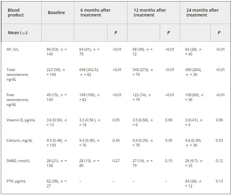
Analysis of the relationship between testosterone and AP at initiation of therapy using stiff linear splines suggested that bone turnover occurs at total testosterone levels of <250 ng/dL. In men with testosterone levels of <250 ng/dL, there was a negative correlation between testosterone and AP (R2 = −0.347, P < 0.001), and no correlation when testosterone levels were between 250 and 350 ng/dL. In the subgroup analysis, the mean (sd) testosterone level was 264 (103) ng/dL initially and 701 (245), 539 (292), and 338 (189) ng/dL at 6, 12, and 24 months, respectively. AP decreased from a mean (sd) of 87 (38) U/L to 57 (12) U/L (P = 0.015), 60 (17) U/L (P < 0.001), and 55 (10) U/L (P = 0.03) at 6, 12, and 24 months, respectively. The BMD increased by a mean (sd) of 20 (39)% (P = 0.003) on DEXA.

CONCLUSION

In hypogonadal men, the decrease in AP is associated with an increase in BMD on DEXA testing. This result suggests the use of AP as a marker of response to therapy.

Editorial: On the Mark? Is AP a surrogate for BMD in hypogonadal men?

The current issue of the BJUI contains a paper by Dubaja et al. [1] that may be of interest to physicians who have patients with hypogonadism. The authors speak to an unappreciated aspect of low testosterone; namely, the loss of bone in men and the possible recovery with treatment. Their retrospective study looked at 140 men with hypogonadism treated with exogenous testosterone replacement or clomiphene citrate testosterone enhancement. These men were also assessed for bone mineral density (BMD) markers at 6, 12 and 24 months after initial treatment. Importantly, dual-energy X-ray absorptiometry (DEXA) was performed a second time after 2 treatment years for a subset of these men. DEXA showed that there was a gain in BMD and a loss of serum alkaline phosphatase (AP) for all the men over time. The loss of AP was rapid but stabilized at 6 months. Testosterone and free testosterone increased as expected but there were no changes in vitamin D, calcium, parathyroid hormone or sex hormone-binding globulin. There was a correlation between AP and testosterone. The authors recognized poor bone density at baseline in those men with testosterone levels

Bone mineral density is the best way to predict osteoporosis and fragility fractures [2]. Women loose BMD after menopause and that is accompanied by many changes, including gains in AP [3]. The decline in serum oestrogens is a factor for women, and oestrogen replacement therapy historically has been used to prevent that loss. Not all men undergo a similar loss, i.e. andropause is not recognized in the same way as menopause. Even though osteoporosis is less common in men, the associated comorbidity may be more significant.

There is an age-related decline in testosterone and an acute loss for some men such that they approach their physicians with symptoms. The underlying cause of bone loss in men and women may be the same: serum oestrogen loss. Men who lose testosterone are also losing oestrogen because testosterone is the precursor via aromatase. Repros Therapeutics is developing a way to treat men with secondary hypogonadism. An ongoing 1-year DEXA study recruited eligible men. Key inclusion factors were age < 60 years and body mass index > 25 kg/m2. Remarkably 24% of men failed the screening test because of osteopenia, despite the fact that few of them were old or underweight.

The present paper by Dubaja et al. suggests that a readily available serum test may be able to monitor men on testosterone therapies for gain in BMD. Given the relatively low incidence of osteoporosis, screening every man by DEXA is not cost-efficient. The 1 year or more needed to find BMD loss by DEXA also wastes time and resources. Quantitative CT is more costly and is accompanied by high radiation exposure. The use of AP, as suggested in the present study, may represent a reasonable alternative.

The paper is not without its weaknesses. There was no indication of whether these men had primary or secondary hypogonadism. Transdermal testosterone should raise testosterone and oestrogen in both groups of men whereas clomiphene citrate works through changes in LH and FSH and requires an intact hypothalamic-pituitary-gonadal axis (useful in men with secondary hypogonadism only). Indeed, the two kinds of treatment will have the opposite effect on LH and FSH [4]. The authors recognized the importance of Leydig cells in producing testosterone, yet the effects of a transdermal testosterone would be to shut down testosterone production. We commend their suggestion that other factors that contribute to both bone and Leydig cell function, insulin-like 3 [5] and osteocalcin [6] should be studied in relation to AP. The loss of subjects throughout the 2 years was troubling, but it is known that men on transdermal treatments discontinue with disturbing frequency despite satisfaction [7]. If those who stayed in the present study were those with the best outcomes in terms of testosterone and BMD, potential bias may exist. If men can be encouraged to continue therapy through positive effects on BMD being detected as early as 6 months, AP monitoring may improve patient compliance. Only DEXA can give that assurance now. The authors noted the need for a larger prospective trial. Nevertheless, their paper provides a rationale for monitoring men on testosterone therapies that can be implemented with minimal cost or the need for new diagnostics.

Read the full article
Martin C. Michel
Department of Pharmacology, Johannes Gutenberg University, Mainz, German

References


2 NIH Consensus Development Panel. Osteoporosis prevention, diagnosis and therapy. JAMA 2001; 285: 785–95

3 BiveE.Use of bone turnover markers in clinical practice. Curr O pin Endocrinol Dia betes Obes 2012; 19: 468–73


5 Ivell R, Anand-Ivell R. Biology of insulin-like factor-3 in humareproduction. Hum Reprod Update 2009; 15: 463–76


7 Kovac J, Rajanahally S, Smith R, Coward R, Lamb D, Lipshultz L. Patient satisfaction with testosterone replacement therapies: the reasons behind the choices. JSexMed2014; 11: 553–62


Video: Hypogonadism and testosterone-enhancing therapy on alkaline phosphatase and BMD

The effect of hypogonadism and testosterone-enhancing therapy on alkaline phosphatase and bone mineral density

Ali A. Dabaja, Campbell F. Bryson, Peter N. Schlegel and Darius A. Paduch


Department of Urology, Weill Cornell Medical College, New York, NY, USA


Read the full article
OBJECTIVE

To evaluate the relationship of testosterone-enhancing therapy on alkaline phosphatase (AP) in relation to bone mineral density (BMD) in hypogonadal men.

PATIENTS AND METHODS

Retrospective review of 140 men with testosterone levels of <350 ng/dL undergoing testosterone-enhancing therapy and followed for 2 years. Follicle-stimulating hormone, luteinising hormone, free testosterone, total testosterone, sex hormone binding globulin, calcium, AP, vitamin D, parathyroid hormone, and dual-energy X-ray absorptiometry (DEXA) scans were analysed. A subgroup of 36 men with one DEXA scan before and one DEXA 2 years after initiating treatment was performed.

RESULTS

Analysis of the relationship between testosterone and AP at initiation of therapy using stiff linear splines suggested that bone turnover occurs at total testosterone levels of <250 ng/dL. In men with testosterone levels of <250 ng/dL, there was a negative correlation between testosterone and AP (R2 = −0.347, P < 0.001), and no correlation when testosterone levels were between 250 and 350 ng/dL. In the subgroup analysis, the mean (sd) testosterone level was 264 (103) ng/dL initially and 701 (245), 539 (292), and 338 (189) ng/dL at 6, 12, and 24 months, respectively. AP decreased from a mean (sd) of 87 (38) U/L to 57 (12) U/L (P = 0.015), 60 (17) U/L (P < 0.001), and 55 (10) U/L (P = 0.03) at 6, 12, and 24 months, respectively. The BMD increased by a mean (sd) of 20 (39)% (P = 0.003) on DEXA.

CONCLUSION

In hypogonadal men, the decrease in AP is associated with an increase in BMD on DEXA testing. This result suggests the use of AP as a marker of response to therapy.

Read more articles of the week
© 2024 BJU International. All Rights Reserved.