Tag Archive for: bladder cancer

Posts

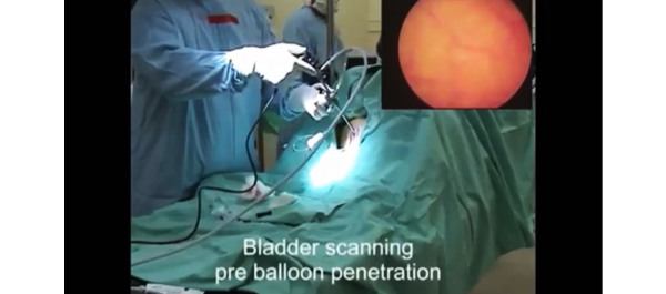
Video: In-Vivo Trials in Sheep


Novel anti-biofilm mechanism for wireless capsule endoscopy in the urinary tract: preliminary study in a sheep model

Amos Neheman*, Claude Schulman and Ofer Yossepowitch†§

*Urology Department, Meir Medical Center, Kfar-Saba, Sackler School of Medicine, Tel-Aviv University, Tel-Aviv, Israel, Department of Urology, University of Brussels, Brussels, Belgium, and §Institute of Urology, Rabin Medical Center, Beilinson, Petah Tikva, Israel

OBJECTIVE

• To develop and test the safety and feasibility of a novel anti-biofilm mechanism configured for wireless capsule endoscopy (WCE) in a sheep bladder model.

MATERIALS AND METHODS

• A WCE mechanism, designed for long-term bladder monitoring, was developed and introduced into a sheep bladder for 5 months.

• The transparency of the surface was assessed by evaluating a resolution target placed inside the capsule at serial intervals using cystoscopy under general anaesthesia.

• Animal behaviour, voiding patterns and urine cultures were monitored throughout the study.

• At study termination, the capsule was extracted and assessed using scanning electron microscopy.

RESULTS

• The resolution target was visualized clearly at all investigation points.

• No notable adverse effects were noted during the entire follow-up period and no urinary tract infection occurred.

• Scanning electron microscopy confirmed the efficacy of the technology to prevent biofilm formation and surface encrustation.

CONCLUSIONS

• We report a novel technology that effectively prevents biofilm formation on the outer surface of foreign objects in the urinary tract.

• Further studies are under way to test the applicability of this technology in bladder WCE to enable high-quality wireless image transmission.


Article of the Week: Better fit than fat when it comes to radical cystectomy for bladder cancer

Every week the Editor-in-Chief selects the Article of the Week from the current issue of BJUI. The abstract is reproduced below and you can click on the button to read the full article, which is freely available to all readers for at least 30 days from the time of this post.

In addition to the article itself, there is an accompanying editorial written by a prominent member of the urological community. This blog is intended to provoke comment and discussion and we invite you to use the comment tools at the bottom of each post to join the conversation.

If you only have time to read one article this week, it should be this one.

Obesity is associated with worse oncological outcomes in patients treated with radical cystectomy

Thomas F. Chromecki1,2*, Eugene K. Cha1*, Harun Fajkovic1,3, Michael Rink1,4, Behfar Ehdaie1, Robert S. Svatek5, Pierre I. Karakiewicz6, Yair Lotan7, Derya Tilki8, Patrick J. Bastian8, Siamak Daneshmand9,Wassim Kassouf10, Matthieu Durand1, Giacomo Novara11, Hans-Martin Fritsche12, Maximilian Burger12, Jonathan I. Izawa13, Antonin Brisuda14, Marek Babjuk14, Karl Pummer2 and Shahrokh F. Shariat1

1Weill Medical College of Cornell University, New York, NY, USA, 2Medical University Graz, Graz, Austria, 3Landeskrankenhaus St Poelten, St Poelten, Austria, 4University Medical Centre Hamburg-Eppendorf, Hamburg, Germany, 5University of Texas Health Science Center San Antonio, San Antonio, TX, USA, 6University of Montréal, Montréal, QC, Canada, 7University of Texas Southwestern Medical Center, Dallas, TX, USA, 8Ludwig-Maximilians-University Munich, Klinikum Grosshadern, Munich, Germany, 9University of Southern California Keck School of Medicine and Norris Comprehensive Cancer Center, Los Angeles, CA, USA, 10McGill University Health Centre, Montréal, QC, Canada, 11University of Padua, Padua, Italy, 12Caritas St Josef Medical Centre, University of Regensburg, Regensburg, Germany, 13University of Western Ontario, London, ON, Canada, and 14Hospital Motol, 2nd Faculty of Medicine, Charles University, Praha, Czech Republic
*These authors contributed equally.

Read the full article
OBJECTIVE

• To investigate the association between body mass index (BMI) and oncological outcomes in patients after radical cystectomy (RC) for urothelial carcinoma of the bladder (UCB) in a large multi-institutional series.

PATIENTS AND METHODS

• Data were collected from 4118 patients treated with RC and pelvic lymphadenectomy for UCB. Patients receiving preoperative chemotherapy or radiotherapy were excluded.

• Univariable and multivariable models tested the effect of BMI on disease recurrence, cancer-specific mortality and overall mortality.

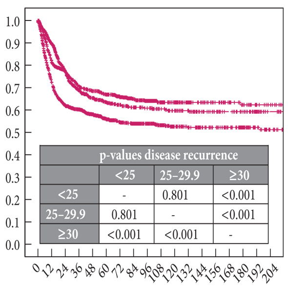
• BMI was analysed as a continuous and categorical variable (<25 vs 25–29 vs 30 kg/m2).

RESULTS

• Median BMI was 28.8 kg/m2 (interquartile range 7.9); 25.3% had a BMI <25 kg/m2, 32.5% had a BMI between 25 and 29.9 kg/m2, and 42.2% had a BMI 30 kg/m2.

• Patients with a higher BMI were older (P < 0.001), had higher tumour grade (P < 0.001), and were more likely to have positive soft tissue surgical margins (P = 0.006) compared with patients with lower BMI.

• In multivariable analyses that adjusted for the effects of standard clinicopathological features, BMI >30 was associated with higher risk of disease recurrence (hazard ratio (HR) 1.67, 95% confidence interval (CI) 1.46–1.91, P < 0.001), cancer-specific mortality (HR 1.43, 95% CI 1.24–1.66, P < 0.001), and overall mortality (HR 1.81, CI 1.60–2.05, P < 0.001). The main limitation is the retrospective design of the study.

CONCLUSIONS

• Obesity is associated with worse cancer-specific outcomes in patients treated with RC for UCB.

• Focusing on patient-modifiable factors such as BMI may have significant individual and public health implications in patients with invasive UCB.


Read Previous Articles of the Week

Editorial: Obesity is associated with worse oncological outcomes in patients treated with radical cystectomy

Michael R. Abern, Stephen J. Freedland and Brant A. Inman

Division of Urologic Surgery, Duke University Medical Center, Durham, NC, USA

Obesity is a worldwide epidemic: it is estimated over 300 million adults are obese and over 1 billion are overweight. As obesity is a risk factor for cancers and is modifiable, the authors of this report retrospectively analyse the association between body mass index (BMI) and outcomes in a large multinational cohort of bladder cancer patients that underwent radical cystectomy. They found that obese patients were older and more likely to have high-grade tumours. Furthermore, obese patients received inferior lymphadenectomies, had more positive margins, and were less likely to receive adjuvant chemotherapy. The end result is an association between obesity and bladder cancer recurrence, and both cancer-specific and overall mortality.

Although these data suggest that obesity is associated with poor radical cystectomy outcomes, this contrasts with evidence showing no link between obesity and bladder cancer mortality in population-based trials such as the Cancer Prevention Study II, which prospectively followed over 900 000 participants. Why the discrepancy? One possible explanation is the presence of confounding factors and one possible confounder is the presence of type 2 diabetes. In population-based studies that considered both BMI and diabetes, people with diabetes were noted to have an increased risk of developing bladder cancer independent of BMI, whereas the converse was not true. Additionally, diabetes has been associated with recurrence and progression of non-muscle invasive bladder cancer whereas obesity has not. The impact of diabetes was not adequately addressed in the current study.

Other limitations also probably affect the results. In the current study, overweight patients (BMI 25–30) had significantly better cancer-specific survival (hazard ratio 0.80, P = 0.01) than those of ‘normal’ weight (BMI < 25). However, a threshold BMI ≥ 30 has been shown to have poor sensitivity for obesity in elderly populations, with over 25% of patients with BMI under 30 qualifying as obese based on body fat. This may result in an overstatement of the effect of obesity. Conversely, the inclusion of underweight patients (BMI < 18.5) in the ‘normal’ group may underestimate the effect between obesity and outcome, as cachexia may be associated with poor outcomes. Another factor mentioned by the authors is the inferior lymphadenectomies performed in obese patients, which introduces a detection bias for lymph node positivity, the strongest predictor after advanced stage for all of their tested outcomes on multivariate analysis (hazard ratio 2.01–2.33, P < 0.001).

Although the true effect of obesity may be hard to quantify with these data, all would agree that maintaining a non-obese bodyweight will help many disease states with little apparent harm. Patients undergoing neoadjuvant chemotherapy before radical cystectomy have a 3-month window to lose weight and exercise more. This could improve surgical outcomes, and possibly tolerance of chemotherapy. Furthermore, if we can prove that obesity leads to increased bladder cancer recurrence or progression, a window of opportunity may exist when a low-risk tumour is diagnosed. Otherwise, we are left with the eighteenth century wisdom of Benjamin Franklin: ‘An ounce of prevention is worth a pound of cure.’

Read the full article

Autologous cytotoxic T lymphocyte therapy was effective for M-VAC-refractory invasive bladder cancer

In the present study, we performed CTL therapy in a patient with metastatic bladder cancer, who had relapsed after M-VAC therapy.


Authors: Kogenta Nakamura MD, PhD1; Kazuhiro Yoshikawa PhD2; Yoshiaki Yamada MD, PhD1; Makoto Sumitomo MD, PhD1

1 Department of Urology, Aichi Medical University School of Medicine
2 Cell Therapy Center, Aichi Medical University Hospital

 
Corresponding Author: Kogenta Nakamura, MD, PhD, Department of Urology, Aichi Medical University School of Medicine. Nagakute-cho, Aichi 480-1195, Japan. Tel: +81-561-62-3311   E-mail address: [email protected]


Abstract
A report on the efficacy of CTL therapy for urogenital cancer already exists in the literature.1 In the present study, we performed CTL therapy in a patient with metastatic bladder cancer, who had relapsed after M-VAC therapy.


Introduction
Standard chemotherapy for invasive bladder cancer with metastases includes M-VAC (methotrexate, vinblastine, doxorubicin, and cisplatin) and GC (gemcitabine and cisplatin).2 Many chemotherapeutic regimens have been used as second-line treatment, but no regimen surpasses M-VAC and GC.  There are high expectations for taxanes and other similar drugs, and the results of a large-scale, randomized trial are awaited.
Autologous cytotoxic T lymphocyte (CTL) therapy using autologous tumor tissue is a technique in which autologous lymphocytes collected from the peripheral blood and an autologous tumor irradiated with γ-rays are cultured in vitro in the presence of interleukin-2 (IL-2), and anti-CD3 for induction and proliferation of killer cells that are expected to be specific to the tumor, and then the killer cells are aseptically returned to the body.  For this therapy, it is essential that MHC-class I antigen is expressed on tumor cells.


Case report
We performed total cystectomy with periaortic and pelvic lymphadenectomy, and bilateral cutaneous ureterostomy in a 61 year old Japanese male patient with a diagnosis of T4N2M0 bladder cancer made 6 years previously.  We chose  to perform urinary diversion to cutaneous ureterostomy instead of ileal conduit, to avoid intestinal complications and also because we wished to start systemic chemotherapy as soon as possible. Pathological examination revealed urothelial carcinoma, grade G3, pT4. The right external iliac lymph nodes, bilateral obturator lymph nodes and aortocaval lymph nodes were positive.  Since the patient subsequently developed enlarged mediastinal, supraclavicular and para-aortic lymph nodes (Figure 1a), three courses of M-VAC therapy were performed in combination with mild hyperthermia, which is known to lead to fewer adverse reactions.3  The efficacy of the chemotherapy was evaluated using the Response Evaluation Criteria In Solid Tumor classification.  CT scan, after completion of three courses of M-VAC therapy, revealed that the mediastinal lymph nodes and the right supraclavicular lymph nodes had disappeared and the para-aortic lymph nodes were reduced in size.  Since CT scan subsequently showed enlarged para-aortic lymph nodes, two further courses of M-VAC therapy were performed.  However, the para-aortic lymph nodes remained unchanged (Figure 1b).
After approval was obtained from the institutional review board of our hospital (# 152), four courses of CTL therapy in combination with IFN-γ and IL-2 administration were initiated (at intervals of two weeks) after surgery.  Two days before administration, 200 mg/body of Endoxan was administered by intravenous infusion.  An average of 22.6  108 cells were administered.  During each course, blood was collected on the day after administration to examine whether the CD8 level had increased or not.  After completion of four courses of treatment, a CT scan was performed and revealed a partial response (PR) of the para-aortic lymph nodes (Figure 1 c).


Figure 1 (a) Computed tomography showing enlargement of para-aortic lymph nodes before M-VAC therapy.


(B) Computed tomography showing para-aortic lymph nodes remaining unchanged after M-VAC therapy.


(C) Computed tomography showing reduced para-aortic lymph node size after CTL therapy.  The lymph node size deceased from 15 to 8 mm.


At present, 71 months after completion of CTL therapy, the patient has had recurrence and has maintained his PR status.


Conclusion
Autologous CTL therapy did not cause any adverse reactions in our patient.  It is suggested that this therapy can be performed safely and should also be considered for cases of progressive bladder cancer with metastasis.


References
 
1 Kawai K, Saijo K, Oikawa T et al : Clinical course and immune response of a renal cell carcinoma patient to adoptive transfer of autologous cytotoxic T lymphocytes. Clin Exp Immunol 2003; 134: 264-269.
2 von der Maase H, Sengelov L, Roberts JT et al : Long-term survival results of a randomized trial comparing gemcitabine plus cisplatin, with methotrexate, vinblastine, doxorubicin, plus cisplatin in patients with bladder cancer. J Clin Oncol 2005; 23: 4602-4608.
3 Yamada Y, Itoh Y, Aoki S et al. Preliminary results of M-VAC chemotherapy combined with mild hyperthermia, a new therapeutic strategy for advanced or metastatic transitional cell carcinoma of the urothelium. Cancer Chemother Pharmacol 2009; 64: 1079-1083.


Date added to bjui.org: 02/02/2012

DOI: 10.1002/BJUIw-2011-059-web
 

Granulomatous Cholangitis as a Complication of Intravesical BCG Administration for Bladder Cancer

We describe a patient who presented with findings suggestive of acute cholecystitis and cholangitis. 


Authors: Hani M. Babiker, MD1, Robyn E. Stiefeld1, MD, and Holenarasipur R. Vikram2, MD

1Department of Hospital Internal Medicine, and
2 Division of Infectious Diseases, Mayo Clinic, Phoenix, Arizona

Corresponding Author: Holenarasipur R. Vikram, MD, Division of Infectious Diseases, 5777 E. Mayo Blvd., Phoenix, AZ 85054. Phone: (480) 342-0115  E-mail: [email protected]


Abstract
 
Intravesical instillation of Bacillus Calmette-Guerin (BCG) remains a first-line treatment for superficial transitional cell carcinoma of the bladder. Although uncommon, clinicians should be aware of major adverse effects and complications resulting from intravesical BCG therapy in order to promptly arrive at the diagnosis and initiate therapeutic measures.
Herein, we describe a patient who presented with findings suggestive of acute cholecystitis and cholangitis. He was started on antibiotics and underwent Endoscopic Retrograde Cholangiopancreatography (ERCP) with sphincterectomy and stent placement. However, his liver function tests remained abnormal. Further inquiry delineated a history of bladder cancer treated with intravesical BCG instillation. A liver biopsy obtained during laparoscopic cholecystectomy confirmed granulomatous cholangitis. The patient received anti-tuberculous therapy and a tapering course of corticosteroids with a successful outcome.


Introduction
  Bacillus Calmette-Guerin (BCG) is a live attenuated vaccine containing Mycobacterium bovis that is administered in many countries to prevent childhood tuberculous meningitis and military tuberculosis. BCG is not utilized in the United States because of the low risk of infection with M. tuberculosis and questionable efficacy. Intravesical BCG administration has been found to be very effective in the treatment in superficial bladder cancer. However, treatment with BCG can be associated with local or systemic complications. Herein, we present a case of granulomatous cholangitis and hepatitis following intravesical BCG therapy. Timely diagnosis and prompt initiation of therapy is essential for ensuring a favorable outcome.


List of Abbreviations
ALT Alanine Aminotransferase
ALK Alkaline Phosphatase
AST Aspartate Aminotransferase
BCG Bacillus Calmette-Guerin
ERCP Endoscopic Retrograde Cholangiopancreatography
LFTs Liver Function Tests
Tbili Total Bilirubin
Dbili Direct Bilirubin


Case Report
A 65 year-old-male with an underlying history of coronary artery disease, hypertension, obstructive airway disease, and bladder cancer presented to our Emergency Department with abdominal discomfort. Computed tomography (CT) of the abdomen was within normal limits. He was discharged once his symptoms abated.
Two weeks later, he presented with fever, jaundice, hypotension, abdominal pain, and jaundice. Abdominal examination revealed normal bowel sounds and mild right upper quadrant tenderness. There was no hepatosplenomegaly, guarding, rebound tenderness, or rigidity. His labs revealed a white blood cell count of 7.8 x 10⁹/L, aspartate aminotransferase (AST) of 278 U/L, alanine aminotransferase (ALT) 305 U/L, alkaline phosphatase (ALK) 227 U/L, total bilirubin (Tbili) of 6.3 mg/dl, direct bilirubin (Dbili) of 3.6 mg/dl, lipase of 30 U/L, and ammonia of 36 mcg/dl. His Hepatitis A, B, and C serologies were negative. Abdominal ultrasound revealed gallbladder wall thickening, biliary sludge, and a normal calibre common bile duct.
He was fluid resuscitated and commenced on ciprofloxacin and metronidazole for a tentative diagnosis of cholangitis and cholecystitis. The next day, his Tbili increased to 7.3 mg/dl and his liver enzymes remained elevated. Repeat abdominal CT scan revealed mild thickening of the gallbladder wall, cholelithiasis, and heterogenous enhancement of the liver parenchyma. ERCP was performed with removal of biliary sludge, stent placement, and sphincterectomy. However, the next day, his Tbili and Dbili increased to 10.2 mg/dl and 8.5 mg/dl, respectively; liver enzymes remained abnormal. An intrahepatic process versus biliary stent stenosis was considered, and repeat ERCP and cholangiogram were planned along with cholecystectomy. Prior to surgery, his liver function    remained abnormal with a TBili of 16.1 mg/dl, DBili 12.8 mg/dl, ALK 507 U/L, AST 135 U/L, and ALT 159 U/L. Further inquiry into his past medical history revealed that he had received a total of three courses of intravesical BCG for superficial bladder cancer within the past 7 months (the last administration was 2 months prior to his current hospitalisation).
A liver biopsy performed at the time of cholecystectomy revealed portal infiltration with non-caseating granulomas centered around the bile ducts. There was focal bile duct proliferation with neutrophilic infiltration consistent with bile duct outflow impairment. Histology of the gall bladder showed acute inflammation with Gram-negative and Gram-positive cocci; granulomas were not evident.


Figure 1. Liver Histopathology


Liver histology demonstrates a granuloma centered around a bile duct ( arrow) and a hepatocyte (arrowhead). Concurrent neutrophilic and eosionphilic infiltration, and bile ductular proliferation resulted in biliary obstruction. Mild fibrosis and marked cholestasis in the surrounding liver was also noted. (Hematoxylin-eosin; ~x400.)


Bacterial, fungal, and mycobacterial smears and cultures were all negative from the liver biopsy specimen. A diagnosis of Mycobacterium bovis granulomatous cholangitis secondary to intravesical BCG administration was entertained. He was commenced on a regimen of isoniazid, rifampin, ethambutol, and vitamin B6 for six months and a 3-week tapering course of corticosteroids. His bilirubin and liver enzymes pursued a downward trend. Isoniazid was discontinued after the first month for transient liver enzyme elevation. His liver enzymes and bilirubin completely normalized within 2 months, and he remained asymptomatic. He completed a 6-month course of rifampin and ethambutol; LFTs remained normal at follow-up 2 months after discontinuing antituberculous medications.


Discussion
Bacillus Calmette-Guerin (BCG) vaccine was introduced in 1910 for protection against tuberculosis (TB). It is a live, attenuated vaccine containing M. bovis. It is utilized in many countries with a high prevalence of TB to prevent childhood tuberculous meningitis and military TB. BCG is not recommended in the United States because of the low risk of infection with M. tuberculosis, its variable and questionable efficacy against adult pulmonary TB, and its interference with tuberculin skin test reactivity. In 1976 Morales and colleagues described a novel utility for BCG – anticancer immunotherapy by intravesical instillation for superficial bladder cancer.  Several studies have subsequently demonstrated its efficacy in treating superficial bladder cancer.   This is now the adjuvant treatment of choice for high grade and recurrent superficial bladder cancer.    Its mechanism of action is incompletely understood; it triggers a local cellular immune response with induction of cytokines that have antiangiogenic activity.
Complications of intravesical BCG treatment can be either local (cystitis) or disseminated, with an early or late presentation. Early manifestations occurs 8 to 12 weeks after BCG instillation, while late manifestations can occur more than a year after BCG therapy.8 Cystitis following BCG administration presents with dysuria, urinary frequency, low-grade fever, and malaise. These symptoms occur in 70% of patients approximately 2 to 4 hours after BCG instillation and resolve within 48 hours. Symptomatic therapy is successful in most instances. For persistent or severe symptoms that last beyond 48 hours, isoniazid should be administered.  Rarely, a sepsis-like syndrome can occur soon after BCG instillation; patients develop fever, hypotension, and respiratory failure. This is thought to represent a hypersensitivity reaction to BCG as opposed to true dissemination.9 Other local complications (<1% each) include hematuria, epididymitis, prostatitis, and ureteral obstruction. Disseminated infection (‘BCGosis’) may result in granulomatous hepatitis, pneumonitis, osteomyelitis, endophthalmitis and prostatitis and other organ involvement.2,5,9 These complications can occur weeks to months after BCG administration in approximately 1% of patients. The most serious complication following BCG therapy is sepsis with hypotension and multiorgan failure in 0.4% of cases and carries a high mortality.5 Other published complications of intravesical BCG therapy include testicular masses, peritonitis, psoas abscess, tuberculous spondylitis, chest wall mass, acute renal failure, rhabdomyolysis, pancytopenia secondary to bone marrow infiltration and ruptured mycotic abdominal aortic aneurysm. 10 In a large study involving 2602 patients who received intravesical BCG, granulomatous hepatitis occurred in 0.7% of patients.9
BCGosis is diagnosed by growth of M. bovis from tissue specimens, or by DNA hybridization. 11 However disseminated M. bovis infection is often paucibacillary, and it is difficult to isolate the pathogen in vitro. Many of the systemic manifestations of BCGosis can be attributed to a hypersensitivity reaction to mycobacterial antigens. Thus, a tentative diagnosis is often made based on the temporal relationship to intravesical BCG administration, clinical manifestations, histopathologic findings, and response to empiric antituberculous therapy. M. bovis is susceptible to first-line anti-tuberculous agents (except pyrazinamide). A 6 to 12 month course of therapy with isoniazid, rifampin, and ethambutol along with vitamin B6 supplementation has been widely utilised in published reports with an excellent response. A tapering dose of corticosteroids is usually included to combat the associated hypersensitivity reaction to mycobacterial antigens.2


Conclusion
We report a case of bacterial cholecystitis wherein liver enzyme and bilirubin levels remained abnormal despite cholecystectomy and antibiotic therapy. Concurrent liver biopsy demonstrated granulomatous cholangitis. LFT abnormalities completely resolved following a tapering course of prednisone and 6 months of antituberculous therapy aimed at M. bovis. This case highlights the importance of considering BCG-induced granulomatous hepatitis and cholangitis as a possible etiology in patients with LFT abnormalities following intravesical BCG administration. To the best of our knowledge, this is the first reported case of BCG-induced granulomatous cholangitis. Awareness of this entity, collection of tissue specimens for histopathologic and microbiologic examination, and prompt initiation of appropriate therapy led to a favorable outcome in our patient.


References
1. Morales A, Eidinger D, Bruce AW. Intracavitary bacillus Calmette-Guerin in the treatment of superficial bladder tumors. J Urol. 1976 Aug: 116:180-183
2. Lamm DL. Complications of bacillus Calmette-Guerin immunotherapy. Urol Clin North Am. 1992 Aug: 19:565-72.
3. Witjes JA, vd Meijden AP, Debruyne FM. Use of intravesical Bacillus Calmette-Guerin in the treatment of superficial transitional cell carcinoma of the bladder: an overview. Urol Int. 1990: 45(3):129-136.
4. Kamat AM, Lamm DL. Immunotherapy for bladder cancer. Curr Urol Rep 2001 Feb: 2(1):62-9.
5. Lamm DL. Efficacy and safety of bacillus Calmette-Guerin immunotherapy in superficial bladder cancer. Clin Infect Dis. 2000 Sep: 31(suppl 3):S86-90.
6. Shelley MD, Wilt TJ, Court J, Coles B, Kynaston H, Madson MD. Intravesical bacillus Calmette-Guerin is superior to mitomycin C in reducing tumor recurrence in high-risk superficial bladder cancer: a meta-analysis of randomized trials. BJU Int. 2004 Mar :93(4):485-90.
7. Bohle A. BCG’s mechanism of action–increasing our understanding. Eur Urol. 2000 :37:Suppl 1:1-8.
8. Gonzales OY, Musher DM, Brar I, et al. Spectrum of Bacille Calmette-Guerin (BCG) Infection after Intravesical BCG Immunotherapy. Clin Infect Dis. 2003 Jan: 36(2):140-8.
9.  Lamm DL, van der Meijden PM, Morales A, et al. Incidence and treatment of complications of bacillus Calmette-Guerin intravesical therapy in superficial bladder cancer. J Urol. 1992 Mar:147(3):596-600.
10.  Safdar N, Abad CL, KauL DR, Jarrard D, Saint S. Clinical problem-solving. An unintended consequence–a 79-year-old man with a 5-month history of fatigue and 20-lb (9-kg) weight loss presented to his local physician. N Engl J Med. 2008 Apr: 358(14):1496-1501.
11. Leebeek FW, Ouwendijk RJ, Kolk AH, et al. Granulomatous hepatitis caused by Bacillus Calmette-Guerin (BCG) infection after BCG bladder instillation. Gut. 1996 Apr: 38(4):616-618.


Date added to bjui.org: 26/10/2011


DOI: 10.1002/BJUIw-2011-093-web


© 2024 BJU International. All Rights Reserved.