Tag Archive for: #BJUI

Posts

Editorial: Contemplate the template: a new prostate biopsy approach

Transperineal magnetic resonance imaging – ultrasound fusion targeted biopsies (MRI-US FTB) of the prostate: the future of prostate diagnostics

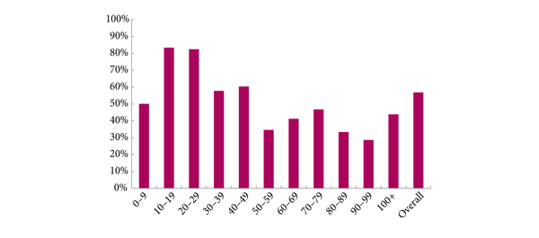
The prostate cancer diagnostic pathway has remained unchanged for 25 years. At best, laterally directed, peripheral zone (PZ) 12-core transrectal biopsies identify cancer in 44% of cases [1] but transrectal biopsies have an inherent sampling error with a risk of misdiagnosis or mischaracterisation of disease. Of those with negative biopsies who undergo transperineal (TP) biopsies, 30% have cancer, most in the anterior PZ. Active surveillance and the promise of less invasive treatment options are becoming popular because of concerns about ‘over treatment’ for low-risk disease.

Saturation transrectal biopsies have been advocated to improve diagnostic yield but do not address the issue of under sampling of the anterior PZ, particularly in the larger gland [2]. TP biopsies can be used to address the issue of under sampling but prostate template-mapping biopsies are labour intensive and require large numbers of biopsies, often between 60 to 90 cores; however, they have been an essential component of focal therapy trials and the evaluation of novel treatment methods [3].

Primary TP biopsy is the subject of the paper published in this edition of the BJUI titled ‘Outcomes of transperineal template-guided prostate biopsy in 409 patients’ [4]. The authors report a single centre experience of primary TP biopsies. The 14-region protocol described is simpler than prostate template-mapping requiring fewer cores (median of 15 and mean of 19 cores) with a comparable primary diagnostic detection rate of 60% and an encouraging side-effect profile. Unfortunately, the approach still has limitations and the authors admit that their limited biopsy protocol may still mischaracterise disease in the larger gland. In a recent paper from the same group, there was a disappointing correlation between their TP biopsy pathology, MRI abnormalities and radical prostatectomy specimens [5]. Uncertainty prevails, the problem is how best to sample the larger gland. The authors [4] and others, often conclude that more biopsies are necessary for larger glands and resort to mapping protocols and many more biopsies. The solution may not be more biopsies but rather better systematic targeting of the PZ. The impact of hyperplasia within the transition zone (TZ) has a profound effect on PZ anatomy. In the smaller prostate, up to 30 mL, there is little TZ and the PZ is much thicker posteriorly than anteriorly, this difference is even more apparent in glands of 30–50 mL. Above 50 mL TZ expansion causes marked attenuation of the PZ, which becomes much thinner, but the overall volume of the PZ does not change. Less than 4% of cancers originate in the TZ [6], consequently biopsies should be concentrated primarily on the PZ.

The future of prostate cancer diagnosis is likely to be a combination of pre-biopsy multiparametric MRI, followed by targeted biopsies of MRI-identified lesions combined with fewer but better systematic targeted biopsies of the PZ. MRI-ultrasound (MRI-US) fusion techniques have been developed in which axial T2 images of the prostate, diffusion-weighted images and/or dynamic contrast-enhanced MRI images are ‘fused’ with the live US images to allow precise targeting of both regions of interest and the PZ. Commercially available biopsy programs, developed from brachytherapy software systems programs allow individual biopsy sites to be recorded and if combined with inking of the specimen can provide precise pathological localisation of disease within the prostate [7].

There are many potential benefits to this approach. Patients who opt for active surveillance will have an archived record of their disease at a given time to facilitate precise replication of further interval biopsies and assess progression. Improved disease management for an individual should be the aim. The suitability or not for focal or targeted therapies, the planning or boosting of identifed lesions with radiotherapy and/or brachytherapy, and the planning of nerve-sparing surgery or wide excisions should be possible. Feedback to the radiologists of both benign and malignant pathology and grade of disease will improve reporting accuracy and provide imaging sciences with the histopathological characteristics of both MRI ‘visible’ and ‘invisible’ cancer to improve MRI interpretation.

MRI–US fusion targeted biopsies are a significant advance in prostate diagnostics and may resolve some uncertainty within the prostate cancer diagnostic pathway. Benefit vs cost is a recurring issue across health care and questions will continue to be asked about the use of increasingly expensive technology in such an indolent disease. The challenge for investigators will be how to prove the benefit of this approach over standard biopsy protocols and integrate this work in to clinical practice.

Richard Popert
Department of Urology, Guy’s Hospital, London, UK

References
  1. Presti JC, O’Dowd GL, Miller MC et al. Extended peripheral zone biopsy schemes increase cancer detection rates and minimize variance in prostate specific antigen and age related cancer rates: results of a community multi-practice study. J Urol 2003; 169:125–129
  2. Stewart CS, Leibovich BC, Weaver AL, Lieber MM. Prostate cancer diagnosis using a saturation needle biopsy technique after previous negative sextant biopsies. J Urol 2001; 166: 86–92
  3. Onik G, Barzell W. Transperineal 3D mapping biopsy of the prostate: an essential tool in selecting patients for focal prostate cancer therapy. Urol Oncol 2008; 26: 506–510
  4. Symons JL, Huo A, Yuen CL et al. Outcomes of transperineal template-guided prostate biopsy in 409 patients. BJU Int 2013; 112: 585–593
  5. Huo AS, Hossack T, Symons JL et al. Accuracy of primary systematic template guided transperineal biopsy of the prostate for locating prostate cancer: a comparison with radical prostatectomy specimens. J Urol 2012; 187: 2044–2050
  6. Patel V, Merrick GS, Allen ZA et al. The incidence of transition zone prostate cancer diagnosed by transperineal template guided mapping biopsy: implications for treatment planning. Urology 2011; 77: 1148–1152
  7. Hadaschik BA, Kuru TH, Tulea C et al. A novel stereotactic prostate biopsy system integrating pre-interventional magnetic resonance imaging and live ultrasound fusion. J Urol 2011; 186: 2214–2220

Annual meeting of the Irish Society of Urology

A wonderful ISU meeting in a stunning setting with exquisite weather in my home county, Wicklow.  Great talks and posters from our Residents, but it was the audience discussion they prompted that made this such a stellar meeting.  Tremendous senior academic input from the Members of the Urological Club of Great Britain and Ireland as well as Guest Speakers Ian Eardley from Leeds and Mike Naslund from the University of Maryland and BAUS President Adrian Joyce.  Excellent back and forth discussion on Surgical Training, Urological Emergencies, Prostate Biopsy Sepsis, Incidentalomas, creating Centres of Excellence for Testis and Penile Cancer and (of course!) the Search for the Truth About Robots.  

Basic Science topics were of a very high standard with Boyce et al promising a blood test of 4 proteins that was far better than the Partin tables!!  Professor Mike Naslund made the complex so simple for us to understand in his talk on Health Care Economics – the take home message being that when you create a system where the patient is not personally out of pocket in accessing health care, you cannot control the costs.  Ian Eardley spoke on “Men vs Health”, enumerated all of the increased risks to the male from Metabolic Syndrome and concluded with the premise that the specialty best suited to drive forth the Men’s Health agenda is Urology.  Most felt that it was one of the best meetings they had been to because it was all about common garden topics they encounter in everyday practice and not the esoteric topics that tend to dominate the larger meetings.  So, come mid-Atlantic in Killarney Co Kerry for the ISU 2014 Meeting on the 25th and 26th of September with Guest Speakers Craig Peters from the US and Prokar Dasgupta from the UK!!!!


Dr David Quinlan
Consultant Urologist, St Vincent’s Hospital,
Senior Lecturer, University College Dublin
Chairman, BJUI

Twitter: @daithiquinlan

Editorial: External validation of Karakiewicz models: do they hold up?

Cancer-specific survival (CSS) in patients with RCC depends on important prognostic factors including specific clinical signs or symptoms, tumour-related factors and various laboratory findings. To better predict prognosis and aid patient counselling, several investigators have developed tools which have greatly enhanced our ability to predict outcomes in patients with RCC. For instance, Kattan et al. [1] have combined manner of presentation, tumour histology, tumour size and pathological stage to develop a nomogram that predicts cancer-free survival after nephrectomy. The stage, size, grade, and necrosis (SSIGN) score is another predominant model that provides individualized information for patients with clear-cell RCC. It incorporates the 1997 TNM stage, tumour size, nuclear grade and presence of tumour necrosis to predict recurrence and survival after radical nephrectomy [2]. The Karakiewicz nomogram [3] was developed to predict CSS based on multi-institutional data. The preoperative nomogram includes patient age, gender, clinical stage, presence of metastases, tumour size and symptom classification. The postoperative one includes TNM stage, tumour size, Fuhrman grade, histological subtype and local symptoms. Tan et al. [4] compared several prognostic systems (the Karakiewicz, Kattan and Sorbellini nomograms, and the Leibovich model) and concluded that in terms of individual counselling, the postoperative Karakiewicz nomogram is likely to be more useful than other models and provides excellently calibrated CSS estimates; however, before a prediction tool becomes popular in clinical use, it is crucial to perform internal and external validation to prove its generalizability. For example, the UCLA integrated staging system (UISS) helps to identify patients with localized or metastatic disease at low, intermediate, and high risk of disease progression and has been validated internally and externally [5].

The present study by Cindolo et al. [6] aims to assess the accuracy and generalizability of the pre- and postoperative Karakiewicz nomograms in predicting CSS. It is a retrospective study involving >3000 patients from multiple European and US centres between 1992 and 2010. They include high-, mid- and low-volume institutes, as well as different populations. This helps to provide a heterogenous study cohort to better reflect the real clinical situation and hence to improve the reproducibility of the nomogram. The preoperative and postoperative models have a good predictive ability with a stratified C-index of 0.784 and 0.842, respectively, and the latter discriminates substantially better. The authors conclude that the Karakiewicz nomograms proved to have excellent accuracy and generalizability.

With more RCC therapeutic options including surveillance, ablation, surgery and systemic therapies, better prediction tools are needed to help clinical decision-making. A wealth of literature now supports the hypothesis that nomograms and artificial neural networks are superior to classic TNM staging systems in risk assessment; therefore, these predictive tools are important to guide the counselling, treatment and follow-up of patients with RCC.

Peggy Chu1 and Ringo Wing-Hong Chu2
1Department of Surgery, Tuen Mun Hospital, and 2Department of Surgery, Kwong Wah Hospital, Hong Kong, China

Read the full article
References
  1. Kattan MW, Reuter V, Motzer RJ et al. A postoperative prognostic nomogram for renal cell carcinoma. J Urol 2001; 166: 63–7
  2. Frank I, Blute ML, Cheville JC et al. An outcome prediction model for patients with clear cell renal cell carcinoma treated with radical nephrectomy based on tumor stage, size, grade and necrosis: the sign score. J Urol 2002; 168: 2395–400
  3. Karakiewicz PI, Briganti A, Chun FK et al. Multi-institutional validation of a new renal cancer-specific survival nomogram. J Clin Oncol 2007; 25: 1316–22
  4. Tan MH, Li H, Choong CV et al. The Karakiewicz nomogram is the most useful clinical predictor for survival outcomes in patients with localized renal cell carcinoma. Cancer 2011; 117: 5314–24
  5. Cindolo L, Chiodini P, Gall C et al. Validation by calibration of the UCLA integrated staging system prognostic model for nonmetastatic renal cell carcinoma after nephrectomy. Cancer 2008; 113: 65–71
  6. Cindolo L, Chiodini P, Brookman-May S et al. Assessing the accuracy and generalizability of the preoperative and postoperative Karakiewicz nomograms for renal cell carcinoma: results from a multicentre European and US study. BJU Int 2013; 112: 578–584.

Editorial: LESS versus laparoscopic nephroureterectomy: the winner is…

In this international multi-institutional study, Park et al. [1] have retrospectively collected and analysed data about 101 patients who underwent laparoendoscopic single-site (LESS) nephroureterectomy (NU) for upper urinary tract (UUT) urothelial carcinoma.

Nowadays, NU represents the standard of care for the surgical treatment of UUT urothelial carcinoma in the majority of patients [2]. Outcomes of such an intervention are strongly improved when lymph node dissection (LND) is performed according to a well-defined template [3].

In recent years, laparoscopy has become an important new approach to reduce the invasiveness of the surgical treatment of UUT urothelial carcinoma. In a multicentre Italian study Porpiglia et al. [4] showed that laparoscopic NU with open ureterectomy was a feasible and safe technique. Oncological results seemed to be similar to those of the traditional open approach, but the laparoscopic approach still has some disadvantages. First, patients who undergo a laparoscopic procedure receive LND with lower frequency. Moreover, the template during a laparoscopic procedure is rarely respected and the number of lymph nodes removed is often suboptimal [3]. Second, there is no consensus in the literature about the pathological stages that could potentially benefit from the bladder-cuff excision step of this procedure [5]. Bladder-cuff excision omission does not seem to undermine survival in patients with low-stage (pT1-2) disease, nevertheless confirmatory recurrence data are required before a NU without bladder-cuff excision may be considered as an option for this patient category.

The present paper shows that advances in surgical technology are being made, but it also underlines the fact that the above-mentioned disadvantages of NU are still under discussion, and these disadvantages are expanded when introducing a newer and challenging technique such as the LESS approach.

In the present study, different devices and instruments were used. Furthermore, the rate of LND reported was very low (27%), as the number of lymph nodes removed (approximately five). LND was often ‘formally’ performed, and no specific template was reported to be used. Bladder-cuff excision was not performed in 20% of cases and, when performed, the technique used was not clearly defined. With regard to oncological efficacy, the recurrence rate of 22% at 11 months is not sufficient to clarify if the LESS approach is oncologically effective [6].

In summary, there are evident limitations to the present paper; some are methodological, such as its retrospective nature and the non-homogeneous datasheets used to collect data, and some are technical and oncological. These limitations are justified by the fact that the technique is in its embryonic stages. Nevertheless, the authors deserve praise for having collected such a large number of cases for their study on LESS NU. Their paper underlines the fact that this technique is feasible and safe, and each surgeon who contributed by insisting on such a challenging and novel approach to NU should be congratulated for their efforts.

Now that the feasibility of the LESS NU technique has been demonstrated, the authors have the task of clarifying whether introducing a LESS approach would or would not compromise oncological outcomes. In any case, it is recommended that surgical oncological principles be respected when a new technique is introduced, especially when dealing with a high-risk cell-seeding tumour such as urothelial carcinoma.

Francesco Porpiglia and Riccardo Bertolo
Department of Clinical and Biological Sciences, San Luigi Hospital, Division of Urology, University of Turin, Orbassano-Turin, Italy

Read the full article
REFERENCES
  1. Park SY, Rha KH, Autorino R et al. Laparoendoscopic single-site nephroureterectomy for upper urinary tract urothelial carcinoma: outcomes of an international multi-institutional study of 101 patients. BJU Int 2013; 112: 610–615
  2. Rouprêt M, Zigeuner R, Palou J et al. European guidelines for the diagnosis and management of upper urinary tract urothelial cell carcinomas: 2011 update. Eur Urol 2011; 59: 584–594
  3. Roscigno M, Brausi M, Heidenreich A et al. Lymphadenectomy at the time of nephroureterectomy for upper tract urothelial cancer.Eur Urol 2011; 60: 776–783
  4. Porpiglia F, Celia A, Luciani L, Terrone C, Guazzoni G, Parma P. Laparoscopic radical nephroureterectomy: results of a multicentric italian study. J Endourol 2009; 23 (Suppl. 1): A109
  5. Lughezzani G, Sun M, Perrotte P et al. Should bladder cuff excision remain the standard of care at nephroureterectomy in patients with urothelial carcinoma of the renal pelvis? A population-based study. Eur Urol 2010; 57: 956–962
  6. Walton TJ, Novara G, Matsumoto K et al. Oncological outcomes after laparoscopic and open radical nephroureterectomy: results from an international cohort. BJU Int 2010; 108: 406–412

UroLift Takes Off From Down Under. The Potential Rewards When Engineers Bring You Into Their Inner Circle

At the American Urological Association meeting in San Antonio in May 2005, I was introduced to a four engineers from a small start up company called NC2 (New Company 2).  It had at that time been recently spun off from the medical device incubator company Exploramed.  They had no product and not even a prototype of a product that could possibly be used in humans but what they did have was a passion to make a difference, incredible ideas and a laptop computer. 

They had thought about the failings of existing mechanical treatments for LUTS/ BPH and the first that comes to your mind is prostatic stents.  No stent conforms perfectly to the shape of the prostatic urethra and there were the issues of encrustation of any elements of stent material that were exposed to the urine.  Rather than throw the baby out with the bathwater, they harnessed what was good about stents, which was the potentially immediate effects they could have on urinary function without associated destruction of tissue and that perhaps tailoring the radial expansion to just a few critical points rather than the entire length of the prostatic urethra could do the trick.

The original idea was that some sort of metallic disc could be placed outside the prostate capsule and one on the urethral side and between them, a non absorbable suture could be placed under tension and therefore draw open the prostatic urethra and defined sites.  How these engineers were to find a way of designing a delivery tool to do this had me a little skeptical at first but there seemed to be no doubt in their minds, even thought they had not yet worked it out, were going to find a way.  Their confidence, intellect and enthusiasm was infectious and you just felt like you wanted to be a part of this project.  It so turned out that the metallic discs would be replaced by linear metallic tabs which logically make for easier delivery.

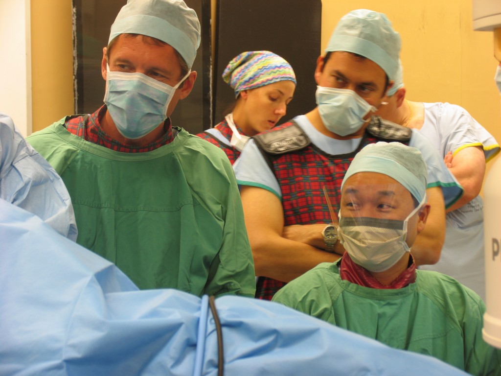
So why involve Australians?  It is difficult to keep things under the radar and one way of doing so is to take the idea where it is less likely to be visible. Additionally, the data needed to be trustworthy and in a place where strong ethic committee governance structures exist. We make no illusion that for once, being Australian, gave us a clinical research opportunity from a company based in the US that would rarely be directed our way.

My Australian colleague, Dr Peter Chin was also brought in on the project.  Over the next few months, we did not hear anything but there was then an urgent call that ‘California was the place we ought to be’ so we literally dropped everything and headed over to Silicon Valley where we had the opportunity to use the first prototype of the device on human cadavers.  Whilst our travel costs were covered by NC2, we received no payment for our time spent during these exercises but remuneration was the last thing on our minds given the exciting path that the idea could potentially take.  Simultaneously, animal studies were being conducted and these demonstrated that the internal metallic tabs of the prosthesis would become covered by urothelium and in combination with the cadaveric work, provided a convincing argument to move forward with human clinical trials.

Putting on a brave face doing the first human Urolift case at Westmead Hospital in Sydney in December 2005

By December 2005, we were ready to conduct the first human trials.  We measured everything that could possibly move and it probably took close to 2 hours to perform the first case.  The initial prototype device used looked like it was literally built in somebody’s garage workshop but it was functional and confirmed proof in principle that a transurethral delivery system could deploy metallic tabs on the capsular side of the prostate and within the urethra that was connected by a tensioned suture. Through this, it created mechanical alteration to the anatomy of the prostatic urethra with positive influence on lower urinary tract symptoms.  From here, multiple clinical trials have been performed by the company that became known as Neotract Inc and as of 13 September 2013, the device received FDA approval.

It is enormous privilege to have played a role in product development from inception of an idea through to FDA approval.  These opportunities are rare and whilst healthy skepticism and caution should be applied to all ideas presented to you, if you are offered such an opportunity to take a side project, it could be a rewarding diversion from your daily clinical practice.  Financially, you will never recoup your time investment but the rewards of making a difference is priceless.

Shared passion for a project can go a long way.   This experience emphasizes the value of engineers interacting with clinicians to achieve a desired outcome and there is certainly room for of such interactions. Opportunities to embrace these relationships are out there and perhaps a good place to start is to become active in the Engineering and Urology Society which as a section of the Endourological Society meets each year at the AUA Annual Meeting.


Henry Woo is an Associate Professor of Surgery at the Sydney Adventist Hospital Clinical School of the University of Sydney in Australia. He has been appointed as the inaugural BJUI CME Editor. He is currently the coordinator of the International Urology Journal Club on Twitter. Follow him on Twitter @DrHWoo

Disclosure: Henry Woo has formerly been an investigator and advisor to Neotract Inc. He holds a small stock investment in the company.

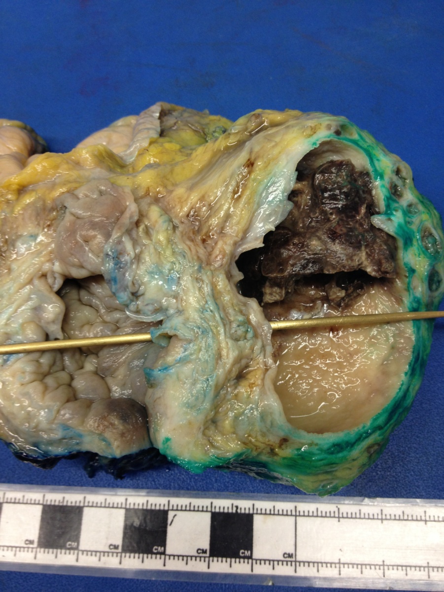
What’s the diagnosis?

Test yourself against our experts with our weekly quiz. You can type your answers here if you want to compare with our answers, or just click the ‘submit’ button below.

[wpsqt name=”Picture Quiz of the Week 13-9-13″ type=”quiz’ label=’

Click here for more Picture Quizzes

If you have a suggestion for a new Picture Quiz please email us.

Heroes

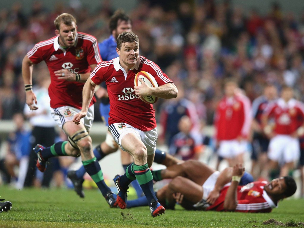
Being an Irishman, and enthusiastic rugby supporter, I, like many other rugby fans, was much vexed when our Irish rugby hero, Brian O’Driscoll, was dropped for the deciding Test for the British and Irish Lions versus Australia this summer.  This vexation was due somewhat to national pride, but more importantly, because he is one of my sporting heroes. This outpouring of emotion on the demotion of someone I’ve never met led me to contemplate the nature of heroism and heroes, how they affect us, and specifically how heroes can have a positive impact on urologists in this day and age of constant cynicism towards the noble deeds of others.

Heroism is defined as conduct as exhibited in fulfilling a high purpose or attaining a noble end. To this end, how does my rugby hero qualify? Is it his display of innate skills that thrill the crowd? Is it his professional attitude to the institution of his sport/profession? Is it his ability to overcome adversity for the good of his team?  (These are all attributes I see in my Urological heroes.)  Personally, I think back to moments where I realised that he had done things that I couldn’t even imagine being able to do, and just marvel at them.  Passing the ball to himself and ghosting past the opposition, scoring a length of the field try and being physically ill afterwards due to a pre-existing virus, being listed as ‘likely to play’ for the following week with a personal injury list of ‘concussion, torn hamstring and lacerated ear’. These are levels of physical and athletic prowess unattainable by most people.


But what of others that I would class as heroes? It is 50 years this June since the late John F. Kennedy made his famous speech in Berlin, immortalised by the phrase “Ich bin ein Berliner’.  However, his greatest segment is when he lists some of the positives that people were attributing to Communism at the time, and extolling his own personal opposition with the repeated statement ‘Let them come to Berlin!’  Watching the reaction of a group of Berliners to this speech 40 years after its occurrence, seeing them moved to tears by his reiteration of the support of the free world to the citizens of Berlin, this alone is enough to convince me of the heroism of this amazing, somewhat flawed, ever impressive man.

Heroism can also be displayed by people using their professional experience to reach extraordinary outcomes in the face of enormous adversity. Captain Chesley ‘Sully’ Sullenberg, who, after a complete engine failure on his commercial jet, in the space of 180 seconds, managed to control and ditch his plane on the Hudson River in New York, saving 155 souls. His wife, on being told that her husband had landed a multi-ton commercial jet in a river, with no harm to anyone, apparently replied laconically “Oh that sounds like Sully, alright”.

But how does this relate to urologists?  It is my personal belief that the ‘heroes’ we have in society today are not fit for purpose, vacuous celebrities of little consequence in general, and that we would all gain much by having a number of personal and professional heroes that we can use as an example when adversity, conflict, or difficult decisions face us as surgeons. Surgeons should, and often do, aim to attain a noble end. I have many heroes in Urology in particular, and often use their example, and sage-like advice in times of difficulty,

It is extremely easy to live life these days in a manner that loses sight of the wonder and awe with which we held medicine when young. It is easy to live life in a manner where much of it seems jaded and worn. It is easy to believe that there are things we cannot do, goals we cannot reach, achievements we cannot achieve. In these situations, having a hero, whose deeds seem somehow beyond what the rest of us can do, can give us a guide, an example to strive to emulate, attempt to equal, maybe even to surpass.  It is this aspect of heroism which can be utilised as something to be aspired to, for the betterment of all.

On a final note, I am often astounded by the heroism of my patients.  For these people to be able to face ill health, their own frailties and mortality and put their trust in us as surgeons, especially if we are recommending a new or unique form of treatment, is to display a level of trust that definitely puts them in the pantheon of heroes for me.  I believe we owe it to them to remain interested, invigorated and willing to sacrifice ourselves to emulate our heroes, for their benefit.  Heroes are great, everyone should have one!


David Bouchier-Hayes is Consultant Urologist and Robotic Surgeon Honoray Clinical Lecturer at the Galway Clinic, Doughiska, Co. Galway, Ireland. Follow him on Twitter @dbh44

Editorial: Regulatory T cells in renal cell carcinoma: additional fuel to the bonfire of debate

In the developing immune system, all T cells are positively selected in the neonatal thymus for the ability to recognize self-antigens, the major histocompatibility complex (MHC) proteins. Thus, the mature T-cell repertoire is trained to ‘see’ foreign pathogens ‘complexed’ with those self-antigens (‘MHC-presentation’). Fundamentally, this requirement predisposes mammalian systems to the development of autoimmune diseases, as all T cells are self-reactive. That such diseases are the exception rather than the rule is attributable to a small population (∼2–5%) of circulating T cells, termed ‘regulatory T cells’ (Tregs), that suppress the activation and function of many other immune cells. The fine balance between Tregs and other pro-inflammatory cells is essential for maintaining self-tolerance while allowing immunological reactivity against danger signals such as foreign antigens (mostly pathogens) and malignant cells. Many pathogens co-evolving alongside the mammalian immune system have learned to ‘hijack’ this balance to propagate disease or to inhibit their own clearance, notably Leishmaniasis, malaria, tuberculosis, HIV, hepatitis C virus and Helicobacter pylori. In these scenarios, an excess of Tregs induced by the pathogens prevents their clearance and establishes infective chronicity.

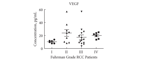
Likewise, in malignant diseases, such as pancreatic and ovarian cancer, an excess of Tregs is thought to contribute to failure of the immune system to clear neoplastic cells. Whether the tumour environment appropriates the regulatory function of Tregs to propagate its own survival in a manner akin to infectious agents, or whether Tregs infiltrate larger tumours in which there is more chronic inflammation is unclear. Nevertheless, a correlation between higher Treg numbers and poorer outcomes is a common feature of malignancies. In this issue of the BJUI Polimeno et al. add evidence to the debate over whether Treg numbers in RCC are associated with worse outcomes. While previous publications both support (Cancer Immunol Immunother 2007, BJU Int 2009) and refute (Clin Cancer Res 2007) this assertion, the data presented by Polimeno et al. identify not only increased circulating Treg numbers in patients with RCC but also find an association betweenTreg numbers, especially those that express the naïve T-cell marker CD45RA, and both larger tumour load and worse prognosis. In the same dataset, as expected, the authors also find that a shorter disease-free survival was evident in patients with lower numbers of tumoricidal natural killer cells. In the serum, patients with RCC had higher concentrations of soluble factors involved in cell growth and movement, such as epidermal growth factor, hepatocyte growth factor, vascular endothelial growth factor and interferon γ-induced protein 10 (also known as CXCL10), and markers of active inflammation, such as interleukins 6 and 8.

These observations suggest several broad possibilities: (i) that the ‘Tregs’ identified in the tumour environment and circulation are not Tregs but are in fact other activated T cells that temporarily express the same surface markers as bona fide Tregs; (ii) that Tregs in the context of RCC are unable to control tumour-associated inflammation; (iii) that Tregs contribute to tumour survival by inhibiting clearance of neoplasms by other immune cells, resulting in chronic inflammation; and/or (iv) that Tregs are actively contributing to the inflammation by converting to pro-inflammatory phenotypes, as has been demonstrated by several groups. These possibilities can be differentiated by isolating Tregs from the tumour environment or local draining lymph nodes and testing their functional characteristics in vitro; however, the fact that CD45RA+ Tregs were independently associated with worse outcomes makes (i) and (ii) less likely, as such cells are less likely to be recently activated and are inherently less plastic than other populations of Tregs.

In our opinion, the clinical value of the data presented in this paper and those of others, even if the underlying biology is poorly understood, should next be determined in a prospective study to see whether the immunological ‘fingerprint’ in peripheral blood can correctly identify those patients who are more likely to do poorly, targeting them for closer monitoring and/or more aggressive therapy.

Behdad Afzali and Giovanna Lombardi
Medical Research Council Centre for Transplantation, King’s College London, King’s Health Partners, Guy’s Hospital, and National Institute for Health Research Biomedical Research Centre at Guy’s and St Thomas’ NHS Foundation Trust and King’s College London, Guy’s Hospital, London, UK

Read the full article
© 2024 BJU International. All Rights Reserved.