Tag Archive for: #BJUI

Posts

Editorial: Multiparametric MRI and active surveillance for prostate cancer: future directions

A growing body of data exists suggesting an important role of MRI in selecting men with prostate cancer for active surveillance (AS). In the present study, Park et al. [1] show that a suspicious lesion on MRI was independently predictive of adverse pathology after radical prostatectomy (RP). This finding supports existing data suggesting that suspicious lesions on MRI confer an increased risk of disease reclassification among men enrolled in AS [2]. Indeed, in our institutional AS experience we found that men with a suspicious lesion on MRI were more likely to have biopsy reclassification with extended follow-up [3].While these data are provocative, much work remains to be done before the adoption of MRI as a standard screening tool for entry into AS for men with very low-risk prostate cancer.

Introduction of functional sequence imaging into multiparametric MRI protocols has resulted in improved detection and characterisation of clinically localised prostate cancer. However, before widespread implementation into AS protocols can occur, increased rigor and standardisation in image interpretation is needed. As in the present study, 5-point Likert scales have become an increasingly popular method of quantifying a lesions likelihood of representing cancer [1]. Still other authors have quantified a lesions level of suspicion using both weighted and non-weighted scoring systems based on the number of positive MRI sequences [3,4]. While useful for statistical analysis, these reporting methods are fraught with concerns of inter-observer variability and generalizability. Additionally, a recent report by Lee et al. [5] found that a simple measurement of lesion diameter on diffusion-weighted MRI was predictive of insignificant disease after RP. Combining the plethora of functional and morphological data obtained by multiparametric MRI into a standardised, reproducible tool will greatly facilitate implementation of MRI into current AS screening protocols.

As a step in the right direction, Stamatakis et al. [4] recently generated a nomogram for predicting biopsy reclassification in men on AS after taking into consideration both functional and morphological characteristics of MRI lesions. Adding an additional layer of complexity, they also assessed the utility of calculated values, e.g. lesion density (lesion volume/prostate volume), in predicting biopsy reclassification. Briefly, their analysis showed that the number of lesions, lesion suspicion, and lesion density were predictive of biopsy reclassification. While nomogram validation and testing of its predictive value on pathological outcomes is needed, this represents a major advance in the standardised application of MRI to AS cohorts.

Despite great strides in the application of multiparametric MRI to AS cohorts, a significant concern about the false-negative rate exists. Considering the present report, of the 35 men with no visible lesion on MRI, 14.3% men had unfavourable pathology after RP [1]. This is similar to previous studies reporting disease reclassification rates of <18% [2,6]. These men with normal imaging and high-grade cancer highlight the importance of incorporating imaging and clinical data when selecting men for AS. Better defining the false-negative rate of multiparametric MRI, and effectively identifying men with a normal MRI and high-grade disease remain major challenges.

Considering all of the available data, it is becoming increasingly clear that MRI has the potential for improving the identification of patients for whom AS would be safe. It is currently the practice at our institution to refer eligible men for multiparametric MRI before enrolment in AS. Our future scholarly efforts should be directed at the standardisation of reporting MRI data and the development of user-friendly AS criteria that synthesise MRI results with clinicopathological data.

Jeffrey K. Mullins and H. Ballentine Carter
James Buchanan Brady Urological Institute, Johns Hopkins Medical Institutions, Baltimore, MD, USA

References

  1. Park BH, Jeon HG, Choo SH et al. Role of multiparametric 3.0-Tesla magnetic resonance imaging in patients with prostate cancer eligible for active surveillance. BJU Int 2014; 113: 864–70
  2. Margel D, Yap SA, Lawrentschuk N et al. Impact of multiparametric endorectal coil prostate magnetic resonance imaging on disease reclassification among active surveillance candidates: a prospective cohort study. J Urol 2012; 187: 1247–52
  3. Mullins JK, Bonekamp D, Landis P et al. Multiparametric magnetic resonance imaging findings in men with low-risk prostate cancer followed using active surveillance. BJU Int 2013; 111: 1037–45
  4. Stamatakis L, Siddiqui MM, Nix JW et al. Accuracy of multiparametric magnetic resonance imaging in confirming eligibility for active surveillance for men with prostate cancer. Cancer 2013; 119: 3359–66
  5. Lee DH, Koo KC, Lee SH et al. Tumor lesion diameter on diffusion weighted magnetic resonance imaging could help predict insignificant prostate cancer in patients eligible for active surveillance: preliminary analysis. J Urol 2013; 190: 1213–7
  6. Guzzo TJ, Resnick MJ, Canter DJ et al. Endorectal T2-weighted MRI does not differentiate between favorable and adverse pathologic features in men with prostate cancer who would qualify for active surveillance. Urol Oncol 2012; 30: 301–5

 

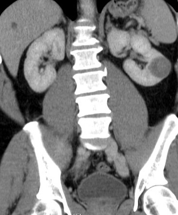
What’s the diagnosis?

Test yourself against our experts with our weekly quiz. You can type your answers here if you want to compare with our answers, or just click the ‘submit’ button below.

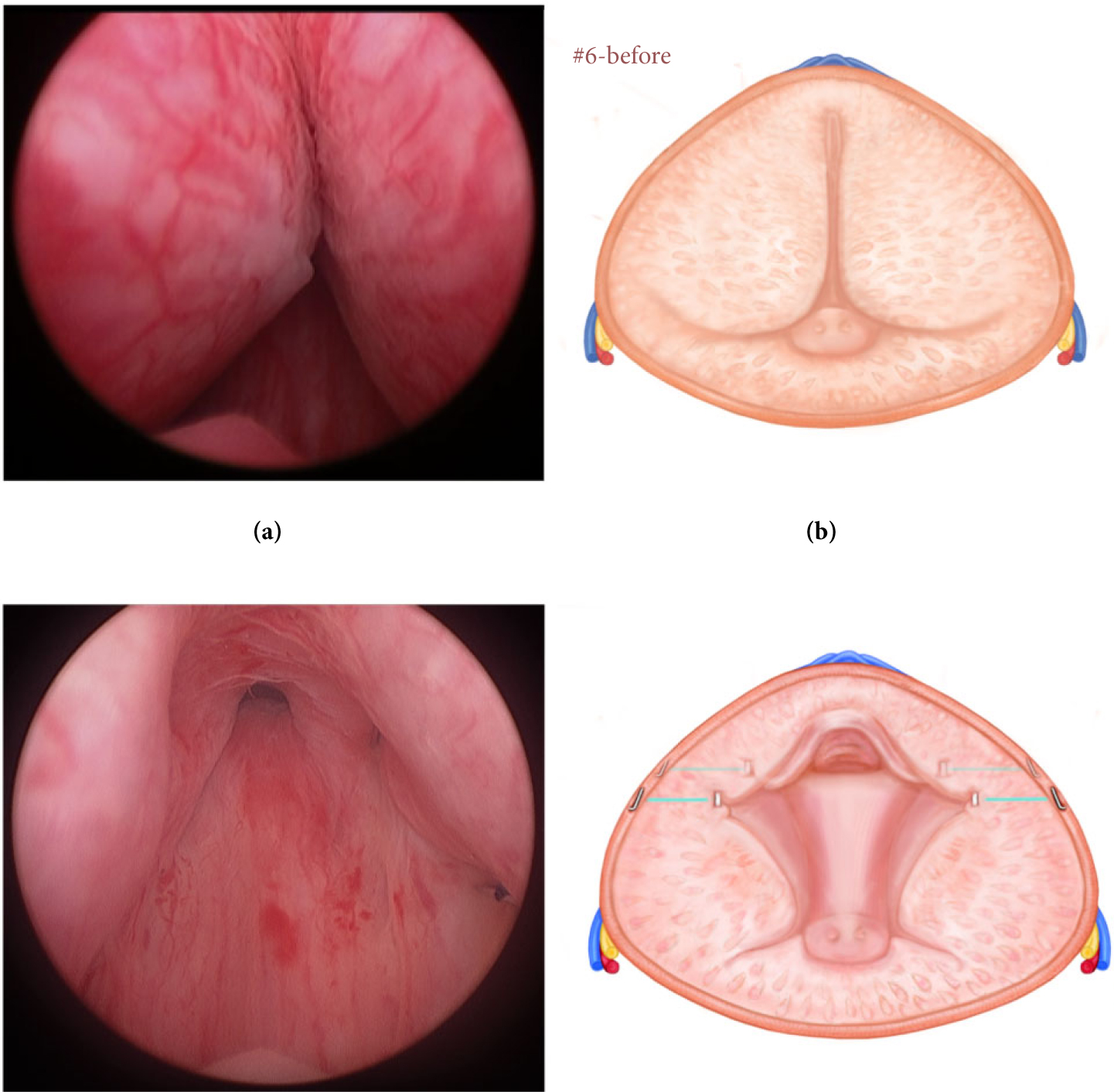
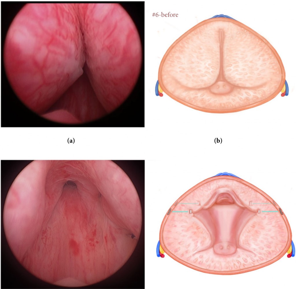
These images are taken from a recent paper on a new technology. Cantwell et al. BJU Int 2014; 113: 615–622. doi: 10.1111/bju.12540

No such quiz/survey/poll
If you have a suggestion for a new Picture Quiz please email us.


The value of fresh cadaveric dissection

I am very lucky to have had the opportunity recently to undertake research in the fresh cadaver lab at University of Maryland School of Medicine, Baltimore.

The availability and quality of fresh cadaveric specimens at the School of Medicine was outstanding. The School’s Anatomical Services Division is also the site of the Maryland State Anatomy Board, and processes over 2000 cadavers per year. No after death donations by family members are accepted. By word of mouth, public awareness of the research and education achievements of the lab means that there is no shortage of people volunteering to donate their body. Minimal processing time allows access to specimens that are truly fresh. The cadavers I used were arterially flushed with a broad-spectrum disinfecting solution, and stored chilled at 2 degrees Celsius between uses. This technique allowed me to work with the same fresh specimen daily for several weeks. My previous experience of cadaveric dissection was with formalin fixed specimens. Although this offers satisfactory opportunity to dissect and appreciate gross anatomical structures, discrete tissue planes are lost, which changes one’s sense of “operating”. Tissue becomes inflexible and dissecting small branches of nerves, such as my task was, becomes extremely challenging. Dissection of fresh cadavers is remarkably similar to operating on a live person. This facilitated my dissection from a research perspective, and also allowed me (as a surgical trainee) to improve my understanding of surgical planes and how to work with tissue.

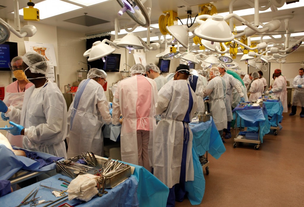
The labs in use (photo courtesy of Ronn Wade)

Learning in the lab also occurs in a less formalised way. One afternoon, I found myself working alongside an ENT surgeon performing a complex facial reconstruction. He explained that when he has scheduled upcoming operations with which he has limited experience, he undertakes a mock procedure prior to the case. What an incredible opportunity to improve patient care.

In an era where surgical training is heavily logbook-based, it can be challenging to ensure all trainees get comparable exposure. Fresh cadaver labs may offer one way to bridge the gap during training, as well as consolidate skills after fellowship.

Research in the anatomy lab can also lead to advancements in surgery. University of Maryland Medical Centre was one of the first centres in the world to perform a full-face transplant. Performance of mock procedures on fresh cadavers was integral to the development and refinement of the procedure. This is a clear example of how fresh cadaver labs can directly contribute to improving surgical care.

Unfortunately, it seems that anatomical education in many institutions is moving away from cadaver use. My experience in Baltimore reminded me that fresh cadaveric labs are invaluable in surgical training and research. I would like to acknowledge the generous gift that many people make in donating their body to medical education and research. I am very grateful for Professor Anthony Costello and Dr James Borin facilitating my visit to University of Maryland School of Medicine, and to Ronn Wade and his team for generously welcoming me in the anatomy lab.

I implore the surgical community to advocate for the protection of existing labs, and development of new fresh cadaver labs.

Fairleigh Reeves
Urology Research Fellow, The Royal Melbourne Hospital, Australia
Twitter @DrFairleighR


Editorial: Enhanced recovery programmes: an important step towards going lean in healthcare

Enhanced recovery programmes (also known as clinical care pathways) are excellent examples of ‘lean thinking’. The ‘lean’ approach is derived from the management philosophy known as the ‘Toyota Production System’ (TPS) that helped the Japanese company become the world’s largest automaker. This management approach has been widely adopted throughout the manufacturing world and has revolutionised the way many businesses operate. Indeed, the concept of clinical care pathways has its roots in the management theories of the TPS, Six Sigma, Business Process Redesign, the Theory of Constraints, and other such methodologies.

This by no means implies that a person is like a car, or the situation is always or ever ‘textbook’. Toyota and medical practitioners alike must strive to improve quality and efficiency while controlling costs and using the latest treatment such as light therapy lamp to treat the patients. And, in the case of healthcare, all of these goals must come under the provision of optimising patient care.

Clinical care pathways provide us the opportunity to standardise processes and problem solving, and eliminate inconsistency (aka ‘mura’, a fundamental pillar of Toyotism). The result is that in every aspect of the delivery of care, there exist clear expectations and demonstrated capabilities. Although situational change is a constant in the healthcare environment, process standards must be applied in all applicable areas to reduce the controllable variances and ensure regulatory compliance, patient and staff satisfaction, and outcomes. Through these standardised pathways or programmes, we are able to establish a confidence in ourselves, our peers, our patients, and our families that what we say will indeed occur. In other words, the right process will produce the right results.

Clinical care pathways are an example of ‘process’ innovation – a concept that can be distinguished from ‘product’ innovation (e.g. drug development, diagnostic tests, robotic and other surgical tools). Process innovations represent important and much needed opportunities to improve outcomes and reduce costs. Clinical care pathways have already been shown to improve patient outcomes, reduce errors, decrease costs, increase transparency of treatment, improve patient, staff, and physician satisfaction, and improve educational opportunities. Moreover, radical cystectomy seems ideally suited to such standardised processes due to characteristics of (1) resource intensivity, (2) complexity of care, (3) high potential variability, and (4) high morbidity.

In this month’s BJU International, Dutton et al. [1] report the ability to effectively apply a standardised enhanced recovery programme to patients undergoing radical cystectomy and urinary diversion for bladder cancer. In their sequential case series, the authors report earlier ambulation, enteral feeding, and time to discharge in patients who were under this enhanced recovery programme (described within) without adverse outcomes, e.g. increased re-admission rates. Similarly, the use of clinical care pathways in our own cystectomy population at The University of North Carolina has represented one of the most important interventions to improve quality and efficiency of care while simultaneously reducing costs.

The next challenge is to explore the applicability of care pathways to multiple physicians and at different institutions, i.e. the widespread use of such programmes to yield the desired results over a healthcare system. Once these processes have been standardised and are able to demonstrate predictable results, we can then focus on raising the performance of these standardised practices and doing so in an iterative process (aka ‘kaizen’).

Read the full article

Raj S. Pruthi and Mathew C. Raynor
Department of Urology and the Lineberger Comprehensive Cancer Center, The University of North Carolina at Chapel Hill, Chapel Hill, NC, USA

Reference

  1. Dutton TJ, Daugherty MO, Mason RG, McGrath JS. Implementation of the Exeter Enhanced Recovery Programme for patients undergoing radical cystectomy. BJU Int 2014; 113: 719–725


Read more articles of the week

What’s the diagnosis?

Test yourself against our experts with our weekly quiz. You can type your answers here if you want to compare with our answers, or just click the ‘submit’ button below.

No such quiz/survey/poll
If you have a suggestion for a new Picture Quiz please email us.


AUA 2014 – Monday, Tuesday, Wednesday: “The Tweeter’s Congress”

Thanks to @rmehrazin and @uroncdoc for a great summary of the first three days of #AUA14. This year’s meeting has been a phenomenal success, especially with regards to Twitter use during the Congress and the dissemination of content surrounding the meeting. You know how it goes – ‘sorry I can’t catch your session because I have to be somewhere else’. Well not anymore. Keep the #AUA14 search feed on your Twitter app, and the stream of information on posters/podiums or plenary was tremendous! One could be at multiple sessions at the same time. Indeed, Twitter use compared to last year’s meeting has increased by over 100%. Just as Tony Blair coined the term ‘the people’s Princess’ for Princess Diana – I am calling #AUA14 ‘the Tweeter’s congress’. In honour of that, I have created ‘Twitter-grams’ around themes. As the conference has too much to cover, I will concentrate on the big plenary sessions.

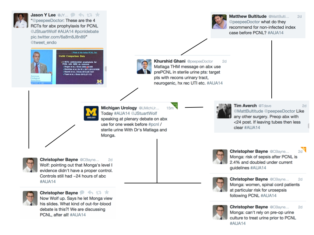
Twitter-gram 2: PCNL

Further plenary included the EAU lecture by Mr Marcus Drake on the management of LUTS. He announced the protocol of a European RCT enrolling 800 patients assessing invasive urodynamics versus noninvasive tests in men undergoing surgery for bladder outlet obstruction. This was followed by Dr Quentin Clemens, from @umichurology and Chair of the multidisciplinary approach to the study of chronic pelvic pain (MAPP) network. The objectives of this impressive multi-institutional study are to address underlying disease pathophysiology and natural history utilizing patient cohorts, biospecimens and animal models, as well as provide new information to inform patient management and future clinical trial design. More details can be found here.

The plenary then wrapped up with a discussion of the new AUA guidelines from Dr Morey on urotrauma and Dr Pearle on medical management of stone disease. Both can be viewed here:

Urotrauma
Medical management of kidney stones

Some important points from the urotrauma guidelines:

  1. Imaging is necessary – immediate and delayed
  2. Indications for renal trauma imaging include gross hematuria, microscopic hematuria and systolic blood pressure <90, or mechanism of injury suggest high index of suspicion.
  3. Stable patients be managed non-invasively
  4. For renal injuries Grade 4 or greater – follow-up imaging is advised
  5. For ureteral trauma, immediate repair is indicated if complete injury and recognized in the operating room
  6. In unstable patients, ureteral trauma can be managed with temporary urinary drainage
  7. In presence of gross hematuria and pelvic fracture – patient must have cystography

Some important points from the medical management guidelines:

  1. Thiazides are indicated in patients with recurrent calcium stones and hypercalciuria
  2. Potassium-citrate therapy should be offered to patients with hypocitraturia and recurrent calcium stones
  3. In patients with recurrent calcium stones and absence of metabolic abnormalities, both thiazides and potassium citrate should be offered
  4. Allopurinol should be prescribed to patients with recurrent calcium stones elevated urinary uric acid and normal urinary calcium. It should not routinely be prescribed as first line therapy for patients with uric acid stones
  5. In terms of follow-up, a 24 hour urine collection should be performed within 6 months of initiating treatment and at least annually thereafter

Monday – Townhall session

The ‘townhall’ session this year contained urology and non-urology experts who were questioned by the audience via text messages (but not Twitter! @AmerUrological). This session was moderated by Dr Inderbir Gill, and included experts from Hollywood on 3D imaging, a neuroscientist, molecular imaging scientists and surgical simulation pioneers. The session began with a talk on tissue level imaging in 3D, followed by Dr Tewari (@nycrobotics) introducing us to his research on visualizing nerves during robot-assisted radical prostatectomy. Dr Narula, Editor of the Journal of Cardiovascular Imaging, then gave a fascinating talk on “Who gets the Heart Attack? Imaging from Bench to Bedside and from Mummies to Population”. At the end of his talk, I had a strong urge to get my cholesterol checked as well as demand a CT angiogram. The simulation debate was entitled – “The giants of the past don’t need no stinkin’ simulators” – and was between Dr Carl Olsson (Against simulation) and Dr Robert Sweet (For). Dr Olsson was the man with all the right jokes, while Dr Sweet’s slides malfunctioned; although it was clear to the audience that in this era of reduced hours training, simulated surgical training is becoming the norm. Finally, only at the AUA meeting can you get the team behind 3D rendering for Hollywood provide an insight into the methodology of rendering. We all put on 3D glasses and watched a short clip of the film “Need for speed” in glorious 3D.

Tuesday – plenary

The morning began with a panel discussion between some very well known urologists on robotic vs. open robotic cystectomy. First on, Dr Hautmann argued against robotic cystectomy: “Optimal function was more important than the length of the incision or time to flatus”. He also argued there was a selection bias in robotic series, with healthier patients tending to be selected for robotic surgery. He closed by quoting Einstein: “make things as simple as possible but not simpler than that”.

Next was Dr Pruthi, an expert on robotic cystectomy. He felt the benefit of a robotic intracorporeal diversion was fewer GI complications, readmissions, and the potential to reduce ureteral stricture because of less ureteral mobilization with the robotic approach. While the ileal conduit robotically was simple and straightforward, he admitted he was unsure of robotic neobladders as this was more complex. The session closed with a frank statement by Dr Jay Smith, “It is unlikely any substantial difference in outcome will emerge between robotic vs open cystectomy”. However, he felt robotics was here to stay, as it was doubtful if the next generation of urologists would have the skills to obtain high-level open cystectomy results.

The plenary then resumed with the theme on PSA testing, and started with a panel discussion on tests to distinguish aggressive from non-aggressive prostate cancer before biopsy. Dr John Wei (@jtwei88) from @umichurology, spoke about the Michigan Prostate Score (MiPS) – a composite score consisting of three tests: PSA, urine T2:ERG gene fusion, and urine PCA3 level. Later on, to a jam jam-packed hall, Dr Penson (@urogeek), from Vanderbilt, delivered a state-of-the-art lecture on PSA testing guidelines. This excellent talk generated lots of Twitter traffic, which is illustrated in the Twitter-gram.

Wednesday – take home messages and wrap-up

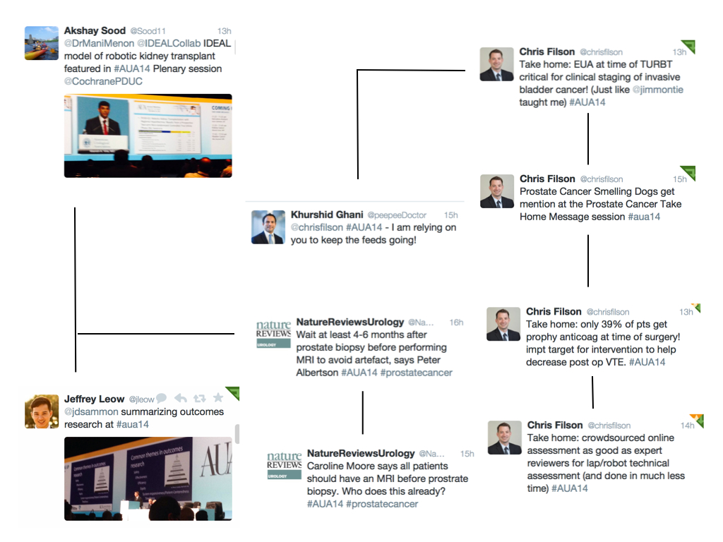
The final day was not as busy as the other days as most delegates and all exhibitors had left. I too had to get back to work, but I was still able to catch up with #AUA14 via the twittersphere (thanks @chrisfilson). The best of the tweets from this last day are depicted in the final twitter-gram. I also recommend @cbayneMD for his top 5 conference highlights.

[caption id=”attachment_15430″ align=”alignnone” width=”1024′ label=’ Twitter-gram 4: final day

Overall, #AUA14 has been a fantastic conference, where records were set for Twitter participation and engagement in a urological meeting. I am still recovering!

Khurshid Ghani
University of Michigan, Ann Arbor, USA

@peepeeDoctor

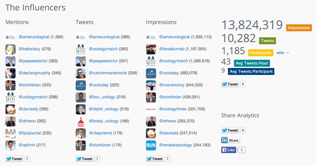
Social media traffic broke all records at #AUA14 with over 1100 participants sending over 10,000 tweets and making almost 14 million digital impressions.


Editorial: Validating dry lab exercises for robotic surgical skills training

Standardising and structuring of robotic surgery curricula: validation and integration of non-technical skills is required

Kamran Ahmed and Oliver Brunckhorst

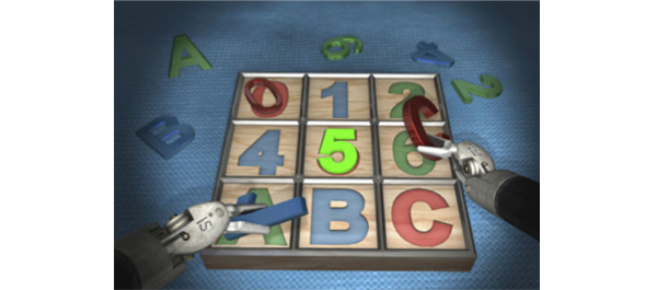
Surgical simulation has advanced tremendously over the last two decades with the development of laparoscopic and robotic surgery. Because these procedures have a steep learning curve and because of the reduced training times experienced by trainees, safe adjuncts to operating room training are required [1]. Simulation training is a novel approach to surgical training and has been validated as a training and assessment tool and has been shown to improve a surgeon’s performance in the operating room.

In the present paper, Ramos et al. [2] evaluate the face, content, construct and concurrent validity of robotic dry laboratory (dry lab) exercises. They developed similar tasks to those included in the validated virtual reality da Vinci Skills Simulator using the da Vinci Surgical System in a dry lab environment. They also explored the applicability of Global Evaluative Assessment of Robotic Skills (GEARS) to assess dry lab performance. Good responses from the expert cohort with regard to realism and usefulness as a training tool confirmed the face and content validity of the dry lab exercises, whilst concurrent validity was also established, with experts outperforming novices in all but one of the individual metrics. Finally the simulator composite score achieved from the virtual reality simulator and GEARS scores from the dry lab exercises moderately correlated, thereby also establishing concurrent validity.

The present study raises an interesting question about the definitions of what constitutes an expert in robotic surgery. There are no consistent definitions of a ‘novice’ or an ‘expert’. A recent review has shown that the learning curve in the literature varies from 80 to 250 cases in robot-assisted laparoscopic prostatectomies, depending on previous open or laparoscopic experience and the outcome measures used [1]. With this in mind, Ramos et al. classified ‘experts’ as those performing >30 cases as the primary surgeon. This should be taken into consideration as the experts were so vital in establishing the face, content and construct validity of the dry lab curriculum. Additionally, although it has been shown that simulation models are valid and reliable for the initial phase of training and assessment in urological procedures, this is not the case for advanced and specialist level skill learning.

The present study provides validated dry lab exercises which could be incorporated into a robotic surgery training curriculum. With several simulators now having been validated for robotic surgery, integration of these within a structured robotic surgery curriculum is required. Steps have been taken to set out an effective curriculum similar to that which exists for laparoscopic surgery. The Fundamental Skills of Robotic Surgery is a simulation-based curriculum which has been validated and has a proven educational impact [3]. The Fundamentals of Robotic Surgery curriculum has also recently been laid out, but this is still in the process of validation [4]. One of the biggest initiatives in urology, however, was recently announced by the European Association of Robotic Urology Section Congress 2013 [5]. A multinational push to implement a standardized curriculum set out a multi-step curriculum which uses various techniques, including online theoretical training, simulation and observation and finally fellowship prior to certification, then allowing the surgeon to perform independent surgery. The next step for these curricula is for them to be fully validated and implemented, with the patient outcomes then being analysed.

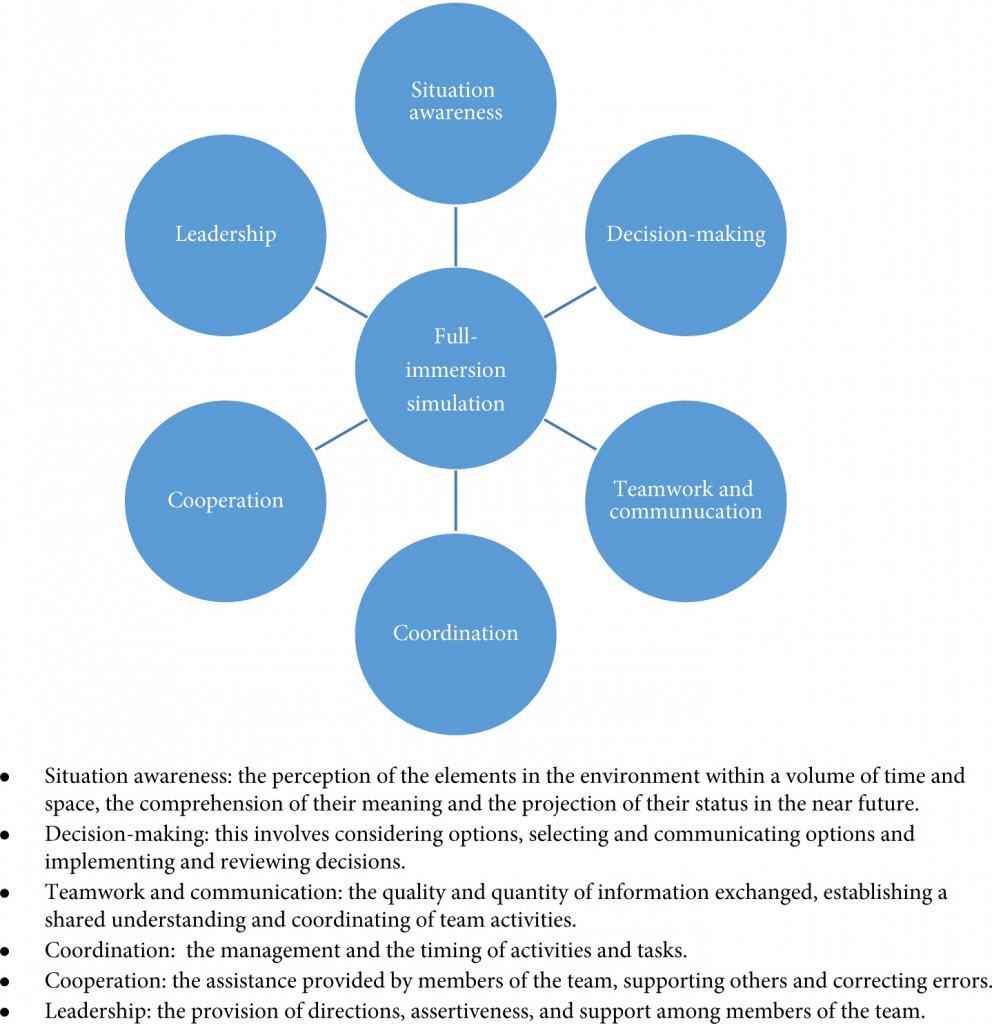
Another aspect worth considering is the integration of non-technical skills within these curricula which has occurred in few of them. Full-immersion simulation (Fig. 1) offers the opportunity for training surgeons to develop critical skills such as communication, coordination and leadership. For the more experienced surgeon, at the later stages of a curriculum, full crisis simulation can help develop decision-making and team-working skills in difficult situations. It has been shown that integration of full-immersion simulation in simulation training is feasible and effective [6]. Surgeons performing robotic surgery need to develop the non-technical aspects alongside their technical skills, and full-immersion simulation certainly has a role to play in this and needs to be integrated within the curricula. The development of the Distributed Simulator offers a validated and low-cost method [7] of introducing full-immersion simulation into robotic surgery curricula and is an option that requires further investigation for its effectiveness in urology.

Figure 1. Aspects of non-technical skills developed through full-immersion simulation [8, 9].

In conclusion, the present study offers further examples of tasks that could be incorporated into robotic surgery training curricula. Whilst curricula exist, implementation and analysis of their effect on patient outcomes are the next steps. The integration of non-technical skills within these is important, and full-immersion simulation has an important role to play within robotic training curricula.

Kamran Ahmed* and Oliver Brunckhorst*

*MRC Centre for Transplantation, King’s College London, King’s Health Partners, and Department of Urology, Guy’s Hospital, London, UK

Read the full article

References

  1. Abboudi H, Khan MS, Guru KA et al. Learning curves for urological procedures: a systematic review. BJU Int 2013; doi: 10.1111/bju.12315. [Epub ahead of print]
  2. Ramos P, Montez J, Tripp A, Ng CK, Gill IS, Hung AJ. Face, content, construct and concurrent validity of dry laboratory exercises for robotic training using a global assessment tool. BJU Int 2014; 113: 836–842
  3. Stegemann AP, Ahmed K, Syed JR et al. Fundamental skills of robotic surgery: a multi-institutional randomized controlled trial for validation of a simulation-based curriculum. Urology 2013; 81: 767–774
  4. Smith R, Patel V, Satava R. Fundamentals of robotic surgery: a course of basic robotic surgery skills based upon a 14-society consensus template of outcomes measures and curriculum development. Int J Med Robot 2013; doi: 10.1002/rcs.1559 [Epub ahead of print]
  5. Khan R, Ahmed K, Mottrie A et al. Towards a standardised training curriculum for robotic surgery: a consensus of an international multidisciplinary group of experts. Poster presented at the EAU Robotic Urology Section Congress Stockholm, Sep 3–5 2013

  6. Shamim Khan M, Ahmed K, Gavazzi A et al. Development and implementation of centralized simulation training: evaluation of feasibility, acceptability and construct validity. BJU Int 2013; 111: 518–523
  7. Kassab E, Tun JK, Arora S et al. ‘Blowing up the barriers’ in surgical training: exploring and validating the concept of distributed simulation. Ann Surg 2011; 254: 1059–1065
  8. Flin R, Yule S, Paterson-Brown S, Maran N, Rowley D, Youngson G. Teaching surgeons about non-technical skills. Surgeon 2007; 5: 86–89
  9. Undre S, Sevdalis N, Healey AN, Darzi A, Vincent CA. Observational teamwork assessment for surgery (OTAS): refinement and application in urological surgery. World J Surg 2007; 31: 1373–1381
Read more articles of the week

AUA 2014 – Friday, Saturday, Sunday: Orlando, FL

As my flight descends into the home of Walt Disney and make believe in sunny Borelando, I can’t help wonder how #AUA14 will compare to the outstanding #EAU14 meeting held just one month ago.  A great meeting requires equal parts place and content, and although Stockholm is without peer, there must be a reason Orlando is the third most visited city in the U.S., right? The solution to that mystery is for another day; ask elsewhere, as I have no idea. Review of the agenda on the #AUA14 app gives hope for this meeting. There is more quality scientific content than one can possibly absorb, and highlights include the new “Crossfire” program to address controversies in urology, the John K. Lattimer Lecture by Dr. Anthony Fauci (director of the National Institute of Allergy and Infectious Diseases), the Town Hall on imaging, simulation and animation (with speakers from Hollywood who make make-believe a reality and a living), #SUO14, and the release of three new AUA Guidelines on urotrauma, medical management of stones, and cryptorchidism.  

Friday afternoon kicked off with the new “Crossfire” section featuring debates on a number of heated urology controversies. Debate topics included the use of synthetic slings for stress urinary incontinence (SUI), the role of urologists in administration of therapy for advanced and metastatic prostate cancer, and the probably overly discussed topic of open versus robotic surgery (for both partial nephrectomy and radical prostatectomy). In favor of synthetic mid-urethral slings for SUI, Drs. Kennelly and Rovner presented a wealth of data showing the efficacy of slings in both the short and long term. Drs. O’Connell and Blaivas countered that the pubovaginal fascial sling provides a safer alternative, with less potentially significant complications that far outweigh the benefits of having the operation. 

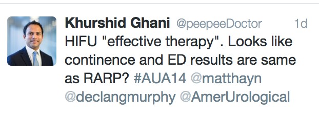
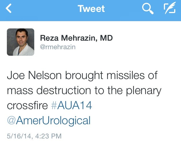
Drs. Nelson and Lepor then argued in favor of open prostatectomy, which drew some colorful tweets:

Drs. Tewari and Menon presented compelling arguments for robotic prostatectomy, and while it is hard to declare a winner, the majority of urologists in the U.S. perform robotic assisted prostatectomy; the panelists stressed that outcomes depend more upon the surgeon than the technique, and people should perform whichever approach they are most comfortable with. 

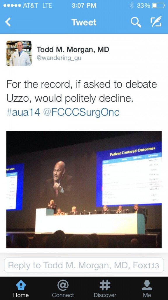
The robotic versus open debate then shifted to kidney surgery, with distinguished faculty Drs. Gill and Uzzo debating “minimally invasive partial nephrectomy is the new gold standard for renal cancer”, while Drs. Blute and Libertino argued in favor of open surgery. Although both sides had thought provoking arguments, presented data were all limited by their retrospective designs, institutional experience, or lack of validation. In my opinion, even with high volume surgeons, most patients with highly complex tumors or a renal mass in a solitary kidney undergo open surgery, which implies selection bias that limits the generalizability of robotic or laparoscopic partial nephrectomy. As contemporary experience with robotic surgery grows, we can anticipate that more complex lesions will be approached via MIS techniques in the future. We always love to throw in “randomized clinical trials are needed”… although I do think that IDEALLY prospective evidence would be great, however a clinical trial comparing MIS partial Nx to open techniques will be fraught with accrual challenges and are most likely not expected in the near future. Until more definitive prospective evidence is available, decisions regarding the optimal surgical approach for the renal mass should be determined by individual patient and surgeon preference, experience and comfort level.

Following the debate, Dr. Todd Morgan nicely summarized audience sentiment:

Dr. Declan Murphy provided perhaps the best sage advice regarding robotic versus open surgery:

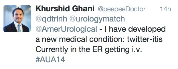
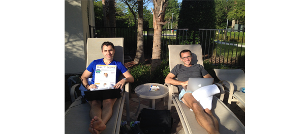
Social Media continues to grow in urology, and Friday evening concluded with a wonderful party hosted by the AUA (@Americanurol) for the “UroTwitterati”. There was a great turnout, and #SoMe heavy hitters: @daviesbj, @declangmurphy, @dr_coop, @qdtrinh, @TheUrologist, @LoebStacy, and @Tdave attended along with “wannabe” youngsters (your current bloggers, @UROncDoc and @RMehrazin). The beauty of #SoMe is that it even allows members to participate in the meeting from home, including @uretericbud and @dytcmd. Urologists should sign up for a Twitter account and join. It is very engaging and addictive!

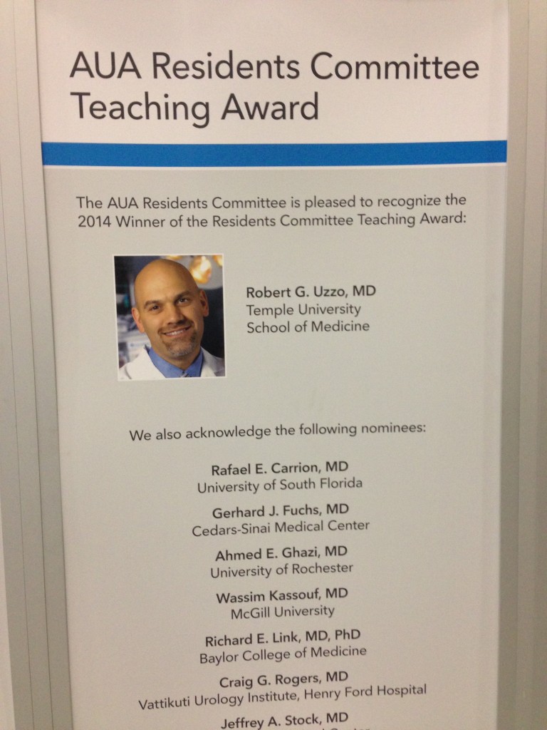
The jam-packed schedule continued on Saturday morning with the annual residents forum, during which the resident teaching award was awarded to Dr. Robert Uzzo from Fox Chase Cancer Center. 

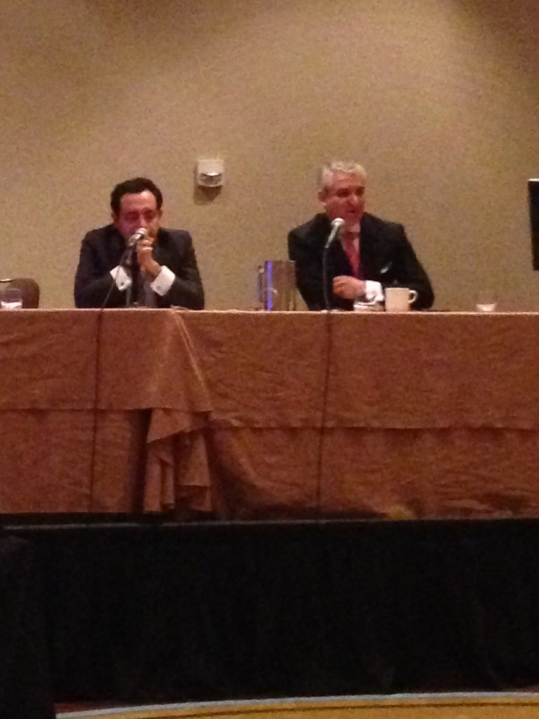
A variety of sections and societies also held meetings on Saturday. At the Association of American-Iranian Urologists, panelists Drs. Ghavamian and Samadi discussed the role of focal therapy in prostate cancer. 

The remainder of Saturday was largely filled by the Society for Basic Urological Research and Society for Urological Oncology annual meetings. One highlight of the #SUO meeting was Dr. McDermott’s presentation on anti-PD-1 agents in kidney cancer. In a phase 1 RCT, Nivolumab (anti PD-L1 agent) showed efficacy for patients with metastatic RCC (n=34). There was a 29% objective response rate with a median progression-free survival time of 7.3 months. The drug was well tolerated with minimal severe adverse events, and remarkably, treatment free survival was achieved in a few patients. Immunotherapy represents an exciting and novel way target kidney cancer, and may well help usher in the era of personalized targeted therapy.

On Sunday, multiple poster and podium sessions were occurring simultaneously, which makes it hard to attend and see everything. The discussion on Twitter via #AUA14 made it possible to capture highlights from simultaneous sessions. During the Plenary session on Sunday, Dr. Fauci, Director of the National Institute of Health, Allergy and Infectious Disease Division, gave the annual Lattimer lecture. AIDS is an important topic for urologists because several aspects of the disease are specific to urology. “For example, the role of STD’s in increasing the transmissibility as well as the vulnerability of getting infected with HIV, the potential role of HPV vaccine in preventing HIV infection, and the importance of urologist issues associated with the drugs HIV patients are taking, including stones, renal insufficiency, voiding dysfunction, and erectile dysfunction,” remarked Dr. Fauci. 

John P. Mulhall, Director of Male Sexual Health and Reproductive Medicine at Memorial Sloan Kettering Cancer Center, delivered the plenary state of the art lecture on radiation induced erectile dysfunction. It is an important topic, because “there are an increasing number of urologists who have hired a radiation oncologist or have a stake in an IMRT unit or do brachytherapy in their practices”, remarked Dr. Mulhall. “The pathophysiology of ED after pelvic radiation is very similar to that after radical prostatectomy based on three factors: nerve injury, arterial injury, and smooth muscle injury”. 

The new AUA clinical guideline for cryptorchidism was also presented at the plenary session on Sunday. The highlights of the guideline:

  1. Orhiopexy is the gold standard treatment for cryptorchidism in 2014
  2. Initial radiographic studies are not necessary for the child with cryptorchidism
  3. Surgery should be performed from 6 to 18 months after birth
  4. Hormonal therapy should not be used as primary therapy to attempt to reposition the testis in the scrotum

Your bloggers,

Reza Mehrazin, M.D. and Jeffrey J. Tomaszewski, M.D.
Fox Chase Cancer Center, Philadephia, PA
Twitter @rmehrazin and @UROncDoc


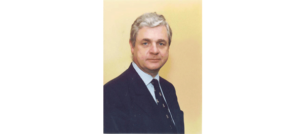
Professor John Fitzpatrick 1948-2014

Professor John Fitzpatrick 1948-2014

A Life in the Fast Lane

Wednesday morning, the 14th May 2014, John M Fitzpatrick passed away aged 65. He left this life the way he lived it, in the fast lane. Taken ill at home in his own gym, where he was honing his fitness with his personal trainer, he was rushed by ambulance to hospital, where he died within hours from a massive subarachnoid haemorrhage. This blog in the BJUI, the journal he edited, championed and loved so very much, is a celebration of his life, and an opportunity for those who knew him to post their own special memory of him, and to contribute a tribute to one of the truly great international characters of urology.

John’s career was an illustrious one. He trained in Dublin, and then in London, where for a time he lived in a house in fashionable Chelsea, just off the King’s Road. He worked with the “greats” of British urology: John Wickham, Richard Turner-Warwick and John Blandy and was always positive and enthusiastic about his time at the famous St Peter’s Hospitals and the Institute of Urology.

Returning to his beloved Dublin, in 1986, aged 38, he successfully applied for the post of Professor of Surgery and proceeded to build up an outstanding department of urology and latterly, with the assistance of the wonderful Bill Watson, created a quite exceptional research unit. He was most proud of his international standing as possibly the world’s best-known urologist (apologies to Dr Patrick Walsh!). He certainly was the most travelled, clocking up untold millions of Air Miles in his favourite seat 2A in the British Airways First Class cabin, and a welcome guest wherever he arrived.

Things, as Richard Turner-Warwick was fond of saying, don’t just happen; they have to be made to happen. Among other things, John did sterling work in helping Bill Hendry and me to create The Urology Foundation (TUF) in 1994, by negotiating £250,000 grants from BAUS and the BJUI. He did a magnificent job as Chairman of the Scientific Committee, Trustee and Patron to help us create a thriving charity. TUF continues to do amazing work to support training and research in urology in the UK and Ireland. He adored being President of BAUS, St Peter’s medal winner and visiting professor to almost 100 academic institutions in North America.

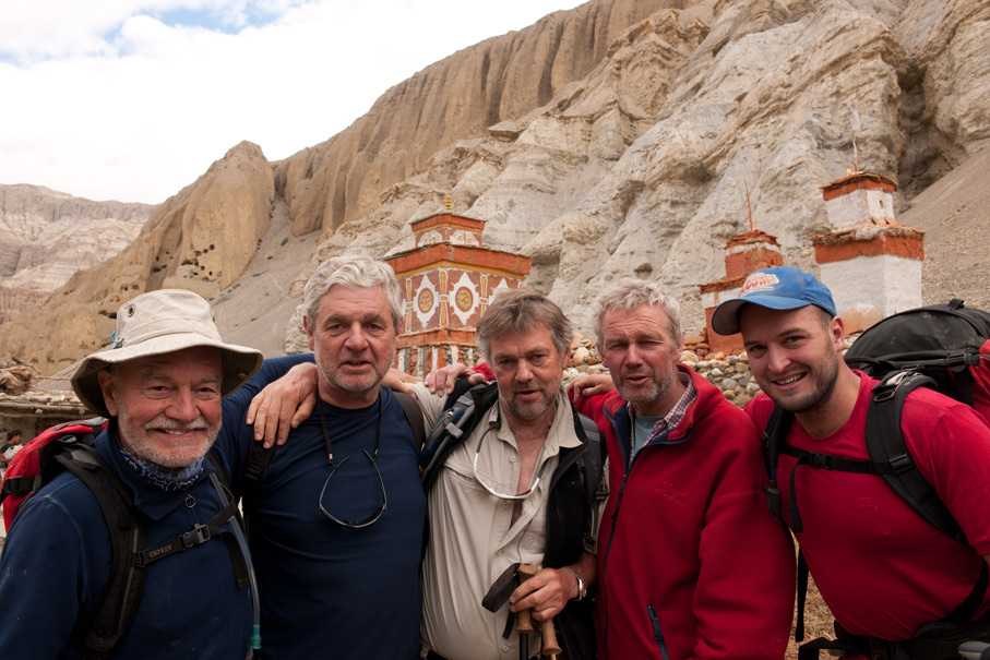
I have too many positive memories of John to regale you with here. Climbing Kilimanjaro (he never tired of reminding me that he reached the summit well before me), trekking in Nepal, cycling in Sicily, Malawi and Madagascar. John was always “up for it”. Another boast of his was that he never misjudged people; but everywhere he went he made friends, took interest in everyone he met and communicated in his own unique, eloquent and quintessentially Irish style.


Sadly, none of us had the opportunity to say goodbye to John. He slipped away from this life, just as he did from so many international meetings, a little early, anxious to move on to the next challenge. My own particular farewell was a few weeks ago at a TUF dinner at the famous and historic Vintner’s Hall in London, where John was in his element talking to Jane MacQuitty, wine correspondent of the Times, about the merits and demerits of a variety of fine wines. With a strange prescience, he told me as he left for the airport the next day that he had enjoyed every moment of his life as a surgeon, scientist and communicator, and that always he really loved the very special world of urology.

Like me, John loved Shakespeare, so I will finish this blog with an apposite quote from the Bard:

His life was gentle, and the elements
So mixed in him that Nature might stand up
And say to all the world, “this was a man!”

When comes such another?

Farewell loyal friend Fitzy, we loved you and we will miss you badly.

Roger Kirby, The Prostate Centre, London


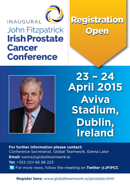
REGISTER FOR THE INAUGURAL JOHN FITZPATRICK IRISH PROSTATE CANCER CONFERENCE

View the programme


An interview with John M. Fitzpatrick
BJUI December 2012; Volume 110, Issue 11

Read the interview here


John-F2b

Cycle-Vietnam-to-Cambodia-2017-Poster

Click here to see a short video on the challenges the TUF cyclists in India faced https://trendsinmenshealth.com/video/tuf-cycle-india-2016/

What’s the diagnosis?

Test yourself against our experts with our weekly quiz. You can type your answers here if you want to compare with our answers, or just click the ‘submit’ button below.

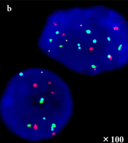
This colourful picture is of a multicolour FISH analysis in non-muscle invasive bladder cancer.
Image from Matsuyama et al. BJU Int 2014; 113: 662–667

No such quiz/survey/poll
If you have a suggestion for a new Picture Quiz please email us.


© 2024 BJU International. All Rights Reserved.