Tag Archive for: #BJUI

Posts

Editorial: Retzius‐sparing robot‐assisted radical prostatectomy

In their commentary in the current issue of BJUI, Stonier et al. [1] examine the potential technical pitfalls and published results of the Retzius‐sparing technique of robotic radical prostatectomy. The authors reviewed three studies from three different groups [2,3], including a study by our group [4], and raised three specific concerns: the oncological efficacy of the procedure; the long learning curve; and the generalizability of the technique to challenging surgical scenarios. We offer a few clarifications and comments.

The first study on Retzius‐sparing robot‐assisted radical prostatectomy came from the Bocciardi group [2]. This was a prospective, single‐arm study of 200 patients. The authors reported a 14‐day continence rate of 90–92%, a 1‐year potency rate of 71–81% (in preoperatively potent patients undergoing bilateral intrafascial nerve‐sparing) and a positive surgical margin rate of 25.5%. The positive surgical margin rate improved in patients with pT2 disease, from 22% to 9% (P = 0.04) over the course of the study (initial 100 vs subsequent 100 patients), while in patients with pT3 disease, it remained stable at ~45%. Lim et al. [3] also noted an improvement in their overall positive surgical margin rate from 20% to 8% when comparing the initial 25 patients with the subsequent 25 patients. In that study, a standard robot‐assisted radical prostatectomy comparator arm was included and there were no differences in overall positive surgical margin rates (14% in both arms), while continence was better with the Retzius‐sparing approach.

Recognizing the potentially technically challenging nature of the Bocciardi approach, we performed a randomized controlled trial to objectively evaluate the technique. Randomized controlled trials are typically designed to answer a single question. Our trial was designed to determine whether there were differences in the rate of return of urinary continence, the primary benefit that previous non‐controlled studies had reported. This our study clearly showed [4].

Once the trial was completed, post hoc analysis of secondary outcomes was performed [5]. One of these outcomes was the positive surgical margin rate. In our trial, we noted an overall positive surgical margin rate of 25% in the Retzius‐sparing arm vs 13% in the control arm, a difference that did not achieve statistical significance (P = 0.11). Stonier et al. [1] suggested that if the sample size of our trial were doubled, then the positive surgical margin rate in each group would be doubled as well, leading to significance. This conclusion is problematic. The likelihood that doubling the sample size would result in the exact doubling of numbers in all four cells of a 2 × 2 contingency table is estimated at <5% using Fisher’s exact test (this calculation is different from the P value). Furthermore, the surgical margins depend as much on the pathological stage as on surgical approach. In our trial, patients were matched preoperatively for risk in the best manner possible for a pragmatic randomized trial. However, it is impossible to predict and control for the final pathological characteristics. Pathological analysis showed that patients undergoing Retzius‐sparing surgery did have significantly more aggressive disease: ≥pT3 disease in 45% vs 23.3% of patients (P = 0.04) [4, 5]. This, by itself, could account for a substantial difference in surgical margin rates.

In writing our paper, we made no judgements as to whether the Bocciardi or posterior technique is fundamentally superior to an anterior or Menon approach, whether it is easier to perform, how generalizable it is [6], or what the learning curve may be. That is best left to the individual surgeon’s training and judgement. We do suggest, however, that surgical margins be interpreted as a function of pathological variables, and not in isolation, and that it is simplistic to assume that identical results will be obtained by doubling sample size. We suggest that such conclusions are hypothesis‐generating, and should best be explored through a separate, purpose‐designed randomized trial.

Authors: Akshay Sood, Firas Abdollah and Mani Menon

References

  1. Stonier T, Simson N, Davis J, Challacombe B. Retzius‐sparing robot‐assisted radical prostatectomy (RS‐RARP) vs standard RARP: it’s time for critical appraisal. BJU Int 2019; 123: 5–10
  2. Galfano A, Di Trapani D, Sozzi F et al. Beyond the learning curve of the Retzius‐sparing approach for robot‐assisted laparoscopic radical prostatectomy: oncologic and functional results of the first 200 patients with >/= 1 year of follow‐up. Eur Urol 2013; 64: 974–80
  3. Lim SK, Kim KH, Shin TY et al. Retzius‐sparing robot‐assisted laparoscopic radical prostatectomy: combining the best of retropubic and perineal approaches. BJU Int 2014; 114: 236–44
  4. Dalela D, Jeong W, Prasad MA et al. A pragmatic randomized controlled trial examining the impact of the Retzius‐sparing approach on early urinary continence recovery after robot‐assisted radical prostatectomy. Eur Urol 2017; 72: 677–85
  5. Menon M, Dalela D, Jamil M et al. Functional recovery, oncologic outcomes and postoperative complications after robot‐assisted radical prostatectomy: an evidence‐based analysis comparing the Retzius sparing and standard approaches. J Urol 2018; 199: 1210–7
  6. Galfano A, Secco S, Bocciardi AM. Will Retzius‐sparing prostatectomy be the future of prostate cancer surgery? Eur Urol 2017; 72: 686–8


Editorial: Reply: RS-RARP vs standard RARP

Since the introduction of robotic surgery in the treatment of patients with prostate cancer (PCa), different surgical innovations have been implemented in order to preserve postoperative functional outcomes while maintaining oncological safety. Sparing the Retzius space during robot‐assisted radical prostatectomy (RARP) was introduced early this decade by Galfano et al [1]. Interestingly, 90% and 96% of patients treated with Retzius‐sparing RARP (RS‐RARP) were continent (no pad/safety pad) at 1 week and 1 year, respectively. Similarly, our group reported a 70% continence rate (no pad) at 1 month after RS‐RARP [2].

The fast urinary continence recovery after RS‐RARP is related to several anatomical factors: the anterior Retzius space is kept intact; the urinary bladder is not dropped; the endopelvic fascia and puboprostatic ligaments are preserved; and there is minimal distortion of the supporting urethral tissues. A recent study reported [3] that less bladder neck descent was observed during postoperative cystogram in patients treated with RS‐RARP than in those treated with standard RARP.

In a recent randomized controlled study, the postoperative continence rate at 1 week was 48% in standard RARP compared with 71% in RS‐RARP (P = 0.01), and this difference was maintained at 3 months (86% standard RARP vs 95% RS‐RARP; P = 0.02). At 1 year, however, the effect on urinary continence difference was muted (93.3% standard RARP vs 98.3% RS‐RARP; P = 0.09) [4]. Similarly, Chang et al. [3] found that the higher continence rate at 1 week (73.3% RS‐RARP vs 26.7% standard RARP; P = 0.000) had vanished at 1 year (100% vs 93.3%; P = 0.15). By contrast, a large recent prospective series showed that the superiority of RS‐RARP in terms of higher early urinary continence was maintained at 1 year (97.5% RS‐RARP vs 68.5% standard RARP) [5].

In addition to a higher early continence rate, RS‐RARP has a lower incidence of postoperative inguinal hernia occurrence compared with standard RARP [6]. Theoretically, RS‐RARP may provide several other potential advantages. It may be advantageous if patients require future surgery necessitating access to the Retzius space and dropping of the bladder, such as an artificial urinary sphincter implantation, an inflatable penile prosthesis insertion, or kidney transplantation. In addition, in patients with previous inguinal hernia repair using mesh, it enables the avoidance of anterior adhesions by accessing the prostate directly from the Douglas pouch. Notably, large‐size glands and/or middle‐lobe, advanced/high‐risk PCa, and patients with previous prostatic surgeries can be managed safely with RS‐RARP in experienced hands.

Undoubtedly, oncological safety is our main concern in treating cancer. To determine the effectiveness of new treatment methods, long‐term follow‐up is warranted. Biochemical recurrence (BCR) is widely used as a primary oncological outcome to assess PCa treatment success. To our knowledge, after radical prostatectomy, ~35% of patients are at risk of developing BCR in the next 10 years. Currently, there are insufficient data regarding the oncological outcomes of RS‐RARP. Only four articles have compared early oncological outcomes between RS‐RARP and standard RARP, and there was no significant difference (Table 1).

More recently, we reported on the mid‐term oncological outcomes of 359 patients who underwent RS‐RARP. The median follow‐up was 26 months. Although this period is not long enough to reach a meaningful conclusion on the oncological safety of RS‐RARP, it is the longest follow‐up period reported in literature. Overall, the positive surgical margin (PSM) rate was 30.6% (14.6% in pT2 and 40.8% in pT3a disease) and the BCR rate was 14.8%. In terms of functional outcomes, the urinary continence rate at 1 year was 93.9% [7]. Interestingly, 164 patients (45.7%) of our cohort had high‐risk PCa. In these patients, the PSM rate was 41.2%, the BCR rate was 22%, and the 3‐year BCR‐free survival (BCRFS) rate was 72%. We compared our results with those in patients with high‐risk PCa treated with standard RARP in the literature. In studies that used the D’Amico criteria the median follow‐up ranged from 12.5 to 37.3 months, the PSM rates were 20.5% to 53.3%, the BCR rates were 17.4% to 31% and the 3‐year BCRFS rates were 41.4% to 86%. In studies that used the National Comprehensive Cancer Network criteria, the median follow‐up ranged from 23.6 to 27 months, the PSM rates were 29% to 38%, the BCR rates were 9.4% to 33%, and the 3‐year BCRFS rates were 55% to 66% [7].

In summary, RS‐RARP is a novel surgical approach which is associated with better urinary continence recovery in the first few months compared with standard RARP [2,3,4,5]. This superiority might be maintained [5] or equalized at 1 year [3,4]. A few studies have compared the early oncological results between RS‐RARP and standard RARP and no significant difference was found [2,3,4,5]. Recently, our group reported the mid‐term oncological outcomes of patients with high‐risk PCa treated with RS‐RARP and these were similar to those of large studies of conventional RARP. This confirms effective and safe mid‐term BCR control after RS‐RARP, while the long‐term oncological results are awaited [7]. Currently, >4 000 cases of RS‐RARP are performed worldwide and more centres are beginning to use and converting to Retzius‐sparing surgery. All centres are experiencing faster recovery of continence. Thanks are due to Drs Galfano and Bocciardi for exploring and sharing this surgical frontier.


References

  1. Galfano A, Di Trapani D, Sozzi F, et al. Beyond the learning curve of the Retzius‐sparing approach for robotassisted laparoscopic radical prostatectomy: oncologic and functional results of the first 200 patients with ? 1 year of follow‐up. Eur Urol 2013; 64: 974‐80
  2. Lim SK, Kim KH, Shin TY et al. Retzius‐sparing robot‐assisted laparoscopic radical prostatectomy: combining the best of retropubic and perineal approaches. BJU Int 2014; 114: 236–44
  3. Chang LW, Hung SC, Hu JC et al. Retzius‐sparing robotic‐assisted radical prostatectomy associated with less bladder neck descent and better early continence outcome. Anticancer Res 2018; 38: 345–51
  4. Menon M, Dalela D, Jamil M et al. Functional recovery, oncologic outcomes and postoperative complications after robot‐assisted radical prostatectomy: an evidence‐based analysis comparing the Retzius sparing and standard approaches. J Urol 2018; 199: 1210–7
  5. Sayyid RK, Simpson WG, Lu C et al. Retzius sparing robotic assisted laparoscopic radical prostatectomy: a safe surgical technique with superior continence outcomes. J Endourol 2017; 31: 1244–50
  6. Chang KD, Abdel Raheem A, Santok GDR et al. Anatomical Retzius‐space preservation is associated with lower incidence of postoperative inguinal hernia development after robot‐assisted radical prostatectomy. Hernia 2017; 21: 555–61
  7. Abdel Raheem A, Kidon C, Alenzi M et al. Predictors of biochemical recurrence after retzius‐sparing robot‐assisted radical prostatectomy: analysis of 359 cases with a median follow‐up of 26 months. Int J Urol 2018; 25: 1006–14


Resident’s podcast: Retzius‐sparing robot‐assisted radical prostatectomy

Maria Uloko is a Urology Resident at the University of Minnesota Hospital. In this podcast she discusses the following BJUI Article of the Week:

Retzius‐sparing robot‐assisted radical prostatectomy (RS‐RARP) vs standard RARP: it’s time for critical appraisal

Thomas Stonier*, Nick Simson*, John Davisand Ben Challacombe


*Department of Urology, Princess Alexandra Hospital, Harlow, Urology Centre, Guy s Hospital, London, UK and Department of Urology, MD Anderson Cancer Center, Houston, TX, USA


Read the full article

Abstract

Since robot‐assisted radical prostatectomy (RARP) started to be regularly performed in 2001, the procedure has typically followed the original retropubic approach, with incremental technical improvements in an attempt to improve outcomes. These include the running Van‐Velthoven anastomosis, posterior reconstruction or ‘Rocco stitch’, and cold ligation of the Santorini plexus/dorsal vein to maximise urethral length. In 2010, Bocciardi’s team in Milan proposed a novel posterior or ‘Retzius‐sparing’ RARP (RS‐RARP), mirroring the classic open perineal approach. This allows avoidance of supporting structures, such as the puboprostatic ligaments, endopelvic fascia, and Santorini plexus, preserving the normal anatomy as much as possible and limiting damage that may contribute to improved postoperative continence and erectile function. There has been much heralding of the excellent functional outcomes in both the medical and the lay press, but as yet no focus or real mention of any potential downsides of this new technique.

Read more Articles of the week


BJUI Podcasts now available on iTunes, subscribe here https://itunes.apple.com/gb/podcast/bju-international/id1309570262


Article of the Month: Use of machine learning to predict early biochemical recurrence after robot‐assisted prostatectomy

Every month, the Editor-in-Chief selects an Article of the Month from the current issue of BJUI. The abstract is reproduced below and you can click on the button to read the full article, which is freely available to all readers for at least 30 days from the time of this post.

In addition to the article itself, there is an accompanying editorial written by a prominent member of the urological community. This blog is intended to provoke comment and discussion and we invite you to use the comment tools at the bottom of each post to join the conversation.

If you only have time to read one article this week, it should be this one.

Use of machine learning to predict early biochemical recurrence after robot‐assisted prostatectomy

Nathan C. Wong , Cameron Lam, Lisa Patterson and Bobby Shayegan
Division of Urology, Department of Surgery, McMaster University, Hamilton, ON, Canada

Read the full article
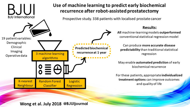
Visual abstract created Rebecca Fisher @beckybeckyfish

Abstract

Objectives

To train and compare machine‐learning algorithms with traditional regression analysis for the prediction of early biochemical recurrence after robot‐assisted prostatectomy.

Patients and Methods

A prospectively collected dataset of 338 patients who underwent robot‐assisted prostatectomy for localized prostate cancer was examined. We used three supervised machine‐learning algorithms and 19 different training variables (demographic, clinical, imaging and operative data) in a hypothesis‐free manner to build models that could predict patients with biochemical recurrence at 1 year. We also performed traditional Cox regression analysis for comparison.

= 0.686) and with a univariate regression model (AUC = 0.865).

Results

K‐nearest neighbour, logistic regression and random forest classifier were used as machine‐learning models. Classic Cox regression analysis had an area under the curve (AUC) of 0.865 for the prediction of biochemical recurrence. All three of our machine‐learning models (K‐nearest neighbour (AUC 0.903), random forest tree (AUC 0.924) and logistic regression (AUC 0.940) outperformed the conventional statistical regression model. Accuracy prediction scores for K‐nearest neighbour, random forest tree and logistic regression were 0.976, 0.953 and 0.976, respectively.

Conclusions

Machine‐learning techniques can produce accurate disease predictability better that traditional statistical regression. These tools may prove clinically useful for the automated prediction of patients who develop early biochemical recurrence after robot‐assisted prostatectomy. For these patients, appropriate individualized treatment options can improve outcomes and quality of life.

Read more Articles of the week

Editorial: Can machine‐learning algorithms replace conventional statistics?

Wong et al. [1] evaluate 19 clinical variables (training data) and three supervised machine‐learning algorithms to predict early biochemical recurrence after robot‐assisted prostatectomy. They further compare the areas under the curve (AUCs) resulting from these algorithms with the AUC of a conventional Cox regression model and conclude that the machine‐learning algorithms can produce accurate disease prognosis, perhaps better than a traditional Cox regression model. As the authors state, predictive models have the potential to better individualize care to patients at highest risk of prostate cancer recurrence and progression.

The authors should be commended for their adoption of machine‐learning algorithms to better interpret the vast volumes of clinical data and assess prognosis after robot‐assisted prostatectomy. This should represent another step forward for the management of prostate cancer, where tailored treatment is now largely based on the clinical risk stratification of the disease [2]. Incidentally, we are also in an era where we are seeing aspects of artificial intelligence (machine learning being a subset of it) vastly transform how we view and process data in everyday life. This has been true in medicine as well, particularly for prostate cancer [3].

While our own research group has also evaluated machine‐learning algorithms to process surgeon performance metrics and predict clinical outcomes after robot‐assisted prostatectomy [4], I want to express a word of caution. Utilization of machine learning does not in itself imply automatic superiority over conventional statistics [5] despite literature that has demonstrated so [3]. The success of predictive models in machine learning still relies on the quality of data introduced and careful execution of the analysis. In our experience, it works best when highly experienced clinicians and data scientists are working hand in hand.

Furthermore, I would argue that the results of this present study do not necessarily show that machine learning is superior to conventional statistics, but rather it highlights an inherent advantage of machine learning. While traditional analyses require the a priori selection of a model based on the available data, machine learning has more flexibility for model fitting [6]. Additionally, inclusion of variables in traditional analyses is constrained by the sample size. In contrast, by design, machine learning models thrive on their ability to consider many variables concurrently, and as such, have the potential to detect underlying patterns that may otherwise be undetectable when data are examined effectively in individual silos.

We look forward to the external validation of the methodology described in the present article. Big and diverse data are critical requirements of machine learning. A multi‐institutional, multi‐surgeon cohort is necessary to confirm the findings in this report. A further step from there is the adoption of such prediction models into clinical use. The ultimate question is how improved prognostic data may influence surgeon and patient decisions.

Conflict of Interest

Dr Hung reports personal fees from Ethicon, Inc, outside the submitted work.

References

  1. Wong NC, Lam C, Patterson L, Shayegan B. Use of machine learning to predict early biochemical recurrence following robotic prostatectomy. BJU Int 2019; 123: 51–7
  2. D’Amico AV, Whittington R, Malkowicz SB et al. Biochemical outcome after radical prostatectomy, external beam radiation therapy or interstitial radiation therapy for clinically localized prostate cancer. JAMA 1998; 280: 969–74
  3. Hung AJ, Chen J, Che Z et al. Utilizing machine learning and automated performance metrics to evaluate robot‐assisted radical prostatectomy performance and predict outcomes. J Endourol 2018; 32: 438–445
  4. Kattan MW. Comparison of Cox regression with other methods for determining prediction models and nomograms. J Urol 2003; 170 (6 Pt 2): S6–9
  5. Hung AJ, Chen J, Gill IS. Automated performance metrics and machine learning algorithms to measure surgeon performance and anticipate clinical outcomes in robotic surgery. JAMA Surg 2018; 153: 770–1

Editorial: Predicting progression in T1 non‐muscle‐invasive bladder cancer: back to histology

Stage pT1 bladder carcinomas (BCs) represent a difficult clinical scenario as they have different outcomes and are associated with a high risk of progression to muscle‐invasive tumours. The optimal therapeutic approach for individual patients in this setting is still unclear: conservative treatment with BCG instillation and intravesical chemotherapy may lead to disease progression and death, while radical cystectomy may represent a mutilating overtreatment for patients with tumours that may have low potential for progression.

The ability to discriminate those patients who will probably progress to carcinoma invading bladder muscle is therefore crucial. Among prognostic factors associated with progression to muscle invasion, tumour grade is one of the most important. In their important paper, van de Putte et al. [1] aimed to compare the prognostic value of the WHO 1973 and 2004 grading systems, the latter being recommended by the AUA guidelines as the most widely accepted in the USA [2], although it has not been proven superior to the other [3].

The authors collected transurethral resections from 601 primary T1 BCs, initially managed conservatively (BCG), from four institutions, and three pathologists reviewed the slides. Importantly, a second transurethral resection was performed if the muscularis propria was absent and/or the initial resection was incomplete. Grade was assigned according to the WHO 1973 (G1–3) and WHO 2004 (low grade [LG] and high grade [HG]) systems. None of the cases was classified as G1. The prognostic value of both grading systems for progression‐free and cancer‐specific survival was then assessed. Notably, the author found WHO1973 G3 to be significantly negatively associated with progression‐free survival and cancer‐specific survival on multivariable analysis, while the WHO 2004 grading system was not. Importantly, intra‐observer variability was assessed in 66 cases and was found to be almost perfect for the WHO 1973 and moderate to substantial for the WHO 2004 system, while inter‐observer variability ranged from moderate to substantial for both systems. One of the reasons for the lack of prognostic potential of the WHO 2004 system, as underscored by the authors, is the fact that the morphological criteria defined in the WHO 2004 system cause an important shift of many cases from the G2 to HG category, rendering it an almost one‐tier system with consequently very few LG tumours. Other studies have assessed the prognostic value of the WHO 1973 and WHO 2004 systems [3] but so far no clear superiority emerged for one system over the other, probably because of relatively low sample sizes.

Other clinical prognostic factors associated with progression to muscle‐invasive tumours include tumour dimension, the presence of multiple lesions, the presence of carcinoma in situ, lymphovascular invasion and level of lamina propria invasion. Regarding the latter prognostic factor, different studies have defined T1 sub‐staging according to invasion above (T1a), within (T1b) or beyond (T1c) the muscularis mucosae and vascular plexus; however, this approach has been found not to be applicable in >40% of cases because of difficulties in identifying the vascular plexus or lack of orientation of the specimens. A more friendly and reproducible method has been proposed by some of the authors of the study, consisting of a categorization of T1 BCs into microinvasive (T1m) and extensively invasive (T1e) tumours, which has been demonstrated to be applicable in 100% of cases and more reproducible [4]. Further study incorporating T1 sub‐staging together with grade may prove very useful.

Different studies have been performed to identify prognostic markers at the molecular level; however, despite huge efforts, no molecular biomarker with prognostic potential is currently suitable for clinical application [5]. Moreover, in six studies that investigated T1 sub‐stage and molecular markers in the same series, T1 sub‐stage showed the highest prognostic value [4]. More recently, subtyping BC into basal‐like and genomically unstable or squamous cell carcinoma‐like tumours has emerged as a promising tool for dividing T1 BCs into low‐ and high‐risk categories [6]; however, such an approach must be combined with the prognostic value of the classic histological variables discussed so far before eventually being integrated into prognostic tools.

In this regard, van de Putte et al. [1] have shown that tumour grade still represents a powerful marker in T1 BC and that the WHO 2004 grading system cannot replace the WHO 1973 system as a prognosticator of T1 BC; therefore, as recommended by the European Association of Urology guidelines, the WHO 1973 grading system categories should always be present in the pathology reports.


References

  1. van de Putte EEF, Bosschieter J, van der Kwast TH et al. The World Health Organization 1973 classification system for grade is an important prognosticator in T1 non‐muscle‐invasive bladder cancer. BJU Int 2018; 122: 978–85
  2. Chang SS, Boorjian SA, Chou R et al. Diagnosis and treatment of non‐muscle invasive bladder cancer: AUA/SUO guideline. J Urol 2016; 196: 1021–93
  3. Babjuk M, Bohle A, Burger M et al. EAU guidelines on non‐muscle‐invasive urothelial carcinoma of the bladder: update 2016. Eur Urol 2017; 71: 447–614
  4. van Rhijn BW, Liu L, Vis AN et al. Prognostic value of molecular markers, sub‐stage and European Organisation for the Research and Treatment of Cancer risk scores in primary T1 bladder cancer. BJU Int 2012; 110: 1169–76
  5. Munari E, Chaux A, Maldonado L et al. Cyclin A1 expression predicts progression in pT1 urothelial carcinoma of bladder: a tissue microarray study of 149 patients treated by transurethral resection. Histopathology 2015; 66: 262–9
  6. Patschan O, Sjodahl G, Chebil G et al. A molecular pathologic framework for risk stratification of stage T1 urothelial carcinoma. Eur Urol 2015; 68: 824–32


Guideline of guidelines: primary monotherapies for localised or locally advanced prostate cancer

Abstract:

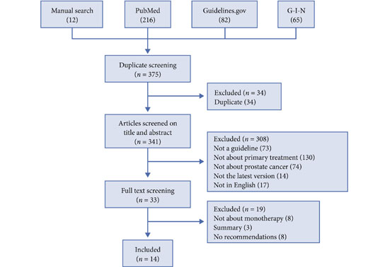
Decisions regarding the primary treatment of prostate cancer depend on several patient‐ and disease‐specific factors. Several international guidelines regarding the primary treatment of prostate cancer exist; however, they have not been formally compared. As guidelines often contradict each other, we aimed to systematically compare recommendations regarding the different primary treatment modalities of prostate cancer between guidelines. We searched Medline, the National Guidelines Clearinghouse, the library of the Guidelines International Network, and the websites of major urological associations for prostate cancer treatment guidelines. In total, 14 guidelines from 12 organisations were included in the present article. One of the main discrepancies concerned the definition of ‘localised’ prostate cancer. Localised prostate cancer was defined as cT1–cT3 in most guidelines; however, this disease stage was defined in other guidelines as cT1–cT2, or as any T‐stage as long as there is no lymph node involvement (N0) or metastases (M0). In addition, the risk stratification of localised cancer differed considerably between guidelines. Recommendations regarding radical prostatectomy and hormonal therapy were largely consistent between the guidelines. However, recommendations regarding active surveillance, brachytherapy, and external beam radiotherapy varied, mainly as a result of the inconsistencies in the risk stratification. The differences in year of publication and the methodology (i.e. consensus‐based or evidence‐based) for developing the guidelines might partly explain the differences in recommendations. It can be assumed that the observed variation in international clinical practice regarding the primary treatment of prostate cancer might be partly due to the inconsistent recommendations in different guidelines.

Michelle Lancee, Kari A.O. Tikkinen, Theo M. de Reijke, Vesa V. Kataja, Katja K.H. Aben and Robin W.M. Vernooij

Access the full article

Article of the Week: NICE Advice – Prolaris Gene Expression Assay

Every Week, the Editor-in-Chief selects an Article of the Week from the current issue of BJUI. The summary is reproduced below and you can click on the button to read the full article, which is freely available to all readers for at least 30 days from the time of this post.

If you only have time to read one article this week, it should be this one.

NICE Advice – Prolaris gene expression assay for assessing long‐term risk of prostate cancer progression

Read the full article

Read more articles of the week

Resident’s Podcast: NICE Guidance – ceftolozane & tazobactam for complicated UTIs

Eleanor Zimmermann is due to start her Urology registrar training in the Southwest this October, and is a BURST Core Surgical Trainee Representative. @BURSTUrology

In this Residents’ Podcast, Eleanor discusses the NICE Guidance on complicated urinary tract infections: ceftolozane/tazobactam


BJUI Podcasts now available on iTunes, subscribe here https://itunes.apple.com/gb/podcast/bju-international/id1309570262


Video: β3‐adrenoceptor agonists inhibit carbachol‐evoked Ca2+ oscillations in murine detrusor myocytes

β3‐adrenoceptor agonists inhibit carbachol‐evoked Ca2+ oscillations in murine detrusor myocytes


Read the full article

Abstract

Objective

To test if carbachol (CCh)‐evoked Ca2+ oscillations in freshly isolated murine detrusor myocytes are affected by β3‐adrenoceptor (β‐AR) modulators.

Materials and Methods

Isometric tension recordings were made from strips of murine detrusor, and intracellular Ca2+ measurements were made from isolated detrusor myocytes using confocal microscopy. Transcriptional expression of β‐AR sub‐types in detrusor strips and isolated detrusor myocytes was assessed using reverse transcriptase‐polymerase chain reaction (RT‐PCR) and real‐time quantitative PCR (qPCR). Immunocytochemistry experiments, using a β3‐AR selective antibody, were performed to confirm that β3‐ARs were present on detrusor myocytes.

Results

The RT‐PCR and qPCR experiments showed that β1‐, β2‐ and β3‐AR were expressed in murine detrusor, but that β3‐ARs were the most abundant sub‐type. The selective β3‐AR agonist BRL37344 reduced the amplitude of CCh‐induced contractions of detrusor smooth muscle. These responses were unaffected by addition of the BK channel blocker iberiotoxin. BRL37344 also reduced the amplitude of CCh‐induced Ca2+ oscillations in freshly isolated murine detrusor myocytes. This effect was mimicked by CL316,243, another β3‐AR agonist, and inhibited by the β3‐AR antagonist L748,337, but not by propranolol, an antagonist of β1‐ and β2‐ARs. BRL37344 did not affect caffeine‐evoked Ca2+ transients or L‐type Ca2+ current in isolated detrusor myocytes.

Conclusion

Inhibition of cholinergic‐mediated contractions of the detrusor by β3‐AR agonists was associated with a reduction in Ca2+ oscillations in detrusor myocytes.

View more videos


© 2024 BJU International. All Rights Reserved.