Tag Archive for: #BJUI

Posts

Editorial: Evidence trumps consensus

We read with great interest the article by Khetrapal et al. [1]. Certain advantages of robotic cystectomy have been shown in retrospective studies and confirmed in the RAZOR trial [2]. Robotic cystectomy has been associated with lower blood loss, lower transfusion rates and a shorter length of stay; however, two randomized trials have shown no difference in complication rates, which was the original reason robotic cystectomy was attempted [2,3]. Khetrapal et al. seem to believe that this was because diversions were performed extracorporeally, and intracorporeal diversion would allow urologists to uncover the true benefit of robotic cystectomy. When the RAZOR trial was being designed (in 2009), intracorporeal diversion was early in development. Even today its use in the USA is restricted to a few centres and the Pasadena consensus statement (2015) acknowledges that only 3% of all diversions were performed intracorporeally [4]. While more commonly performed in Europe, intracorporeal diversions still form the minority of all urinary diversions. To date there are no reliable prospective data to convince us that intracorporeal diversion is superior, and the low quality of available evidence has been acknowledged in the Pasadena statement [4]. The iROC trial is a step in the right direction and we await its results with interest [5].

We agree with the authors that cost analysis is essential in evaluating the exact role of robotic cystectomy. It is also worth factoring in the indirect costs of the two procedures, given that most patients undergoing robotic cystectomy will have a shorter hospital stay and fewer blood transfusions, although robotic cystectomies may take longer to perform. We anticipate that as newer robotic systems are introduced the direct surgical costs may be reduced.

There is no universally accepted learning curve for performing a cystectomy based on prospective studies. Ten cystectomies in the preceding year before enrolment in the RAZOR trial was the lowest number of cystectomies permitted for the surgeon to be eligible to participate [2]. All surgeons were fellowship-trained with high-volume bladder cancer practices, and the majority had performed significantly more than 10 cystectomies. The high quality of surgical surrogates for both approaches that we reported, namely, lymph node yield, positive margins and complication rates, are testament to this. We believe that the authors’ statement that novice surgeons may have operated on trial patients is simply inaccurate. It is largely self-serving to fit the results of the RAZOR trial into their own narrative about their beliefs in the advantages of robotic surgery. The iROC trial requires surgeons to have carried out 30 or more intracorporeal diversions in their entire career, with accredited surgeons being required to perform more than 10 cystectomies per year for the last 2 years as primary surgeon, which does not seem remarkably different from the RAZOR trial criteria for surgeon participation [5].While it is clear that large volumes are associated with better outcomes, the magic number is unclear. The Pasadena Consensus Statement cites the National Institute for Health and Care Excellence (NICE) guidelines in the UK, which mandate a minimum of five cystectomies per year per surgeon as adequate surgical volume [4].

Operating time in RAZOR was defined as the time from patient entry to the time the patient exited the operating theatre [2]. In most instances, the time for positioning and anaesthesia (preparation and induction) before making any incision and the time after closure for extubation and leaving the room is generally ~60–80 min. The Pasadena Consensus statement recommends that experienced surgeons should aim to complete robotic cystectomies within 5–6 h, depending on the type of diversion, basing their recommendation on three available studies [4]. Of those papers, Hayn et al. (overall mean operating time 386 min and mean operating time after 50th case 339 min) and Richards et al. (mean operating time 449 min after 40th case of learning curve) defined operating time in their papers as incision to closure time [6,7]. The paper by Collins et al. does not define operating time; however, the mean operating time for cystectomy with intracorporeal diversion for both surgeons in that study was 438 min, and 87.5% of the cases selected in this study had ≤pT2 disease, suggesting a significant selection bias [8]. This institution is a part of the International Robotic Cystectomy Consortium (IRCC) which defines operating time as incision to closure time, leading us to believe that this was the probably the definition they used [8]. A recent study from the IRCC reported a mean operating time (incision to closure) of 364 min in 2134 patients [9]. All these data suggest that operating times in RAZOR were extremely competitive if not actually faster, once again attesting to the proficiency of the participating surgeons. Khetrapal et al. would have reached a different conclusion about the RAZOR trial results had they accurately interpreted the scientific data from the above-mentioned studies.

The RAZOR trial provided level 1 evidence proving the oncological efficacy of robotic cystectomy and confirming advantages such as reduced blood loss and length of stay [2]. We agree that the true place for robotic cystectomy will be determined once a cost–benefit analysis can be performed, and after we obtain high-level prospective data about intracorporeal diversions. To this end, we look forward to the successful completion of the iROC trial and await its publication. Until such time, we suggest more reliance on high-level evidence than on consensus statements and narratives.

by Vivek Venkatramani and Dipen J. Parekh on behalf of RAZOR trial investigators

References

  1. Khetrapal P, Kelly J, Catto J, Vasdev N. Does the robot have a role in radical cystectomy? BJU Int 2019; 123(3): 380-2.
  2. ParekhDJ, Reis IM, Castle EP et al. Robot-assisted radical cystectomy versus open radical cystectomy in patients with bladder cancer (RAZOR): an openlabel, randomised, phase 3, non-inferiority trial. Lancet 2018; 391: 2525–36
  3. Bochner BH, Dalbagni G, Sjoberg DD et al. Comparing open radical cystectomy and robot-assisted laparoscopic radical cystectomy: a randomized clinical trial. Eur Urol 2015; 67: 1042–50
  4. Wilson TG, Guru K, Rosen RC et al. Best practices in robot-assisted radical cystectomy and urinary reconstruction: recommendations of the Pasadena Consensus Panel. Eur Urol 2015; 67: 363–75
  5. Catto JWF, Khetrapal P, Ambler G et al. Robot-assisted radical cystectomy with intracorporeal urinary diversion versus open radical cystectomy (iROC): protocol for a randomised controlled trial with internal feasibility study. BMJ Open 2018; 8: e020500
  6. Hayn MH, Hussain A, Mansour AM et al. The learning curve of robot- assisted radical cystectomy: results from the international robotic cystectomy consortium. Eur Urol 2010; 58(2): 197–202
  7. Richards KA, Kader K, Pettus JA et al. Does initial learning curve compromise outcomes for robot-assisted radical cystectomy? A critical evaluation of the first 60 cases while establishing a robotics program. J Endourol 2011; 25(9): 1553–8
  8. Collins JW, Tyritzis S, Nyberg T et al. Robot-assisted radical cystectomy (RARC) with intracorporeal neobladder – what is the effect of the learning curve on outcomes? BJU Int 2014; 113(1): 100-7
  9. Hussein AA, May PR, Ahmed YE et al. Development of a patient and institutional-based model for estimation of operative times for robot-assisted radical cystectomy: results from the international robotic cystectomy consortium. BJU Int 2017; 120(5): 695–701

Video: Does the robot have a role in radical cystectomy?

Does the robot have a role in radical cystectomy

Read the full article

Abstract

Between 2014 and 2015, 3742 radical cystectomies (RCs) were performed in the UK. The majority of these were open RCs (ORCs), and only 25% were performed with robot assistance. These data contrast starkly with the picture in radical prostatectomy (RP), for which most operations are robot assisted (79.4% of the 7673 in 2016). Given that most pelvic surgeons have access to robotic facilities (as shown by the RP trends) and urologists are typically early adopters, one must question why many surgeons have yet to be convinced by robot‐assisted RC (RARC). This question is particularly perplexing given that RC is a more morbid operation than RP and most patients with bladder cancer are considerably less fit than the average man with prostate cancer, and therefore, reductions in morbidity are especially rewarding in this cohort.

View more videos


Article of the week: Prognostic evaluation of perinephric fat, renal sinus fat, and renal vein invasion for patients with pathological stage T3a clear‐cell RCC

Every week, the Editor-in-Chief selects an Article of the Week from the current issue of BJUI. The abstract is reproduced below and you can click on the button to read the full article, which is freely available to all readers for at least 30 days from the time of this post.

In addition to the article itself, there is an editorial written by a prominent member of the urological community. These are intended to provoke comment and discussion and we invite you to use the comment tools at the bottom of each post to join the conversation. 

If you only have time to read one article this week, it should be this one.

Prognostic evaluation of perinephric fat, renal sinus fat, and renal vein invasion for patients with pathological stage T3a clear‐cell renal cell carcinoma

Paras H. Shah*, Timothy D. Lyon*, Christine M. Lohse, John C. Cheville,
Bradley C. Leibovich*, Stephen A. Boorjian* and R. Houston Thompson*
 
*Department of Urology, Department of Health Sciences Research, and
Department of Pathology and Laboratory Medicine, Mayo Clinic, Rochester, MN, USA


Read the full article

Abstract

Objective

To investigate the prognostic significance of various patterns of extrarenal extension that comprise pathological stage T3a clear‐cell renal cell carcinoma (ccRCC) amongst patients undergoing nephrectomy for non‐metastatic disease.

Patients and Methods

A retrospective review of 563 patients who underwent radical nephrectomy for pathologically confirmed T3aN0/NxM0 ccRCC between 1970 and 2011 was performed. All pathological slides were re‐reviewed by one urological pathologist. Associations of patterns of extrarenal extension (perinephric fat [PF], renal sinus fat [SF], and renal vein [RV], in isolation or in any combination) with disease progression, cancer‐specific mortality (CSM), and all‐cause mortality were evaluated on multivariable analyses.

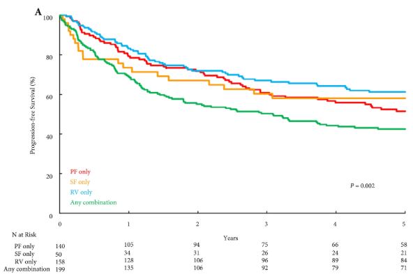
Fig. 1. Progression-free survival stratified by type of extrarenal extension

Results

Overall, PF invasion, renal SF invasion, and RV tumour thrombus were present in 144 (26%), 51 (9%), and 163 (29%) patients, respectively, with multiple patterns of extrarenal extension identified in 205 (36%) patients. There were no significant differences in survival outcomes for isolated involvement of PF, renal SF, or RV. However, patients with multiple patterns of extrarenal extension were at significantly increased risk of disease progression (hazard ratio [HR] 1.31, 95% confidence interval [CI] 1.04–1.65; P = 0.020), CSM (HR 1.64, 95% CI 1.27–2.12; P < 0.001), and all‐cause mortality (HR 1.32, 95% CI 1.08–1.61; P = 0.008).

Conclusions

The presence of multiple patterns of extrarenal extension is associated with a higher risk of disease progression and cancer‐related death after radical nephrectomy compared to isolated involvement of the PF, renal SF, or RV, which carry similar prognostic weight. If validated, these findings may help refine risk stratification of non‐metastatic T3a RCC by distinguishing patients with multiple vs one pattern of extrarenal extension.

Read more Articles of the week


Editorial: Does knowing the risk of relapse in localized renal cell carcinoma matter?

Shah et al. [1] report a retrospective analysis from the Mayo Clinic investigating the prognostic significance of different patterns of pathological T3a clear‐cell RCC in patients who underwent radical nephrectomy for localized disease. There was no difference in disease progression, cancer‐specific mortality or all‐cause mortality when comparing isolated perinephric fat invasion vs isolated renal sinus fat invasion vs isolated renal vein invasion. Multiple sites of extra‐renal extension compared with one site, however, was independently associated with an increased risk of disease progression (hazard ratio [HR] 1.31, P = 0.02), death from RCC (HR 1.64, P < 0.001) and all‐cause mortality (HR 1.32, P = 0.008) when adjusting for multiple key variables including age, tumour size, grade, presence of coagulative tumour necrosis and sarcomatoid differentiation. The authors incorporated multiple sites of extra‐renal extension vs one site into three RCC prognostic models: SSIGN score, UISS and MSKCC nomogram. After controlling for these three predictive tools independently, multiple sites of extra‐renal disease predicted progression, death from RCC and all‐cause death. These data suggest that risk stratification for pT3aN0MO clear‐cell RCC is improved by differentiating multiple vs one site of extra‐renal extension.

Does an improved ability to predict recurrence and mortality increase the likelihood of cure in high‐risk localized RCC patients in 2018? Unfortunately, the answer is no. Ideally, prognostic models would identify patients at sufficient risk to consider adjuvant therapy, which would increase cure rates by eradicating micro‐metastatic disease with an acceptable toxicity. Regrettably, in RCC management there are no well‐established post‐surgical therapies that improve cure rates. The deficiency of established adjuvant therapies is not attributable to a lack of investigative trials. In the era before vascular endothelial growth factor receptor (VEGFR) targeting, adjuvant vaccines, immunotherapies and other systemic therapies failed to demonstrate improved recurrence‐free (RFS) or overall survival (OS) [2]. The efficacy of VEGFR‐targeted therapies in the metastatic setting re‐energized the hope for adjuvant therapy in patients with high‐risk localized RCC after surgical resection in the past two decades. The results to date have been disappointing. To date, three trials (ASSURE, PROTECT and S‐TRAC) have been completed, comparing oral VEGFR tyrosine kinase inhibitors with placebo in high‐risk localized clear‐cell RCC, with disease‐free survival (DFS) as the primary endpoint [3,4,5]. ASSURE and PROTECT showed no difference in RFS or OS [3,4,5]. S‐TRACT demonstrated an improvement in DFS but not in OS [4]. A pooled analysis of these three trials also failed to demonstrate improved DFS or OS with adjuvant VEGFR‐targeted therapy [6]. Significant side effects with discontinuation of adjuvant therapy occurred in 28–45% of patients as a result of drug‐related toxicity [6]. Trials investigating immune checkpoint inhibitors have yet to be published and, with the established efficacy of these drugs in the metastatic setting, hope still remains for adjuvant therapy in resected high‐risk localized RCC.

If the current literature does not support adjuvant therapy for resected high‐risk RCC, does knowing the risk of relapse alter surveillance? National Comprehensive Cancer Network guidelines for resected stage III RCC recommend chest and abdominal imaging within 3–6 months, along with subsequent chest and abdominal imaging every 3–6 months for 3 years, and then annually up to 5 years. Although the ideal schedule for surveillance imaging is unknown, further characterizing of the risk of relapse in high‐risk localized RCC would not be likely to affect this schedule significantly.

Although knowing the risk of relapse in high‐risk localized RCC does not help management in 2018, there is still a value to enhancing our prognostic tools. For one, our prognostic tools help clinicians counsel patients appropriately about their risk of recurrence. In addition, enhanced prognostic tools will assist in selecting appropriate patients with high‐risk localized RCC for future clinical trials of adjuvant therapy and also help us understand the results when comparing cohorts within and between trials.

References

  1. Shah PH, Lyon TD, Lohse CM. Prognostic evaluation of perinephric fat, renal sinus fat, and renal vein invasion for patients with pathologic stage T3a clear cell renal cell carcinoma. BJU Int 2019; 123: 270–6
  2. Scherr AJO, Lima JPSN, Sasse EC et al. Adjuvant therapy for locally advanced renal cell cancer: a systematic review with meta‐analysis. BMC Cancer 2011; 11: 115–21
  3. Haas N, Manola J, Uzzo R et al. Adjuvant sunitinib or sorafenib for high‐risk, non‐metastatic renal‐cell carcinoma (ECOG‐ACRIN E2805): a double‐blind, placebo‐controlled, randomised, phase 3 trial. Lancet 2016; 387: 2008–16
  4. Ravaud A, Motzer RJ, Pandha HS et al. Adjuvant sunitinib in high‐ risk renal‐cell carcinoma after nephrectomy. N Engl J Med 2016; 375: 2246–54
  5. Motzer RJ, Haas NB, Donskov F et al. Randomized phase III trial of adjuvant pazopanib versus placebo after nephrectomy in patients with localized or locally advanced renal cell carcinoma. J Clin Oncol 2017; 35: 3916–23
  6. Sun M, Marconi L, Eisen T et al. Adjuvant vascular endothelial growth factore‐targeted therapy in renal cell carcinoma. Eur Urol 2018; 74: 611–20


#RudeFood: Foodporn for a purpose

The Internet is full of weird and wonderful things. Of course, we all know what is most frequently viewed and shared online. That’s right – food! Nonetheless, when celebrity chef Manu Fieldel posted a photo of his latest creation, it certainly made people look long and hard!


Soon it became clear that this naughty creation had a noble purpose – supporting a campaign to raise awareness of the so-called #BelowTheBelt cancers. While most people may have heard of prostate and bladder cancers, being relatively common, other #BelowTheBelt cancers such as penile and testicular cancers are rarer and relatively unknown. To make matters worse, these cancers affect men either exclusively or predominantly – and we all know how reluctant men can be to go to the doctors.

Hence, the #RudeFood campaign was developed by the Australian and New Zealand Urogenital and Prostate (ANZUP) Cancer Trials Group. ANZUP is the peak co-operative trials group for #BelowTheBelt cancers in Australia and New Zealand. ANZUP has and continues to develop and run many significant clinical trials, including the Enzamet and Enzarad trials for prostate cancer, the Phase III accelerated BEP trial for germ-cell tumours, the sequential BCG-mitomycin trial for bladder cancer and the Eversun and Unison trials in kidney cancer.

The week started with things heating up at ANZUP as they brought #RudeFood to the unsuspecting world!

Manu’s phallic creation was also matched by Ainsley Harriot, Sonia Meffadi and Monty Kulodrovic.

To counterpoint the raunch, there were also poignant personal connections from Simon Leong and Scott Gooding who both described family members who had suffered from prostate cancer.


Over the week, #RudeFood has certainly drawn some attention, including from media outlets such as Mamamia, news.com.au and GOAT. 

A poetic contribution on #RudeFood caught the eye of @UroPoet across the seas. Let us hope this campaign will also lead to greater awareness of #BelowTheBelt cancers and improved outcomes for those affected by them.


Shomik Sengupta is Professor of Surgery at the EHCS of Monash University and visiting urologist & Uro-Oncology lead at Eastern Health. Shomik has particular interests in prostate cancer, including open and robotic prostatectomy, as well as bladder cancer, including cystectomy with neobladder diversion. Shomik is the current leader of the UroOncology SAG within USANZ, and the past chair of Victorian urology training.  Shomik is a Board member and scientific advisory member of the ANZUP Cancer trials group and is heavily involved in numerous clinical trials in GU oncology.

Twitter: @shomik_s 


Article of the week: Adjuvant radiation with androgen‐deprivation therapy for men with lymph node metastases after radical prostatectomy

Every week, the Editor-in-Chief selects an Article of the Week from the current issue of BJUI. The abstract is reproduced below and you can click on the button to read the full article, which is freely available to all readers for at least 30 days from the time of this post.

In addition to the article itself, there is an editorial written by a prominent member of the urological community. These are intended to provoke comment and discussion and we invite you to use the comment tools at the bottom of each post to join the conversation. 

If you only have time to read one article this week, it should be this one.

Adjuvant radiation with androgen‐deprivation therapy for men with lymph node metastases after radical prostatectomy: identifying men who benefit

Mohit Gupta*, Hiten D. Patel*, Zeyad R. Schwen*, Phuoc T. Tran*† and Alan W. Partin*


*Department of Urology, James Buchanan Brady Urological Institute, and Department of Radiation Oncology and Molecular Radiation Sciences and Oncology, The Sidney Kimmel Comprehensive Cancer Center, Johns Hopkins Medical Institutions, Baltimore, MD, USA


Read the full article

Abstract

Objectives

To perform a comparative analysis of three current management strategies for patients with lymph node metastases (LNM; pN1) following radical prostatectomy (RP): observation, androgen‐deprivation therapy (ADT), and external beam radiation therapy (EBRT) + ADT.

Patients and Methods

Patients with LNM after RP were identified using the National Cancer Database (2004–2013). Exclusion criteria included any use of radiation therapy or ADT before RP, clinical M1 disease, or incomplete follow‐up data. Patients were categorised according to postoperative management strategy. The primary outcome was overall survival (OS). Kaplan–Meier curves and adjusted multivariable Cox proportional hazards models were employed. Sub‐analyses further evaluated patient risk stratification and time to receipt of adjuvant therapy.

Results

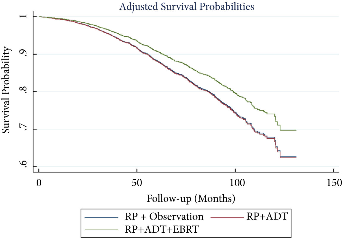
A total of 8 074 patients met the inclusion criteria. Postoperatively, 4 489 (55.6%) received observation, 2 065 (25.6%) ADT, and 1 520 (18.8%) ADT + EBRT. The mean (median; interquartile range) follow‐up was 52.3 (48.0; 28.5–73.5) months. Patients receiving ADT or ADT + EBRT had higher pathological Gleason scores, T‐stage, positive surgical margin rates, and nodal burden. Adjusted multivariable Cox models showed improved OS for ADT + EBRT vs observation (hazard ratio [HR] 0.77, 95% confidence interval [CI] 0.64–0.94; P = 0.008) and vs ADT (HR 0.76, 95% CI: 0.63–0.93; P = 0.007). There was no difference in OS for ADT vs observation (HR 1.01, 95% CI: 0.87–1.18; P = 0.88). Findings were similar when restricting adjuvant cohorts for timing of adjuvant therapy. There was no difference in OS between groups for up to 2 549 (31.6%) patients lacking any of the following adverse features: ≥pT3b disease, Gleason score ≥9, three or more positive nodes, or positive surgical margin.

Conclusions

For patients with LNM after RP, the use of adjuvant ADT + EBRT improved OS in the majority of patients, especially those with adverse pathological features. Conversely, adjuvant therapy did not confer significant OS benefit in up to 30% of patients without high‐risk features, who may be managed with observation and forego the morbidity associated with immediate ADT or radiation.

Read more Articles of the week

Editorial: Postoperative radiation and hormonal therapy for men with node‐positive prostate cancer: a new standard?

The best management strategy for men with pathologically node‐positive (pN+) prostate cancer after radical prostatectomy (RP) has been debated for decades [1]. In the 1990s, the Radiation Therapy and Oncology Group (RTOG) initiated the RTOG 9608 trial to test the impact of radiotherapy (RT) and androgen‐deprivation therapy (ADT) in this setting. However, due to the rise in PSA screening and the practice of treating high‐risk prostate cancer with primary RT, the incidence of pN+ disease fell. Consequently, the trial closed due to poor accrual and the question faded in prominence. Today, both trends have reversed. PSA screening is less common and men with high‐risk prostate cancer are more frequently opting for RP. As such, physicians increasingly face the dilemma of pN+ disease. Guidelines provide little assistance, as they support everything from observation to multimodal treatment with RT and ADT. Patients and providers want to know, is there a standard treatment for all patients, and if not, how should one choose between such disparate options?

To answer these questions, one must start with the little randomised data that exist in this setting. The seminal trial by Messing et al. [1] randomised men with pN+ prostate cancer to ADT or observation with initiation of ADT after the development of symptomatic progression or distant metastases. ADT clearly improved overall survival and prostate cancer‐specific survival. However, critics noted the relatively poor outcomes in the observation group and the small sample size. Later, retrospective studies called the benefit of immediate ADT into question [2].

Against this backdrop, it is interesting that Gupta et al. [3] found the most common management approach in the USA National Cancer Database (NCDB) was observation rather than immediate ADT. Despite the randomised data, the cumulative side‐effects from lifelong ADT in a cohort of patients with no disease‐related symptoms and a median survival of well over 10 years are unappealing. Ultimately, many men do not appear to be willing to endure the diminished quality of life in exchange for a small improvement in quantity of life.

In contrast to the non‐curative nature of ADT, the possibility exists that the combination of postoperative RT and ADT could provide durable disease control, perhaps even without lifelong ADT. The data reported by Gupta et al. [3] in this edition of the BJUI provide support for this paradigm. These data add to a growing body of literature [4] that tells a consistent story with two common themes: (i) postoperative RT with ADT appears to be associated with improved survival in men with pN+ prostate cancer, and (ii) RT appears to convey the largest benefit to men with certain high‐risk pathological features. Should this body of literature lead us to eschew the old standard and advise observation for low‐risk men and RT with ADT for men at higher risk?

Before a new standard is declared, the limitations of retrospective population‐based research must be addressed. The authors performed a sophisticated analysis to reduce the impact of selection bias. However, due to the limitation of the available data, the authors were not able to account for possibly the most important variable: the postoperative PSA. One study showed that men with pN+ disease with a persistent PSA had an 8‐year clinical recurrence rate of 69% vs 12% for those with undetectable PSA [5].

It is likely that men with persistent PSA in the NCDB would have received immediate ADT with or without RT rather than observation. As such, one must be cautious of the similar survival between the observation and ADT group, especially in light of contradictory randomised data. That being said, it is reasonable for some men to conclude that the side‐effects of ADT outweigh the potential benefit, especially those with low‐risk features such as an undetectable postoperative PSA, low Gleason score, and limited lymph node involvement.

As RT with ADT appears superior to either observation or ADT alone, should more men receive RT? Probably. Of the men with high‐risk features, only 22% actually received postoperative RT. Should postoperative RT now be considered the standard for all men? Probably not. Whilst it appears that some men may indeed benefit from RT, the possibility of selection bias driving this result is real. Even if there is a true effect, identifying which patients harbour residual local disease, but do not already have subclinical distant metastatic disease is challenging. RT for all would lead to unnecessary side‐effects for men that would not benefit from the treatment. Ultimately, a randomised trial will be required to establish the benefit of RT and to define subgroups of men that may or may not benefit. Until then, we will continue to rely on excellent work like the accompanying paper from Gupta et al. [3] to identify men who may benefit from postoperative RT and ADT.

References

  1. Messing EM, Manola J, Sarosdy M, Wilding G, Crawford ED, Trump D. Immediate hormonal therapy compared with observation after radical prostatectomy and pelvic lymphadenectomy in men with node‐positive prostate cancer. N Engl J Med 1999341: 1781–8
  2. Wong YN, Freedland S, Egleston B, Hudes G, Schwartz JS, Armstrong K. Role of androgen deprivation therapy for node‐positive prostate cancer. J Clin Oncol 200927: 100–5
  3. Gupta M, Patel HD, Schwen ZR, Tran PT, Partin AW. Adjuvant radiation with androgen deprivation therapy for men with lymph node metastases following radical prostatectomy: identifying men who benefit. BJU Int 2019123: 252–60
  4. Abdollah F, Karnes RJ, Suardi N et al. Impact of adjuvant radiotherapy on survival of patients with node‐positive prostate cancer. J Clin Oncol 201432: 3939–47
  5. Bianchi L, Nini A, Bianchi M et al. The role of prostate‐specific antigen persistence after radical prostatectomy for the prediction of clinical progression and cancer‐specific mortality in node‐positive prostate cancer patients. Eur Urol 201669: 1142–8


#UroPoet – restoring our humanity with creative writing and poetry

The global urology community on Twitter

— Todd M. Morgan, MD (@wandering_gu) February 9, 2019


Over the past several years, many urologists have gravitated to Twitter. Through Twitter we have shared information and experience, created relationships, and built community. Twitter has brought us together in many ways never thought possible before. Some great examples include #UroSoMe, #prostateJC#CUAJC, and the grandfather of them all, #urojc.

Behind the screens

Behind our screens, however, many of us face significant challenges, both professional and personal. Urologists around the world find themselves spending more and more time typing on their keyboards and less and less time in face-to-face conversation with patients.

Growing rates of burnout in urology are being reported in the United States. There is also a burgeoning trend toward consolidation, mergers, and loss of autonomy in healthcare. When you add in the current global political and cultural turmoil, even Twitter starts to lose its luster and become divisive.


The power of creative writing and poetry

Recently, at the invitation of my friend Pam Ressler, I had the opportunity to participate in a January haiku challenge. To be honest, I was really busy in January, and initially, wasn’t all that excited about it.

However, I quickly began to realize that the discipline of writing a daily haiku made me feel better. Over the course of that month, I developed a new sense of gratitude. By spending just a few minutes, here and there, thinking about the next poem I might write, the recurrent annoyances of each day became fewer and smaller.

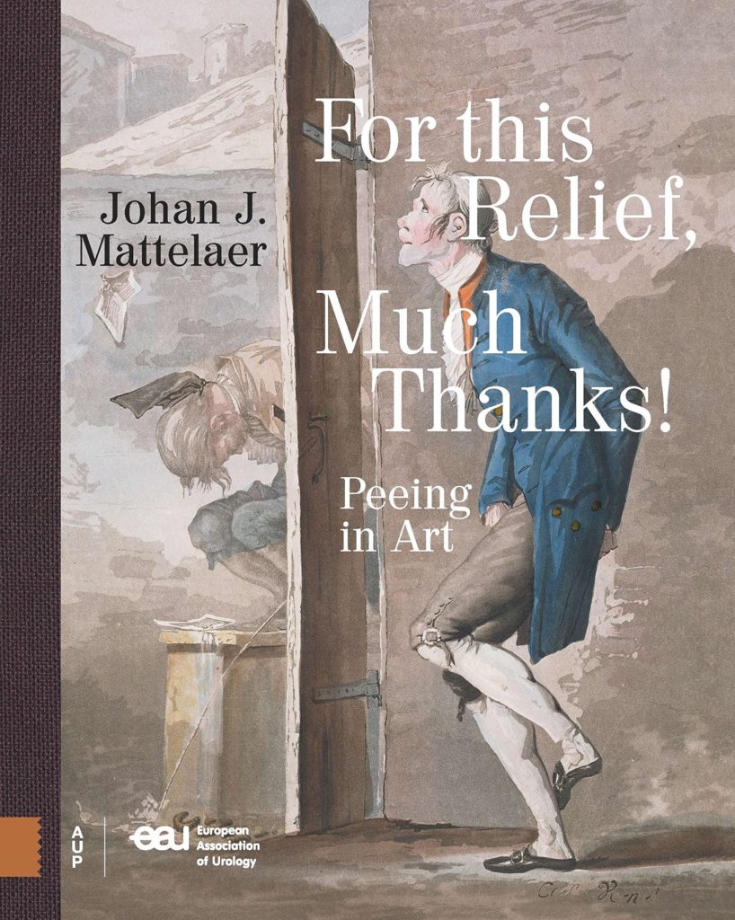
Humankind has a rich history of storytelling with prose. Poems about ‘pee’ were written long before urology, as exemplified in Dr. Johan Mattelaer’s wonderful book, “For this Relief, Much Thanks!”

Restoring our humanity

In the spirit of friendship, I invite you to join me in celebrating life, and our noble profession of urology, with the power of creative writing and poetry on Twitter at #UroPoet. My hope is that everyone will feel welcome to use this hashtag, responsibly, and to share the things they love most about our profession, our patients, our families, and life itself through the use of creative writing and poetry.

— Dr. Brian Stork (@StorkBrian) February 3, 2019

In the short time the hashtag has been active, topics ranging from research to prolapse have been posted in the form of limericks, essays, song lyrics, poems and haiku. I hope you will take a moment to at least follow along and consider making a regular or one-time post of your own – adding the hashtag #UroPoet.

I’ll be posting regularly from a second Twitter account @UroPoet where I will also be retweeting #UroPoet tweets. If the spirit moves you, you can also follow me @StorkBrian.


The beginning of #UroSoMe

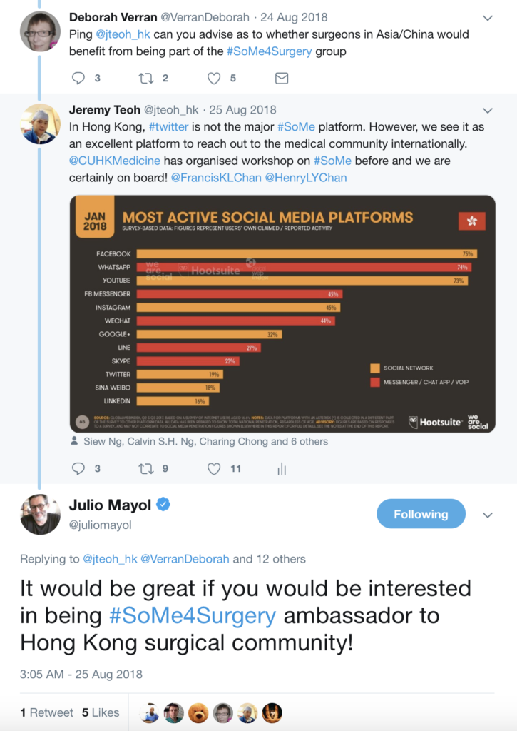
I had been using Twitter for a while but I never experienced the true power of this social media platform. It was a cold call from @VerranDeborah and @juliomayol when I started to notice the hashtag #SoMe4Surgery. I was pleasantly surprised and impressed by the active engagement of the #SoMe4Surgery participants. After participating in a #SoMe4Surgery live conversation event on #surgicalinfection, I finally realized the potential impact of a simple hashtag.

While I was amazed by how #SoMe4Surgery brought the surgical community together, many of the topics being discussed were not entirely relevant or specific to a urologist per se. I felt the need of a hashtag specific to Urology, and I quickly started to conceptualize and plan ahead in building up the #UroSoMe community. The #UroSoMe twitter account was officially registered in August 2018.

#UroSoMe stands for ‘Urology Social Media’. My initial thought about #UroSoMe was simple. I wanted to develop a hashtag specific to urology. I wanted to increase public awareness about different urological conditions. Most importantly, I wanted to bring the urology community closer together through this social media platform. I believe there is so much for us to learn from each other, and such interactions should never be bounded by physical or geographical restrictions. Coincidentally, I was invited to talk about social media at the 27th Malaysian Urological Conference 2018, and I decided to take this opportunity to introduce #UroSoMe to the urology community.

The initial response from the audience was promising. Even after the meeting, many urologists came to me for in-depth discussions about the opportunities and applications of social media in urology. I felt that #UroSoMe might really work and it was time to gather more people to establish the community. The first invitation sent in on 14 December 2018, which I often regard as the ‘start date’ of the #UroSoMe community.

By inviting and encouraging people around to use a common hashtag, the #UroSoMe community keeps growing. With increasing momentum, the first #LiveCaseDiscussions was planned. It was a pre-planned event for urologists to get ‘online’ and discuss about some posted cases. A polling had been held in advance, and the topic to be discussed was chosen to be ‘Stone’.

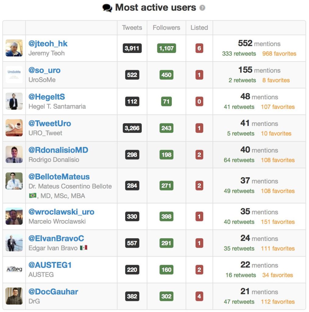
The #LiveCaseDiscussions was on air at 4pm (CET) on 5 January 2019. A total of 9 cases had been presented and discussed. Hosting this event was overwhelming with vigorous discussions among the participants. It took approximately 2 hours to ‘complete’ the event, but the conversations went on for the next few days. Special thanks must be given to the most active users. #UroSoMe and the first #LiveCaseDiscussions would never be successful without their tremendous support.

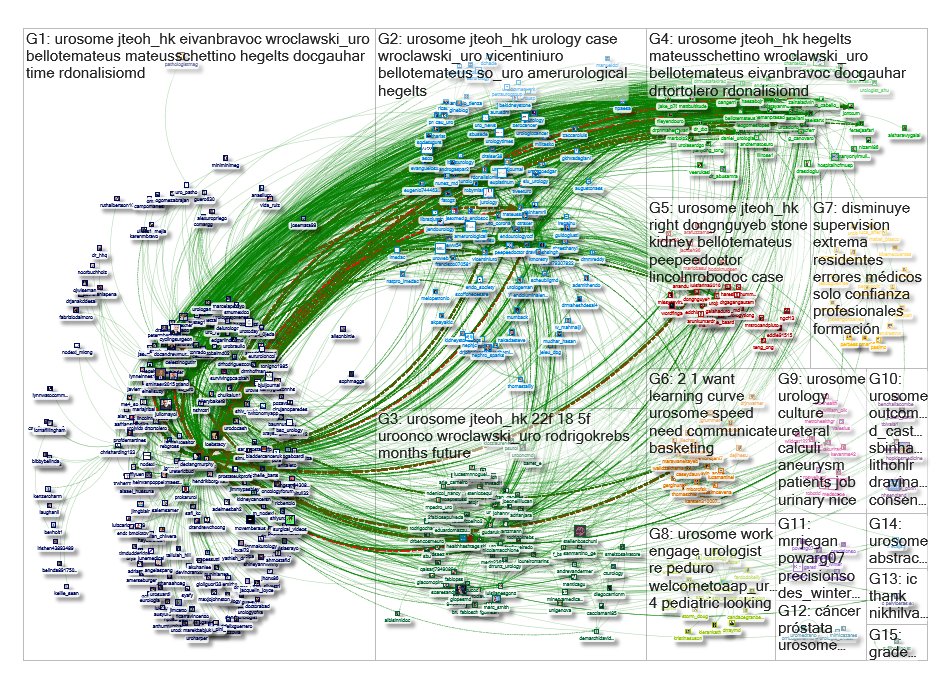
The immediate effect of the #UroSoMe #LiveCaseDiscussions event was overwhelming. This graph represents a network of 515 twitter users whose tweets contained the hashtag #UroSoMe. 6692 mentions, 1044 retweets and 617 replies were recorded within a 10-day period from 27 December 2018 to 6 January 2019. From a social science point of view, this picture represents a ‘tight crowd’, in which discussions are characterized by highly interconnected people with few isolated participants. I guess this is exactly how we feel about the urology community!

Apart from #LiveCaseDiscussions, the #UroSoMe working group is also keen to host events including #LiveForum, #LiveJournalClub and #LiveTeaching. This is only the beginning of #UroSoMe and we believe there is huge potential to be explored. It is only with your support that #UroSoMe can continue to grow. We look forward to meeting you on Twitter and, hopefully, at #EAU19 and #AUA19 as well!

P.S. I must thank @juliomayol for the inspiration of #UroSoMe, @gmacscotland for his teaching on social media analytics, and @marc_smith for his support in NodeXL.

About the author:

Jeremy Teoh (@jteoh_hk) is a Urologist based in Hong Kong, China.

The #UroSoMe working group:

@jteoh_hk, @adelmesbah2, @BelloteMateus, @DocGauhar, @DrTortolero, @D_Castellani, @EdgarLindenMD, @EIvanBravoC, @HegeltS, @JontxuM, @gudaruk, @MarcelaPelayo, @RdonalisioMD, @Urologeman, @wroclawski_uro and @zainaladwin.


© 2024 BJU International. All Rights Reserved.