Tag Archive for: #BJUI

Posts

Article of the week: A machine learning‐assisted decision‐support model to better identify patients with PCa requiring an extended pelvic lymph node dissection

Every week, the Editor-in-Chief selects an Article of the Week from the current issue of BJUI. The abstract is reproduced below and you can click on the button to read the full article, which is freely available to all readers for at least 30 days from the time of this post.

In addition to the article itself, there is an editorial written by a prominent member of the urology community and a video prepared by the authors; we invite you to use the comment tools at the bottom of each post to join the conversation. 

If you only have time to read one article this week, it should be this one. Merry Christmas!

A machine learning‐assisted decision‐support model to better identify patients with prostate cancer requiring an extended pelvic lymph node dissection

Ying Hou*, Mei-Ling Bao, Chen-Jiang Wu*, Jing Zhang*, Yu-Dong Zhang* and Hai-Bin Shi*

*Department of Radiology and Department of Pathology, The First Affiliated Hospital with Nanjing Medical University, Nanjing, Jiangsu Province, China

Abstract

Objectives

To develop a machine learning (ML)‐assisted model to identify candidates for extended pelvic lymph node dissection (ePLND) in prostate cancer by integrating clinical, biopsy, and precisely defined magnetic resonance imaging (MRI) findings.

Patients and Methods

In all, 248 patients treated with radical prostatectomy and ePLND or PLND were included. ML‐assisted models were developed from 18 integrated features using logistic regression (LR), support vector machine (SVM), and random forests (RFs). The models were compared to the Memorial Sloan Kettering Cancer Center (MSKCC) nomogram using receiver operating characteristic‐derived area under the curve (AUC) calibration plots and decision curve analysis (DCA).

Results

A total of 59/248 (23.8%) lymph node invasions (LNIs) were identified at surgery. The predictive accuracy of the ML‐based models, with (+) or without (−) MRI‐reported LNI, yielded similar AUCs (RFs+/RFs: 0.906/0.885; SVM+/SVM: 0.891/0.868; LR+/LR: 0.886/0.882) and were higher than the MSKCC nomogram (0.816; P < 0.001). The calibration of the MSKCC nomogram tended to underestimate LNI risk across the entire range of predicted probabilities compared to the ML‐assisted models. The DCA showed that the ML‐assisted models significantly improved risk prediction at a risk threshold of ≤80% compared to the MSKCC nomogram. If ePLNDs missed was controlled at <3%, both RFs+ and RFs resulted in a higher positive predictive value (51.4%/49.6% vs 40.3%), similar negative predictive value (97.2%/97.8% vs 97.2%), and higher number of ePLNDs spared (56.9%/54.4% vs 43.9%) compared to the MSKCC nomogram.

Conclusions

Our ML‐based model, with a 5–15% cutoff, is superior to the MSKCC nomogram, sparing ≥50% of ePLNDs with a risk of missing <3% of LNIs.


Editorial: A better way to predict lymph node involvement using machine learning?

In their study in this issue of BJUIHou et al. [1] use machine‐learning algorithms to evaluate several preoperative clinical variables (highlighting specific MRI findings of locally advanced prostate cancer) to determine whether lymph node involvement (LNI) could be present during radical prostatectomy, which would justify an extended pelvic lymph node dissection (PLND). This is a well‐designed study with scientific rigour, providing evidence‐based justifications and definitions (i.e. of relevant MRI findings). The authors successfully illustrate a practical application of using artificial intelligence (AI) methods to augment clinical decision‐making prior to and during surgery compared to today’s ‘gold standard’ (nomograms).

For many years, the Memorial Sloan Kettering Cancer Centre (MSKCC) nomogram, among a number of predictive models, has been used to determine the probability of LNI. The output of these tools has assisted surgeons in determining whether to perform a PLND, and if so, to what extent [2,3,4]. The authors hypothesize that, with additional MRI parameters not previously used, machine‐learning algorithms can better select which patients are more likely to have LNI and will therefore require extended PLND. In fact, the authors report that the MSKCC nomogram and conventional MRI reporting of LNI consistently underestimated LNI risk compared to the machine‐learning‐assisted models presented in their study. The outputs of the present models would allow a higher number of extended PLNDs to be spared compared to reliance on the MSKCC nomogram alone. It was appropriate to use several existing AI models in this study, as it is never readily apparent initially which existing predictive model may perform best with a given dataset. In fact, all the models used – logistic regression (LR), support vector machine (SVM) and random forest (RF) – while similar in performance to each other, outperformed the MSKCC nomogram (P < 0.001). Many adjustments were probably performed for each model to tailor it to the dataset and optimize prediction performance.

Criticisms of the study are that: (i) cases for which PLND was not performed were excluded, which could have created a selection bias; (ii) the model would only be applicable when the patient has undergone MRI; (iii) the study was conducted at a single institution in a small sample (AI methods thrive on big and diverse datasets).

This study by Hou et al. is a great example of a machine‐learning application that may positively impact clinical practice. For many years, we have relied on nomograms, but with increasing use of MRI, additional factors should also be included, as Hou et al. have done. Machine‐learning is particularly adept at simultaneously examining numerous variables to elicit which ones may contribute best to a particular outcome. As BJUI has evaluated many manuscripts examining machine‐learning methods for clinical decision‐making in the past year, we have encouraged authors to use present‐day gold standard methods, such as the MSKCC nomogram, as controls [5]. As we embrace AI methods, we must keep one eye on the tried and tested conventional ways. This ensures that we do not take backward steps but rather take forward steps responsibly. Similarly to recent AI studies published in the BJUI, the sample size in this study was relatively small. External validation in a multicentre study on larger datasets is highly recommended.

by Andrew J. Hung

References

  1. Hou YBao MWu CJZhang JZhang YDShi HBA machine learning‐assisted decision support model with mri can better spare the extended pelvic lymph node dissection at cost of less missing in prostate cancerBJU Int 2019124972– 83
  2. Briganti ALarcher AAbdollah F et al. Updated nomogram predicting lymph node invasion in patients with prostate cancer undergoing extended pelvic lymph node dissection: the essential importance of percentage of positive cores. Eur Urol 201261480– 7
  3. Memorial Sloan Kettering Cancer CenterDynamic prostate cancer nomogram: coefficients. Accessed April 2018
  4. Tosoian JJChappidi MFeng Z et al. Prediction of pathological stage based on clinical stage, serum prostate-specific antigen, and biopsy Gleason score: Partin Tables in the contemporary era. BJU Int 2017119676– 83
  5. Hung AJCan machine‐learning algorithms replace conventional statistics? BJU Int 20181231


Video: Machine learning‐assisted decision‐support model to identify PCa patients requiring an extended PLND

A machine learning‐assisted decision‐support model to better identify patients with prostate cancer requiring an extended pelvic lymph node dissection

Abstract

Objectives

To develop a machine learning (ML)‐assisted model to identify candidates for extended pelvic lymph node dissection (ePLND) in prostate cancer by integrating clinical, biopsy, and precisely defined magnetic resonance imaging (MRI) findings.

Patients and Methods

In all, 248 patients treated with radical prostatectomy and ePLND or PLND were included. ML‐assisted models were developed from 18 integrated features using logistic regression (LR), support vector machine (SVM), and random forests (RFs). The models were compared to the Memorial SloanKettering Cancer Center (MSKCC) nomogram using receiver operating characteristic‐derived area under the curve (AUC) calibration plots and decision curve analysis (DCA).

Results

A total of 59/248 (23.8%) lymph node invasions (LNIs) were identified at surgery. The predictive accuracy of the ML‐based models, with (+) or without (−) MRI‐reported LNI, yielded similar AUCs (RFs+/RFs: 0.906/0.885; SVM+/SVM: 0.891/0.868; LR+/LR: 0.886/0.882) and were higher than the MSKCC nomogram (0.816; P < 0.001). The calibration of the MSKCC nomogram tended to underestimate LNI risk across the entire range of predicted probabilities compared to the ML‐assisted models. The DCA showed that the ML‐assisted models significantly improved risk prediction at a risk threshold of ≤80% compared to the MSKCC nomogram. If ePLNDs missed was controlled at <3%, both RFs+ and RFs resulted in a higher positive predictive value (51.4%/49.6% vs 40.3%), similar negative predictive value (97.2%/97.8% vs 97.2%), and higher number of ePLNDs spared (56.9%/54.4% vs 43.9%) compared to the MSKCC nomogram.

Conclusions

Our ML‐based model, with a 5–15% cutoff, is superior to the MSKCC nomogram, sparing ≥50% of ePLNDs with a risk of missing <3% of LNIs.

View more videos

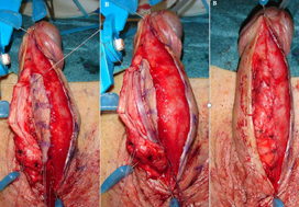
What’s the diagnosis?

These images are taken from Barbagli et al, BJUI 2019 which shows in a step-by-step guide the details and outcomes of a modified technique.

No such quiz/survey/poll

Article of the week: The role of extended venous thromboembolism prophylaxis for major urological cancer operations

Every week, the Editor-in-Chief selects an Article of the Week from the current issue of BJUI. The abstract is reproduced below and you can click on the button to read the full article, which is freely available to all readers for at least 30 days from the time of this post.

In addition to the article itself, there is an editorial written by a prominent member of the urology community, a video prepared by the authors and a visual abstract; we invite you to use the comment tools at the bottom of each post to join the conversation. 

If you only have time to read one article this week, it should be this one.

The role of extended venous thromboembolism prophylaxis for major urological cancer operations

Rishi Naik*, Indrajeet Mandal*, Alexander Hampson, Tim Lane, Jim Adshead, Bhavan Prasad Rai and Nikhil Vasdev†§

*Faculty of Medical Sciences, UCL Medical School, University College London, London, Department of Urology, Lister Hospital, Stevenage, Department of Urology, Freeman Hospital, Newcastle upon Tyne and §School of Life and Medical Sciences, University of Hertfordshire, Hatfield, UK

Rishi Naik and Indrajeet Mandal are joint first authors.

Read the full article

Abstract

Objectives

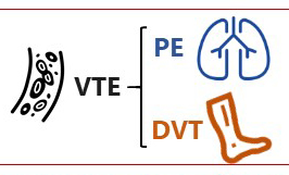
Venous thromboembolism (VTE), consisting of both pulmonary embolism (PE) and deep vein thromboses (DVT), remains a well‐recognised complication of major urological cancer surgery. Several international guidelines recommend extended thromboprophylaxis (ETP) with LMWH, whereby the period of delivery is extended to the post‐discharge period, where the majority of VTE occurs. In this literature review we investigate whether ETP should be indicated for all patients undergoing major urological cancer surgery, as well as procedure specific data that may influence a clinician’s decision.

Methods

We performed a search of six databases (PubMed, Cochrane, EMBASE, Cumulative Index to Nursing and Allied Health Literature (CINAHL), PsycINFO, and British Nursing Index (BNI)) from inception to June 2019, for studies looking at adult patients who received VTE prophylaxis after surgery for a major urological malignancy.

Results

Eighteen studies were analysed. VTE risk is highest in open and robotic Radical Cystectomy (RC) (2.6–11.6%) and ETP demonstrates a significant reduction in risk of VTE, but not a significant difference in Pulmonary Embolism (PE) or mortality. Risk of VTE in open Radical Prostatectomy (RP) (0.8–15.7%) is comparable to RC, but robotic RP (0.2–0.9%), open partial/radical nephrectomy (1.0–4.4%) and robotic partial/radical nephrectomy (0.7–3.9%) were lower risk. It has not been shown that ETP reduces VTE risk specifically for RP or nephrectomy.

Conclusion

The decision to use ETP is a fine balance between variables such as VTE incidence, bleeding risk and perioperative morbidity/mortality. This balance should be assessed for each specific procedure type. While ETP still remains of net benefit for open RP as well as open and robotic RC, the balance is closer for minimally invasive RP as well as radical and partial nephrectomy. Due to a lack of procedure specific evidence for the use of ETP, adherence with national guidelines remains poor. Therefore, we advocate further studies directly comparing ETP vs standard prophylaxis, for specific procedure types, in order to allow clinicians to make a more informed decision in future.

Editorial: How long is long enough for pharmacological thromboprophylaxis in urology?

Each year, millions of patients who undergo urological surgery incur the risk of deep vein thrombosis and pulmonary embolism, together referred to as venous thromboembolism (VTE), and major bleeding. Because pharmacological prophylaxis decreases the risk of VTE, but increases the risk of bleeding, and because knowledge of the magnitude of these risks remains uncertain, both clinical practice and guideline recommendations vary widely [1]. One of the uncertainties is the recommended duration of pharmacological thromboprophylaxis.

In this issue of the BJUI, Naik et al. [2] provide an up‐to‐date review that summarises the articles that examined extended thromboprophylaxis in patients with cancer who underwent radical prostatectomy (RP), radical cystectomy (RC) or nephrectomy. The outcomes on which they focussed include risks of VTE, bleeding, renal failure and mortality – all potentially influenced by whether or not patients receive extended prophylaxis.

After screening >3500 articles, the authors included 18 studies, none of them randomised controlled trials (RCTs) [2]. They found that VTE risk is highest in open and robot‐assisted RC, and that, based on observational studies, extended thromboprophylaxis significantly reduces the risk of VTE relative to shorter duration prophylaxis. Evidence suggested that robot‐assisted RP, as well as both open and robot‐assisted partial and radical nephrectomies, incur lower VTE risk than RCs or open RP. They did not find studies comparing extended prophylaxis to standard prophylaxis for RPs or nephrectomies [2].

Overall, these findings are consistent with systematic reviews that estimated the procedure‐ and patient risk factor‐specific risks for 20 urological cancer procedures [3]. As these reviews suggested substantial procedure‐specific differences in the VTE risk estimates, the European Association of Urology (EAU) Guidelines provided separate recommendations for each procedure [4]. For urological (as well as gastrointestinal and gynaecological) patients, the National Institute for Health and Care Excellence (NICE) Guidelines suggest to ‘consider extending pharmacological VTE prophylaxis to 28 days postoperatively for people who have had major cancer surgery in the abdomen’ [5]. Because of variation in both bleeding and thrombosis risks across procedures, this advice is appropriate for some procedures and misguided for others. For instance, the procedure‐specific EAU Guidelines recommend extended VTE prophylaxis for open RC but not for robot‐assisted RP without lymphadenectomy [4].

The review by Naik et al. [2] identified the lack of urology‐specific studies comparing the in‐hospital‐only prophylaxis to extended prophylaxis. The few included studies were observational with considerable limitations (e.g. limited adjustment for possible confounders).

A recent update of a Cochrane review compared the impact of extended thromboprophylaxis with low‐molecular‐weight heparin (LMWH) for at least 14 days to in‐hospital‐only prophylaxis in abdominal or pelvic surgery procedures [6]. The authors identified seven RCTs (1728 participants) evaluating extended thromboprophylaxis with LMWH and generated pooled estimates for the incidence of any VTE (symptomatic or asymptomatic) after major abdominal or pelvic surgery of 13.2% in the control group compared with 5.3% in the patients receiving extended out‐of‐hospital LMWH (odds ratio [OR] 0.38, 95% CI 0.26–0.54).

Most events were asymptomatic, although the incidence of symptomatic VTE was also reduced from 1.0% in the in‐hospital‐only group to 0.1% in patients receiving extended thromboprophylaxis (OR 0.30, 95% CI 0.08–1.11). The authors reported no persuasive difference in the incidence of bleeding complications within 3 months of surgery (defined as major or minor bleeding according to the definition provided in the individual studies) between the in‐hospital‐only group (2.8%) and extended LMWH (3.4%) group (OR 1.10, 95% CI 0.67–1.81).

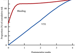
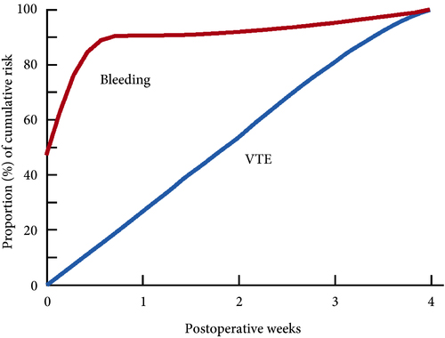
These findings are consistent with our own modelling study that demonstrated an approximately constant hazard of VTE up to 4 weeks after surgery [7]. That study also found that bleeding risk, by contrast, is concentrated in the first 4 days after surgery [7] (Fig.1). Using these findings, the EAU Guidelines suggest for patients in whom pharmacological prophylaxis is appropriate, extended pharmacological prophylaxis for 4 weeks [4]. Consistent with these recommendations, Naik et al. [2] found that 15 studies of 18 included in their review recommended extended prophylaxis.

Fig.1 Proportion of cumulative risk (%) of venous thromboembolism (VTE) and major bleeding by week since surgery during the first 4 postoperative weeks. Reproduced from: Tikkinen et al. [7].

(This is an Open Access article distributed under the terms of the Creative Commons Attribution License (https://creativecommons.org/licenses/by/4.0/), which permits unrestricted use, distribution, and reproduction in any medium, provided the original work is properly credited. The Creative Commons Public Domain Dedication waiver (https://creativecommons.org/publicdomain/zero/1.0/) applies to the data made available in this article, unless otherwise stated.)


Overall, as shown also by this review [2], the evidence base for urological thromboprophylaxis is limited. Although current evidence supports extended prophylaxis, definitively establishing the optimal duration of thromboprophylaxis will require large‐scale RCTs. Other unanswered key questions include: baseline risks of various procedures, timing of prophylaxis, patient risk stratification, as well as effectiveness of direct oral anticoagulants. In the meanwhile, suggesting extended duration to patients whose risk of VTE is sufficiently high constitutes a reasonable evidence‐based approach to VTE prophylaxis.

by Kari A.O. Tikkinen and Gordon H. Guyatt


References

  1. Violette PDCartwright RBriel MTikkinen KAGuyatt GH Guidelines of guidelines: thromboprophylaxis for urological surgery. BJU Int 2016118351– 8
  2. Naik RMandal IHampson A et al. The role of extended venous thromboembolism prophylaxis for major urological cancer operations. BJU Int 2019; 124: 935-44
  3. Tikkinen KACraigie SAgarwal A et al. Procedure‐specific risks of thrombosis and bleeding in urological cancer surgery: systematic reviews and meta‐analyses. Eur Urol 201873242– 51
  4. Tikkinen KACartwright RGould MK et al. EAU Guidelines on Thromboprophylaxis in Urological Surgery, 2017. European Association of Urology, 2018. Accessed November 2019
  5. National Institute for Health and Care Excellence (NICE)Venous Thromboembolism in over 16s: reducing the risk of hospital‐acquired deep vein thrombosis or pulmonary embolism. NICE guideline [NG89]. London: NICE, 2018. Accessed November 2019
  6. Felder SRasmussen MSKing R et al. Prolonged thromboprophylaxis with low molecular weight heparin for abdominal or pelvic surgery. Cochrane Database Syst Rev 20193CD004318
  7. Tikkinen KAAgarwal ACraigie S et al. Systematic reviews of observational studies of risk of thrombosis and bleeding in urological surgery (ROTBUS): introduction and methodology. Syst Rev 201423150. DOI: 10.1186/2046‐4053‐3‐150.


Video: Role of extended venous thromboembolism prophylaxis for major urological cancer operations

The role of extended venous thromboembolism prophylaxis for major urological cancer operations

Read the full article

Abstract

Objectives

Venous thromboembolism (VTE), consisting of both pulmonary embolism (PE) and deep vein thromboses (DVT), remains a well‐recognised complication of major urological cancer surgery. Several international guidelines recommend extended thromboprophylaxis (ETP) with LMWH, whereby the period of delivery is extended to the post‐discharge period, where the majority of VTE occurs. In this literature review we investigate whether ETP should be indicated for all patients undergoing major urological cancer surgery, as well as procedure specific data that may influence a clinician’s decision.

Methods

We performed a search of six databases (PubMed, Cochrane, EMBASE, Cumulative Index to Nursing and Allied Health Literature (CINAHL), PsycINFO, and British Nursing Index (BNI)) from inception to June 2019, for studies looking at adult patients who received VTE prophylaxis after surgery for a major urological malignancy.

Results

Eighteen studies were analysed. VTE risk is highest in open and robotic Radical Cystectomy (RC) (2.6–11.6%) and ETP demonstrates a significant reduction in risk of VTE, but not a significant difference in Pulmonary Embolism (PE) or mortality. Risk of VTE in open Radical Prostatectomy (RP) (0.8–15.7%) is comparable to RC, but robotic RP (0.2–0.9%), open partial/radical nephrectomy (1.0–4.4%) and robotic partial/radical nephrectomy (0.7–3.9%) were lower risk. It has not been shown that ETP reduces VTE risk specifically for RP or nephrectomy.

Conclusion

The decision to use ETP is a fine balance between variables such as VTE incidence, bleeding risk and perioperative morbidity/mortality. This balance should be assessed for each specific procedure type. While ETP still remains of net benefit for open RP as well as open and robotic RC, the balance is closer for minimally invasive RP as well as radical and partial nephrectomy. Due to a lack of procedure specific evidence for the use of ETP, adherence with national guidelines remains poor. Therefore, we advocate further studies directly comparing ETP vs standard prophylaxis, for specific procedure types, in order to allow clinicians to make a more informed decision in future.

View more videos

Visual abstract: The role of extended venous thromboembolism prophylaxis for major urological cancer operations

See more infographics

90 years of BJU International

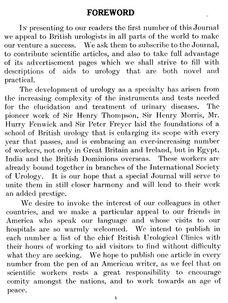
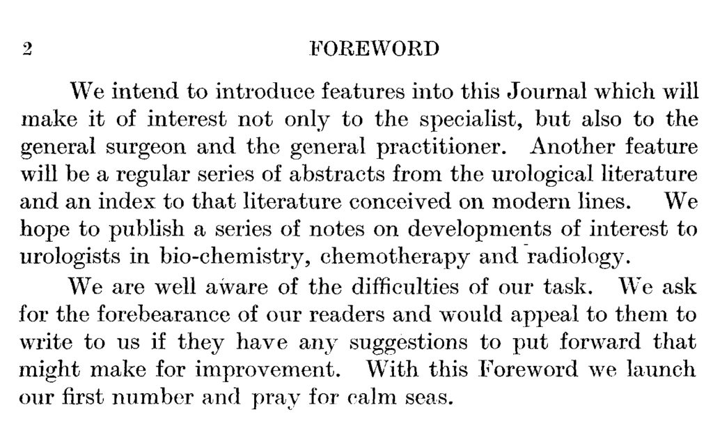
This blog marks the launch of the British Journal of Urology 90 years ago.  Since then the Journal has undergone many changes as urology and the publishing environment have changed. The first Editors outlined their vision in the Foreword of the first issue, which includes an introduction to the speciality, the locations and opening hours of Urology Clinics and a plea for peace:

The British Journal of Urology Vol.1 1929

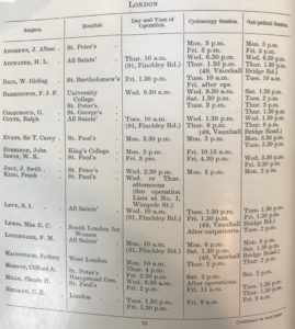
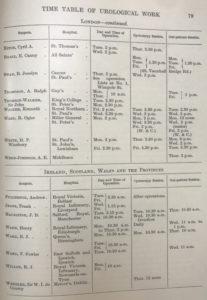
From the outset, the Editors placed emphasis on seeking collaboration and contributions internationally. From 1929 up to 1942 there was even a section in the Journal advising urologists visiting London where to find details of urological clinics which they could visit.

This time-table is published in each number of the Journal for the guidance of travelling medical practitioners who may wish to visit Urological Clinics.

Visitors to London are advised to call at No.1 Wimpole St, W.1, where daily bulletins of operations and lectures issued by the Fellowship of Medicine are available for inspection….”


Advertisements


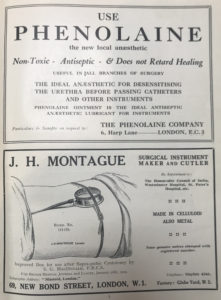
A sample of the advertisements included in the first edition shows the kit available at the time.


Articles

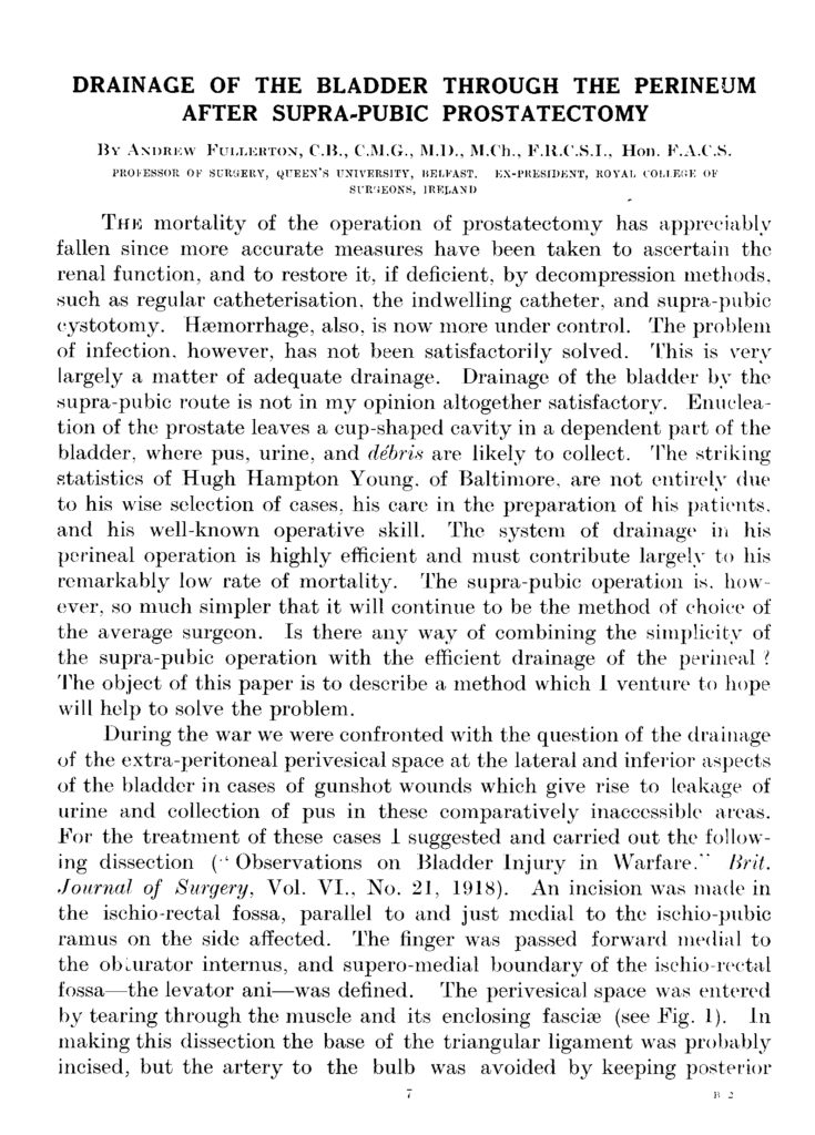
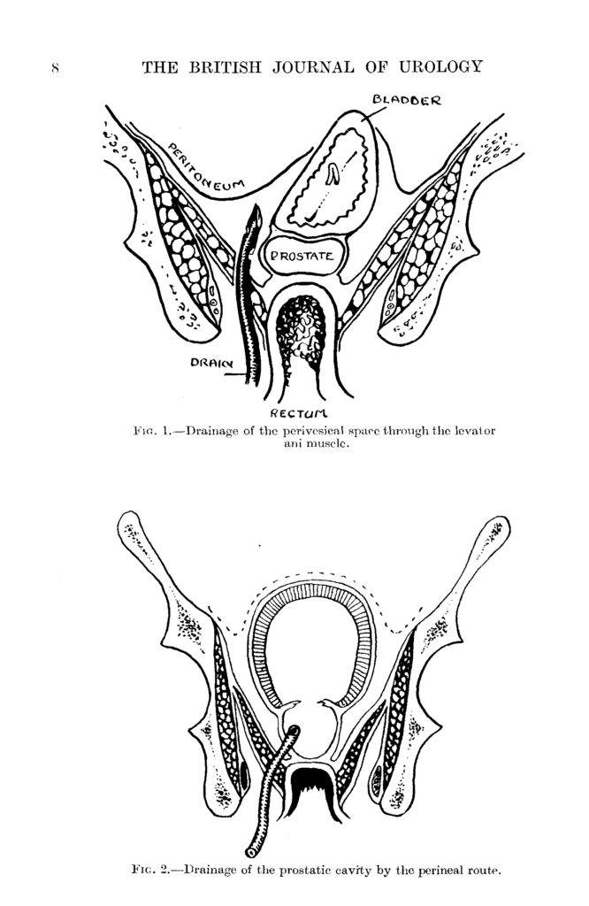
The first two pages of the very first article are reproduced here and can be accessed via Wiley Online. Note the detailed hand-drawn diagrams. It is also interesting to note the lack of articles on prostate cancer – very different from a urological journal of today.

Collaboration

In 1946 the Journal included a report on “… the Inaugural Scientific Meeting of the British Association of Urological Surgeons and acknowledged the importance of taking on “…the role of official organ of the B.A.U.S….”

The Journal is proud to continue to be an official journal of BAUS and to be the official journal of, or to be affiliated with, many urological associations around the world including:

Urological Society of Australia and New Zealand

Urological Society of India

Caribbean Urological Association

Hong Kong Urological Association

Irish Society of Urology

Swiss Continence Foundation

Korean journal, Investigative and Clinical Urology

Indonesian Urological Association

International Alliance of Urolithiasis

Malaysian Urological Association.

The Journal was, of course, solely a paper-based one from inception until 1994 when content was made available on CD ROM.  This innovation was soon superseded by the advent of the internet and the Journal was first published online in 1997. This signal event resulted in a transformation of the procedure for submitting and processing papers through peer review from a paper-based, postal one to an online one. All articles published in the Journal since 1929 have been digitised so that they are accessible online. Over the last two decades, the demand for print subscriptions has waned.

In 1999 the name of the Journal was changed to BJU International, reflecting the Journal’s British heritage and the importance of the contributions from the UK and internationally.

BJUI Editors 1929-2019

Responsibility for the Journal has been held by a succession of Editors and their Editorial Teams. There have been 10 editors during the first 90 years, some of whom served as co-editors (early version of a job-share?).

1929-1933

Frank Kidd & HP Winsbury White

1933-1948

HP Winsbury White

1949-1966

David Band

1967-1972

JD Fergusson

1972-1977

W Keith Yeates

1978-1993

GD Chisholm

1994-1996

Hugh Whitfield & Bill Hendy

1997-2002

Hugh Whitfield

2003-2012

John Fitzpatrick

2013-2020

Prokar Dasgupta

Thanks to Jonathan Goddard for the photos of the editors

Our current Editor-in-Chief, Prokar Dasgupta, is supported by an internationally based team of Associate Editors and Consulting Editors. In August next year, he will hand on the Editorship to Freddie Hamdy.

Thank you to our Editorial Teams, authors and peer reviewers for all their hard work in contributing to the success of the Journal.


IP4-CHRONOS is launched

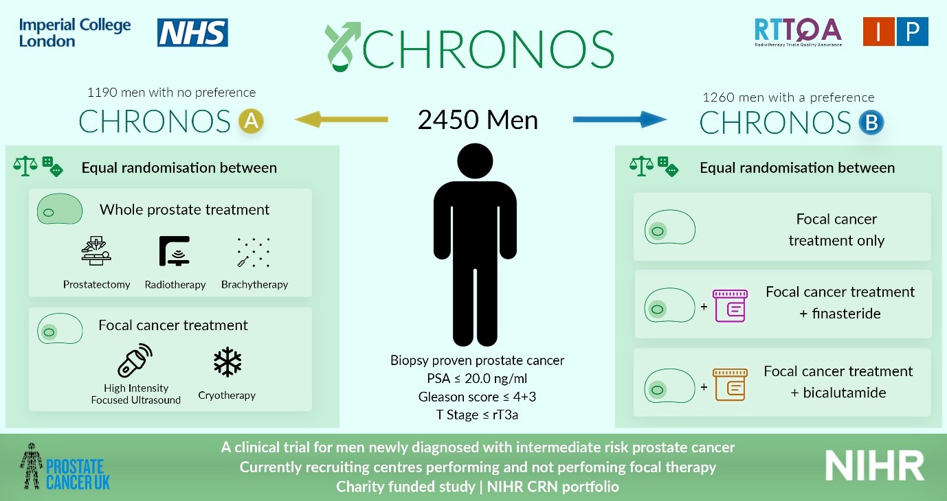
IP4- CHRONOS is open! CHRONOS is a phase II randomised control trial, that will review the outcomes (including oncological, functional, quality of life and cost-effectiveness) of focal therapy against those from radical therapy, in men with newly diagnosed localised clinically significant prostate cancer.


All men newly diagnosed with low-intermediate risk prostate cancer, confined to the prostate, with a life expectancy of at least 10 years will be screened for eligibility. Men must be well enough to undergo the interventions outlined in the trial prior to being enrolled.

Men will then have a choice of enrolling into CHRONOS A or CHRONOS B. CHRONOS A will randomise men to having radical whole gland treatment (radiotherapy, brachytherapy or prostatectomy), or focal therapy (HIFU or cryotherapy). CHRONOS A will answer the question, ‘is focal therapy equivalent in cancer control as radical therapy?’ CHRONOS B will randomise men to having focal therapy with or without additional neoadjuvant treatment and will answer the question: ‘can the success of focal therapy be improved by using neoadjuvant treatment?’ Randomisation will be stratified by disease characteristics.

All men will undergo intervention as they would within the NHS, however by doing so in a trial setting, we can directly compare the results of such treatments against each other. As the follow up mimics that of standard of care, the extra burden of treatment within the trial is minimal.

60 men will be recruited into both CHRONOS A and CHRONOS B (total 120) over a 1-year period, during the pilot, and if recruitment is successful the aim is to continue to a larger study assessing 2450 patients over 5 years, with a minimum follow up of 3 years. The primary outcome measures will be progression free survival in CHRONOS A, and failure free survival in CHRONOS B. The CHRONOS pilot will open in 12 UK hospital sites, aiming to open across the UK and Europe within the larger study.

CHRONOS is entirely funded by the Prostate Cancer UK charity, and available on the NIHR CRN portfolio. If you would like to join the main phase of CHRONOS as a site, please contact Miss Deepika Reddy ([email protected]) or visit our website for further information www.imperialprostate.org.uk/CHRONOS

Prof Hashim U. Ahmed (CHRONOS PI&CI)

Mr Taimur T. Shah (CHRONOS sub-investigator, Urology SpR & Research Fellow)

Miss Deepika Reddy (CHRONOS Clinical Research Fellow)


© 2025 BJU International. All Rights Reserved.