Tag Archive for: biopsy

Posts

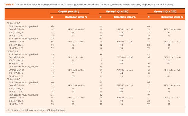
Article of the Week: Evaluation of targeted and systematic biopsies using MRI and US image-fusion guided transperineal prostate biopsy

Every Week the Editor-in-Chief selects an Article of the Week from the current issue of BJUI. The abstract is reproduced below and you can click on the button to read the full article, which is freely available to all readers for at least 30 days from the time of this post.

In addition to the article itself, there is an accompanying editorial written by a prominent member of the urological community. This blog is intended to provoke comment and discussion and we invite you to use the comment tools at the bottom of each post to join the conversation.

If you only have time to read one article this week, it should be this one.

Multicentre evaluation of targeted and systematic biopsies using magnetic resonance and ultrasound image-fusion guided transperineal prostate biopsy in patients with a previous negative biopsy


Nienke L. Hansen*†‡, Claudia Kesch§, Tristan Barrett, Brendan Koo, Jan P. Radtke§**, David Bonekamp** , Heinz-Peter Schlemmer**, Anne Y. Warren‡††, Kathrin Wieczorek‡‡Markus Hohenfellner§, Christof Kastner§§ and Boris Hadaschik§


*Department of Diagnostic and Interventional Radiology, University Hospital RWTH Aachen, Aachen, Germany, CamPARI Clinic, Addenbrookes Hospital and University of Cambridge, Cambridge, UK, Department of Diagnostic and Interventional Radiology, University Hospital Cologne, Cologne§Department of Urology, University Hospital Heidelberg, Heidelberg, Germany, Department of Radiology, Addenbrookes Hospital and University of Cambridge, Cambridge, UK, **Department of Radiology, DKFZ, Heidelberg, Germany, ††Department of Pathology, AddenbrookeHospital and University of Cambridge, Cambridge, UK, ‡‡Institute of Pathology, University of Heidelberg, Heidelberg, Germany, and §§Department of Urology, Addenbrookes Hospital and University of Cambridge, Cambridge, UK


Abstract

Objectives

To evaluate the detection rates of targeted and systematic biopsies in magnetic resonance imaging (MRI) and ultrasound (US) image-fusion transperineal prostate biopsy for patients with previous benign transrectal biopsies in two high-volume centres.

Patients and Methods

A two centre prospective outcome study of 487 patients with previous benign biopsies that underwent transperineal MRI/US fusion-guided targeted and systematic saturation biopsy from 2012 to 2015. Multiparametric MRI (mpMRI) was reported according to Prostate Imaging Reporting and Data System (PI-RADS) Version 1. Detection of Gleason score 7–10 prostate cancer on biopsy was the primary outcome. Positive (PPV) and negative (NPV) predictive values including 95% confidence intervals (95% CIs) were calculated. Detection rates of targeted and systematic biopsies were compared using McNemar’s test.

Results

The median (interquartile range) PSA level was 9.0 (6.7–13.4) ng/mL. PI-RADS 3–5 mpMRI lesions were reported in 343 (70%) patients and Gleason score 7–10 prostate cancer was detected in 149 (31%). The PPV (95% CI) for detecting Gleason score 7–10 prostate cancer was 0.20 (±0.07) for PI-RADS 3, 0.32 (±0.09) for PI-RADS 4, and 0.70 (±0.08) for PI-RADS 5. The NPV (95% CI) of PI-RADS 1–2 was 0.92 (±0.04) for Gleason score 7–10 and 0.99 (±0.02) for Gleason score ≥4 + 3 cancer. Systematic biopsies alone found 125/138 (91%) Gleason score 7–10 cancers. In patients with suspicious lesions (PI-RADS 4–5) on mpMRI, systematic biopsies would not have detected 12/113 significant prostate cancers (11%), while targeted biopsies alone would have failed to diagnose 10/113 (9%). In equivocal lesions (PI-RADS 3), targeted biopsy alone would not have diagnosed 14/25 (56%) of Gleason score 7–10 cancers, whereas systematic biopsies alone would have missed 1/25 (4%). Combination with PSA density improved the area under the curve of PI-RADS from 0.822 to 0.846.

Conclusion

In patients with high probability mpMRI lesions, the highest detection rates of Gleason score 7–10 cancer still required combined targeted and systematic MRI/US image-fusion; however, systematic biopsy alone may be sufficient in patients with equivocal lesions. Repeated prostate biopsies may not be needed at all for patients with a low PSA density and a negative mpMRI read by experienced radiologists.

Article of the Week: Profiling microRNA from nephrectomy and biopsy specimens

Every Week the Editor-in-Chief selects an Article of the Week from the current issue of BJUI. The abstract is reproduced below and you can click on the button to read the full article, which is freely available to all readers for at least 30 days from the time of this post.

In addition to the article itself, there is an accompanying editorial written by a prominent member of the urological community. This blog is intended to provoke comment and discussion and we invite you to use the comment tools at the bottom of each post to join the conversation.

If you only have time to read one article this week, it should be this one.

Profiling microRNA from nephrectomy and biopsy specimens: predictors of progression and survival in clear cell renal cell carcinoma


Casey G. Kowalik*, Drew A. Palmer*, Travis B. Sullivan, Patrick A. TeebagyJohn M. Dugan, John A. Libertino*, Eric J. Burks, David Canes* and Kimberly M. Rieger-Christ


Departments of *Urology, Translational Research Ian C. Summerhayes Cell and Molecular Biology Laboratory, and Pathology, Lahey Hospital and Medical Center, Burlington, MA, USA


Read the full article

Abstract

Objective

To identify microRNA (miRNA) characteristic of metastatic clear cell renal cell carcinoma (ccRCC) and those indicative of cancer-specific survival (CSS) in nephrectomy and biopsy specimens. We also sought to determine if a miRNA panel could differentiate benign from ccRCC tissue.

Materials and Methods

RNA was isolated from nephrectomy and kidney biopsy specimens (n = 156 and n = 46, respectively). Samples were grouped: benign, non-progressive, and progressive ccRCC. MiRNAs were profiled by microarray and validated by quantitative reverse transcription-polymerase chain reaction. Biomarker signatures were developed to predict cancer status in nephrectomy and biopsy specimens. CSS was examined using Kaplan–Meier and Cox proportional hazards analyses.

Results

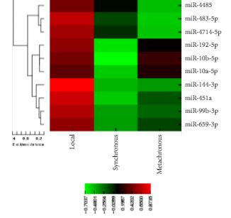
Microarray analysis revealed 20 differentially expressed miRNAs comparing non-progressive with progressive tumours. A biomarker signature validated in nephrectomy specimens had a sensitivity of 86.7% and a specificity of 92.9% for differentiating benign and ccRCC specimens. A second signature differentiated non-progressive vs progressive ccRCC with a sensitivity of 93.8% and a specificity of 83.3%. These biomarkers also discriminated cancer status in biopsy specimens. Levels of miR-10a-5p, -10b-5p, and -223-3p were associated with CSS.

Conclusion

This study identified miRNAs differentially expressed in ccRCC samples; as well as those correlating with CSS. Biomarkers identified in this study have the potential to identify patients who are likely to have progressive ccRCC, and although preliminary, these results may aid in differentiating aggressive and indolent ccRCC based on biopsy specimens.

Editorial: The utility of microRNAs as biomarkers in predicting progression and survival in patients with clear-cell renal cell carcinoma

RCC constitutes a diverse group of malignancies, yet the clear-cell subtype comprises ~80% of all diagnosed RCC cases [1]. The widespread use of abdominal imaging and subsequent stage migration has resulted in improved RCC 5-year cancer-specific survival. However, the overall mortality of RCC remains largely unchanged [2] and one-third of the patients have metastatic disease at the time of presentation [3]. Accordingly, the ability to precisely predict patient outcome has become an increasingly significant question in the management of these patients with RCC.

Accruing evidence suggests that changes in various biomarkers and their consequent downstream pathways affect cancer initiation and progression. Therefore, accurate prediction of the outcome and prognosis after treatment is necessary [4]. MicroRNAs (miRNAs) are small non-coding RNA molecules that can have significant functions in tumorigenesis [5]. Because of their ability in post-transcriptional regulation of gene expression, tumour-specific genetic defects in miRNA biogenesis and production correlate with development of human cancers. Thus, the differential expression of specific miRNA signatures in different tumours might become an important tool to help in directing cancer diagnosis and treatment [5].

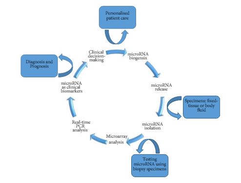
As such, Kowalik et al. [6] report on profiling miRNA to identify biomarker signatures predictive of clear-cell RCC (ccRCC) progression and survival. The authors used 202 formalin-fixed paraffin-embedded samples to isolate RNA from nephrectomy and biopsy specimens (n = 156 and n = 46, respectively) (Fig. 1).

Figure 1. Schematic diagram of miRNA-based biomarkers and potential utility in clinical decision-making approach for targeted therapy and RCC personalised treatment.

The primary analysis of their study [6] focused on the identification of miRNA signatures capable of differentiating between benign and ccRCC, as well as discerning those patients with a non-progressive ccRCC from a progressive clear-cell subtype. The secondary outcome examined the association of miRNA profiles discovered on cancer-specific survival.

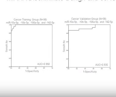
In their initial microarray screening 20 differentially expressed miRNAs, comparing non-progressive with progressive tumours, were identified. The authors found four miRNA panels (10a-5p, 10b-5p, 106a-5p, and 142-5p) as a potential biomarker signature. This model was validated in nephrectomy specimens and resulted in a sensitivity of 86.7%, a specificity of 92.9%, and an area under the curve (AUC) of 0.930 for detecting ccRCC. Further analysis revealed a second signature of two biomarkers (miR-10a-5p and -223-3p) with 93.8% sensitivity, 83.3% specificity, and an AUC of 0.932 when validated for detecting progressive ccRCC. Similarly, the differential expression of these biomarkers could delineate cancer status in biopsy specimens. For correlation of miRNA expression levels with cancer-specific survival, higher expression levels of (miR-10a-5p and miR-10b-5p) and a lower expression level of (miR-223-3p) were significantly associated with survival (P< 0.001), and the median survival times were not reached.

In conclusion, the lack of precise prediction tools has led the authors to explore the potential utility of miRNAs as biomarkers to detect disease presence, biological aggressiveness, and prognosis in ccRCC. However, until future multicentre large prospective studies validate the results of the present work, the transition of miRNA from bench to bedside is emerging on the horizon and has encouraged urologists and scientists to pursue intense translational research in the field. The ability to use miRNAs as biomarkers might be promising for diagnostic and prognostic purposes. These biomarkers may exemplify different aspects of RCC pathogenesis and may potentially have important therapeutic implications to help in a clinical decision-making approach for targeted therapy and RCC personalised treatment.

Firas G. Petrosand Christopher J.D. Wallis†‡
*Department of Urology, The University of Texas MD Anderson Cancer Center, Houston, TX, USADivision of Urology, Department of Surgery, University of Toronto, Toronto, ON, Canada and Division of Urology, Department of Surgery, Sunnybrook Health Sciences Centre, Toronto, ON, Canada


Read the full article


References


1 Reuter VE, Presti JC Jr. Contemporary approach to the classication of renal epithelial tumors. Semin Oncol 2000; 27: 12437


2 Hollingsworth JM, Miller DC, Daignault S, Hollenbeck BK. Rising incidence of small renal masses: a need to reassess treatment effect. J Natl Cancer Inst 2006; 98: 13314


3 Gupta K, Miller JD , Li JZ, Russell MW, Charbonneau C. Epidemiologic and socioeconomic burden of metastatic renal cell carcinoma (mRCC): literature review. Cancer Treat Rev 2008; 34: 193205


5 Esquela-Kerscher A, Slack FJ. Oncomirs microRNAs with a role in cancer. Nat Rev Cancer 2006; 6: 25969


Video: Profiling microRNA from nephrectomy and biopsy specimens

Profiling microRNA from nephrectomy and biopsy specimens: predictors of progression and survival in clear cell renal cell carcinoma


Read the full article

Abstract

Objective

To identify microRNA (miRNA) characteristic of metastatic clear cell renal cell carcinoma (ccRCC) and those indicative of cancer-specific survival (CSS) in nephrectomy and biopsy specimens. We also sought to determine if a miRNA panel could differentiate benign from ccRCC tissue.

Materials and Methods

RNA was isolated from nephrectomy and kidney biopsy specimens (n = 156 and n = 46, respectively). Samples were grouped: benign, non-progressive, and progressive ccRCC. MiRNAs were profiled by microarray and validated by quantitative reverse transcription-polymerase chain reaction. Biomarker signatures were developed to predict cancer status in nephrectomy and biopsy specimens. CSS was examined using Kaplan–Meier and Cox proportional hazards analyses.

Results

Microarray analysis revealed 20 differentially expressed miRNAs comparing non-progressive with progressive tumours. A biomarker signature validated in nephrectomy specimens had a sensitivity of 86.7% and a specificity of 92.9% for differentiating benign and ccRCC specimens. A second signature differentiated non-progressive vs progressive ccRCC with a sensitivity of 93.8% and a specificity of 83.3%. These biomarkers also discriminated cancer status in biopsy specimens. Levels of miR-10a-5p, -10b-5p, and -223-3p were associated with CSS.

Conclusion

This study identified miRNAs differentially expressed in ccRCC samples; as well as those correlating with CSS. Biomarkers identified in this study have the potential to identify patients who are likely to have progressive ccRCC, and although preliminary, these results may aid in differentiating aggressive and indolent ccRCC based on biopsy specimens.

View more videos

Article of the Week: ERSPC risk calculators significantly outperform the PCPT 2.0 in the prediction of PCa

Every Week the Editor-in-Chief selects an Article of the Week from the current issue of BJUI. The abstract is reproduced below and you can click on the button to read the full article, which is freely available to all readers for at least 30 days from the time of this post.

In addition to the article itself, there is an accompanying editorial written by a prominent member of the urological community. This blog is intended to provoke comment and discussion and we invite you to use the comment tools at the bottom of each post to join the conversation.

If you only have time to read one article this week, it should be this one.

European Randomised Study of Screening for Prostate Cancer (ERSPC) risk calculators significantly outperform the Prostate Cancer Prevention Trial (PCPT) 2.0 in the prediction of prostate cancer: a multi-institutional study

Robert W. Foley*,, Robert M. Maweni, Laura Gorman, Keefe Murphy§,, Dara J. Lundon Z*,,**, Garrett Durkan††,‡‡, Richard Power§§, Frank OBrien¶¶, Kieran J. OMalley**, David J. Galvin,**,***, T. Brendan Murphy§,¶ and R. William Watson*,


*UCD School of Medicine, University College Dublin, UCD Conway Institute of Biomolecular and Biomedical Research, University College Dublin, Dublin, Ireland, Croydon NHS Trust, Croydon University Hospital, London, UK, §UCD School of Mathematical Sciences, University College Dublin, Insight Centre for Data Analytics, University College Dublin, **Department of Urology, Mater Misericordiae University Hospital, Dublin, ††Department of Urology, University Hospital Galway, Galway, ‡‡Department of Urology, University Hospital Limerick, Limerick, §§Department of Urology, Beaumont Hospital, Dublin, ¶¶Department of Urology, University Hospital Waterford, Waterford, and ***Department of Urology, St. Vincents University Hospital, Dublin, Ireland


Read the full article

Objective

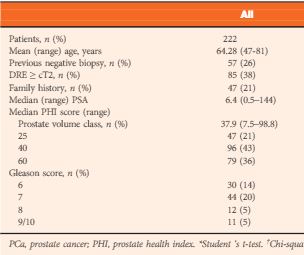
To analyse the performance of the Prostate Cancer Prevention Trial Risk Calculator (PCPT-RC) and two iterations of the European Randomised Study of Screening for Prostate Cancer (ERSPC) Risk Calculator, one of which incorporates prostate volume (ERSPC-RC) and the other of which incorporates prostate volume and the prostate health index (PHI) in a referral population (ERSPC-PHI).

Patients and Methods

The risk of prostate cancer (PCa) and significant PCa (Gleason score ≥7) in 2001 patients from six tertiary referral centres was calculated according to the PCPT-RC and ERSPC-RC formulae. The calculators’ predictions were analysed using the area under the receiver-operating characteristic curve (AUC), calibration plots, Hosmer–Lemeshow test for goodness of fit and decision-curve analysis. In a subset of 222 patients for whom the PHI score was available, each patient’s risk was calculated as per the ERSPC-RC and ERSPC-PHI risk calculators.

aotwnov2

Results

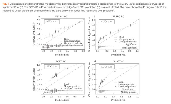
The ERSPC-RC outperformed the PCPT-RC in the prediction of PCa, with an AUC of 0.71 compared with 0.64, and also outperformed the PCPT-RC in the prediction of significant PCa (P<0.001), with an AUC of 0.74 compared with 0.69. The ERSPC-RC was found to have improved calibration in this cohort and was associated with a greater net benefit on decision-curve analysis for both PCa and significant PCa. The performance of the ERSPC-RC was further improved through the addition of the PHI score in a subset of 222 patients. The AUCs of the ERSPC-PHI were 0.76 and 0.78 for PCa and significant PCa prediction, respectively, in comparison with AUC values of 0.72 in the prediction of both PCa and significant PCa for the ERSPC-RC (P = 0.12 and P = 0.04, respectively). The ERSPC-PHI risk calculator was well calibrated in this cohort and had an increase in net benefit over that of the ERSPC-RC.

Conclusions

The performance of the risk calculators in the present cohort shows that the ERSPC-RC is a superior tool in the prediction of PCa; however the performance of the ERSPC-RC in this population does not yet warrant its use in clinical practice. The incorporation of the PHI score into the ERSPC-PHI risk calculator allowed each patient’s risk to be more accurately quantified. Individual patient risk calculation using the ERSPC-PHI risk calculator can be undertaken in order to allow a systematic approach to patient risk stratification and to aid in the diagnosis of PCa.

Editorial: Prostate cancer risk calculators – still much work ahead

Several risk calculators (RCs) have been developed to predict prostate cancer (PCa) diagnosis at prostate biopsy. These multivariable tools have constantly been shown to be superior to risk prediction using PSA testing alone. Their use in personalized clinical decision-making is thus increasingly recommended to reduce overdiagnosis and overtreatment of PCa [1]. Foley et al. [2] conducted a multi-institutional external validation of the most recent versions of the European Randomised Study of Screening for Prostate Cancer Risk Calculator (ERSPC-RC) and the Prostate Cancer Prevention Trial Risk Calculator (PCPT-RC) in a large cohort of patients from six different Irish tertiary referral centres. The study showed that the two RCs performed moderately well. Both RCs performed less optimistic compared with their original reports. The ERSPC-RC showed superior discrimination (area under the curve of 0.74 vs 0.69 for high grade PCa) and a greater net benefit in decision-curve analysis (DCA) than the PCPT-RC; however, although the ERSPC-RC was superior to the PCPT-RC in this well-conducted study, neither RC can be recommended for PCa risk prediction in this specific Irish cohort.

The authors chose to perform DCAs, which are of great value for further assessing the utility of a risk prediction model using visualization of the clinical net benefit and net harm. The benefit threshold of >30%, as shown in the DCA of the ERSPC-RC for high grade PCa, is too high for a clinically meaningful prediction tool. Below this threshold the RC did not provide further benefit compared with a strategy of performing a biopsy on everybody. It is questionable whether clinicians or patients would opt to use an RC which only provides a benefit if a risk of 30% as the lowest acceptable threshold for high grade disease is accepted.

What are the reasons for the suboptimum performance of the RCs in the Irish cohort? It is well known that RC performance is often less optimistic in external validations [3]. Differences in cohort characteristics, biopsy strategies and screening recommendations between RC development cohorts and the tested cohorts, but also changes in clinical practice over time, are potential reasons. Although the RCs have constantly been modified to establish their role as a general one-size-fits-all risk prediction model, their performance varied significantly in different cohorts. We recently evaluated the same RCs in a large Swiss single-centre cohort and found similar discrimination but better calibration, a greater net benefit and a lower and thus clinically useful benefit threshold in DCAs compared with the present Irish study [4]. The cohort in the present study was unique because it consisted of a highly preselected group of patients. This is attributable to the specific referral practice for prostate biopsies in Ireland and is reflected in the high number of patients with a positive DRE (47% in the group diagnosed with PCa) or a positive family history (11%). Accordingly, the overall PCa detection rate (58%) and the detection rate of high grade disease (35%) were higher than usually expected. From a scientific point of view, the Irish cohort is not the optimum cohort to validate these RCs. Far more importantly, however, from a clinical point of view, the evaluation showed that these RCs are not really useful in the specific Irish health system.

What can be done to improve the performance RCs in the future? It is obvious that specific characteristics of the tested cohorts will affect RC performance. These local or regional characteristics usually cannot be changed. Thus modifications of available RCs according to local patient practice might be necessary. This concept has recently been examined by Strobl et al. [5]. They were able to show that recalibration of the static PCPT-RC according to local cohort and practice characteristics can improve its accuracy. Additionally, RCs developed from contemporary clinical cohorts that were, for example, diagnosed using current state-of-the-art biopsy strategies (i.e. 12-core biopsies) instead of historical cohorts from, for example, randomized clinical trials might also result in better RC performance in clinical practice. Furthermore, the inclusion of novel variables in the RC might be useful. Results from imaging studies, such as multiparametric prostate MRI, or promising new biomarkers might increase the overall performance of PCa RCs. The study by Foley et al. shows that the inclusion of novel markers can be of benefit. The ERSPC-PHI RC, which includes the Prostate Health Index (PHI) as an additional variable, was investigated in a subset of patients in their study and was superior to the conventional ERSPC-RC; however, when novel variables are integrated, their potential clinical harm (e.g. unpleasant or costly investigations) has to be balanced against their potential benefit.

The work of Foley et al. nicely illustrates the limitations of current PCa RCs. Locally tailored static RCs, RCs based on contemporary clinical cohorts, or RCs including novel variables need to be developed to assess whether overall RC performance can be improved in the future. There is still much work to do!

Read the full article
Cedric Poyet and Thom as Hermanns 


Department of Urology, University Hospital Zurich, University of Zurich, Zurich, Switzerland


References


Article of the Week: TRUS-Guided RB PCa Detection – Reasons for Targeted Biopsy Failure

Every Week the Editor-in-Chief selects an Article of the Week from the current issue of BJUI. The abstract is reproduced below and you can click on the button to read the full article, which is freely available to all readers for at least 30 days from the time of this post.

In addition to the article itself, there is an accompanying editorial written by a prominent member of the urological community. This blog is intended to provoke comment and discussion and we invite you to use the comment tools at the bottom of each post to join the conversation.

Finally, the third post under the Article of the Week heading on the homepage will consist of additional material or media. This week we feature a video from Hannes Cash and Patrick Asbach, discussing their paper.

If you only have time to read one article this week, it should be this one.

Prostate cancer detection on transrectal ultrasonography-guided random biopsy despite negative real-time magnetic resonance imaging/ultrasonography fusion-guided targeted biopsy: reasons for targeted biopsy failure

Hannes Cash*, Karsten Gunzel*, Andreas Maxeiner*, Carsten Stephan*, Thomas Fischer, Tahir Durmus, Kurt Miller*, Patrick Asbach, Matthias Haas† and Carsten Kempkensteffen*


*Department of Urology, and Department of Radiology, ChariteUniversity of Medicine Berlin, Berlin, Germany M. H. and C.K. contributed equally to the study.


Read the full article

Objective

To examine the value of additional transrectal ultrasonography (TRUS)-guided random biopsy (RB) in patients with negative magnetic resonance imaging (MRI)/ultrasonography (US) fusion-guided targeted biopsy (TB) and to identify possible reasons for TB failure.

Patients and Methods

We conducted a subgroup analysis of 61 men with prostate cancer (PCa) detected by 10-core RB but with a negative TB, from a cohort of 408 men with suspicious multiparametric magnetic resonance imaging (mpMRI) between January 2012 and January 2015. A consensus re-reading of mpMRI results (using Prostate Imaging Reporting and Data System [PI-RADS] versions 1 and 2) for each suspicious lesion was performed, with the image reader blinded to the biopsy results, followed by an unblinded anatomical correlation of the lesion on mpMRI to the biopsy result. The potential reasons for TB failure were estimated for each lesion. We defined clinically significant PCa according to the Epstein criteria and stratified patients into risk groups according to the European Association of Urology guidelines.

JulAOTW3Results

Results

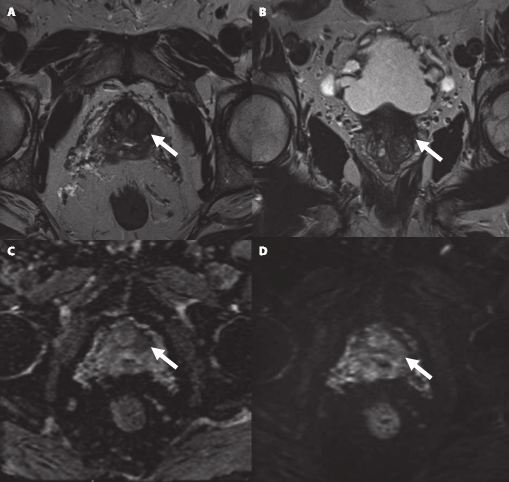
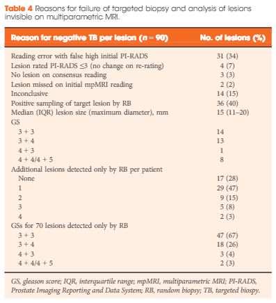
Our analysis showed that RB detected significant PCa in 64% of patients (39/61) and intermediate-/high-risk PCa in 57% of patients (35/61). The initial mpMRI reading identified 90 suspicious lesions in the cohort. Blinded consensus re-reading of the mpMRI led to PI-RADS score downgrading of 45 lesions (50%) and upgrading of 13 lesions (14%); thus, negative TB could be explained by falsely high initial PI-RADS scores for 32 lesions (34%) and sampling of the target lesion by RB in the corresponding anatomical site for 36 out of 90 lesions (40%) in 35 of 61 patients (57%). Sampling of the target lesion by RB was most likely for lesions with PI-RADS scores of 4/5 and Gleason scores (GS) of ≥7. A total of 70 PCa lesions (67% with GS 6) in 44 patients (72%) were sampled from prostatic sites with no abnormalities on mpMRI.

Conclusion

In cases of TB failure, RB still detected a high rate of significant PCa. The main reason for a negative TB was a TB error, compensated for by positive sampling of the target lesion by the additional RB, and the second reason for TB failure was a falsely high initial PI-RADS score. The challenges that arise for both MRI diagnostics and prostate lesion sampling are evident in our data and support the integration of RB into the TB workflow.

Editorial: MRI-Fusion Biopsy – Behind the Scenes

MRI information of the prostate is increasingly used for improving the diagnostic yield of prostate biopsies [1]. However, increasing complexity of a procedure makes it prone to errors at multiple technical and human levels. Incorporating MRI information and ultrasonography (US) images for MRI-fusion biopsies is a technically challenging task. It involves various steps such as the acquisition and fusion of MRI and US images, the needle guidance during biopsy, and the diligence of the pathological evaluation of biopsy specimens. These different steps and interfaces between different medical professions influence the diagnostic performance of MRI-fusion biopsies.

For example, in daily clinical practice, MRIs from different institutions still harbour a great variance of sequences and reporting, despite the European Society of Urogenital Urology (ESUR) recently introducing acquisition and imaging protocols and a new and advanced version of the Prostate Imaging Reporting and Data System (PIRADS) version 2.0 [2]. The usefulness of such reporting schemes is evidenced by a moderate-to-good interobserver agreement between uro-radiologists for tumour lesion interpretation and corresponding κ values ranging from 0.55 to 0.80 [3]. Important pitfalls of image interpretation are benign lesions such as prostatitis, BPH and fibrosis, which might score similarly to prostate cancer lesions. This problem is further aggravated by a high proportion of patients that receive their first multiparametric MRI (mpMRI) of the prostate in the repeat-biopsy setting with a high burden of post-biopsy artefacts (haemorrhage, capsular irregularity) and lower overall cancer detection rate. Also, during MRI-fusion biopsy patient movement, prostate deformation by the US probe, and mismatch of image planes can lead to a biopsy error exceeding 4 mm. Moreover, targeting error might be aggravated by MRI underestimation of the tumour volume compared with final pathology [4]. After various authors reported the advantages and accuracy of MRI/US-fusion biopsy approaches, Cash et al. [5] address potential reasons for targeted biopsy failure to detect prostate cancer compared with random biopsy. Within their analyses the authors address potential limitations and technical considerations. Based on different technical biopsy strategies (with the patient placed within the MRI scanner (‘in-bore’) vs outside) and different technical approaches, these considerations are very important.

In contrast to cognitive fusion, most MRI/US platforms allow needle tracking by archiving the needle orientation, either by an electromagnetic, image-based or stepper-based mechanism [1]. However, lesion targeting by needle guidance is highly dependent on the dimensions of the primary lesion, numbers of relevant lesions, localisation, and overall prostate volume, making MRI-US fusion and cognitive fusion more error prone (i.e. aiming off the mark with the needle) than in-bore biopsies. Moreover, different technical fusion approaches provide different degrees of manual/automated adjustment tools, with for example either rigid or elastic image transformation to facilitate MRI/US image alignment.

In their analyses, Cash et al. [5] found that 34% of negative targeted biopsies could be explained by initially too high estimated PIRADS scores that were downgraded at re-reading. Interestingly, the remaining lesions were without an mpMRI correlate but within this group 92.9% showed a primary Gleason 3 pattern in biopsy pathology, suggesting a high degree of invisibility on mpMRI. Subanalyses did not show an association of targeted biopsy failures in the ventral location. Therefore, the study by Cash et al. [5] is an important precursor for further analyses to address other underlying reasons for targeted biopsy failure. Moreover, it reveals the need for a tight collaboration of radiologists, urologists, and pathologists as interdisciplinary partners involved in MRI-fusion biopsy. Consequently, the optimal diagnostic performance of MRI-fusion biopsies can only be achieved through standardised MRI performance, reading and reporting of MRI findings, as well as final correlation of MRI findings with histopathological work up.

Read the full article
Lars Budaus and Sami-Ramzi Leyh-Bannurah
Martini-Clinic University Hospital Hamburg-Eppendorf, Hamburg, Germany


References


Video: TRUS-Guided RB Prostate Cancer Detection – Reasons for Targeted Biopsy Failure

Prostate cancer detection on transrectal ultrasonography-guided random biopsy despite negative real-time magnetic resonance imaging/ultrasonography fusion-guided targeted biopsy: reasons for targeted biopsy failure

Hannes Cash*, Karsten Gunzel*, Andreas Maxeiner*, Carsten Stephan*, Thomas Fischer, Tahir Durmus, Kurt Miller*, Patrick Asbach, Matthias Haas† and Carsten Kempkensteffen*


*Department of Urology, and Department of Radiology, ChariteUniversity of Medicine Berlin, Berlin, Germany M. H. and C.K. contributed equally to the study.


Read the full article

Objective

To examine the value of additional transrectal ultrasonography (TRUS)-guided random biopsy (RB) in patients with negative magnetic resonance imaging (MRI)/ultrasonography (US) fusion-guided targeted biopsy (TB) and to identify possible reasons for TB failure.

Patients and Methods

We conducted a subgroup analysis of 61 men with prostate cancer (PCa) detected by 10-core RB but with a negative TB, from a cohort of 408 men with suspicious multiparametric magnetic resonance imaging (mpMRI) between January 2012 and January 2015. A consensus re-reading of mpMRI results (using Prostate Imaging Reporting and Data System [PI-RADS] versions 1 and 2) for each suspicious lesion was performed, with the image reader blinded to the biopsy results, followed by an unblinded anatomical correlation of the lesion on mpMRI to the biopsy result. The potential reasons for TB failure were estimated for each lesion. We defined clinically significant PCa according to the Epstein criteria and stratified patients into risk groups according to the European Association of Urology guidelines.

JulAOTW3Results

Results

Our analysis showed that RB detected significant PCa in 64% of patients (39/61) and intermediate-/high-risk PCa in 57% of patients (35/61). The initial mpMRI reading identified 90 suspicious lesions in the cohort. Blinded consensus re-reading of the mpMRI led to PI-RADS score downgrading of 45 lesions (50%) and upgrading of 13 lesions (14%); thus, negative TB could be explained by falsely high initial PI-RADS scores for 32 lesions (34%) and sampling of the target lesion by RB in the corresponding anatomical site for 36 out of 90 lesions (40%) in 35 of 61 patients (57%). Sampling of the target lesion by RB was most likely for lesions with PI-RADS scores of 4/5 and Gleason scores (GS) of ≥7. A total of 70 PCa lesions (67% with GS 6) in 44 patients (72%) were sampled from prostatic sites with no abnormalities on mpMRI.

Conclusion

In cases of TB failure, RB still detected a high rate of significant PCa. The main reason for a negative TB was a TB error, compensated for by positive sampling of the target lesion by the additional RB, and the second reason for TB failure was a falsely high initial PI-RADS score. The challenges that arise for both MRI diagnostics and prostate lesion sampling are evident in our data and support the integration of RB into the TB workflow.

Article of the Week: DSNB for Penile Cancer

Every Week the Editor-in-Chief selects an Article of the Week from the current issue of BJUI. The abstract is reproduced below and you can click on the button to read the full article, which is freely available to all readers for at least 30 days from the time of this post.

In addition to the article itself, there is an accompanying editorial written by a prominent member of the urological community. This blog is intended to provoke comment and discussion and we invite you to use the comment tools at the bottom of each post to join the conversation.

If you only have time to read one article this week, it should be this one.


Dynamic sentinel lymph node biopsy for penile cancer: a comparison between 1- and 2-day protocols

Panagiotis Dimopoulos*, Panagiotis Christopoulos*, Sam Shilito, Zara Gall*, Brian Murby§, David Ashworth§, Ben Taylor, Bernadette Carrington, Jonathan Shanks**, Noel Clarke*, Vijay Ramani*, Nigel Parr*, Maurice Lau* and Vijay Sangar*


Departments of *Urology, §Nuclear Medicine, Radiology , **Pathology, The Christie Hospital, ManchesterMedical School, University of Manchester, Manchester, and Department of Urology, Royal Bolton Hospital, Bolton Lancashire, UK
Read the full article

Objective

To determine the outcome of clinically negative node (cN0) patients with penile cancer undergoing dynamic sentinel node biopsy (DSNB), comparing the results of a 1- and 2-day protocol that can be used as a minimal invasive procedure for staging of penile cancer.

Patients and Methods

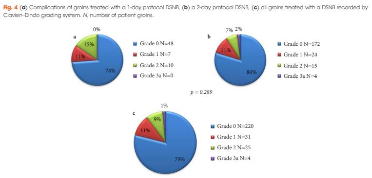
This is a retrospective analysis of 151 cN0 patients who underwent DSNB from 2008 to 2013 for newly diagnosed penile cancer. Data were analysed per groin and separated into groups according to the protocol followed. The comparison of the two protocols involved the number of nodes excised, γ-counts, false-negative rates (FNR), and complication rates (Clavien–Dindo grading system).

JuneAOTW3

Results

In all, 280 groins from 151 patients underwent DSNB after a negative ultrasound ± fine-needle aspiration cytology. The 1-day protocol was performed in 65 groins and the 2-day protocol in 215. Statistically significantly more nodes were harvested with the 1-day protocol (1.92/groin) compared with the 2-day protocol (1.60/groin). The FNRs were 0%, 6.8% and 5.1%, for the 1-day protocol, 2-day protocol, and overall, respectively. Morbidity of the DSNB was 21.4% for all groins, and 26.2% and 20.1% for the 1-day and 2-day protocols, respectively. Most of the complications were of Clavien–Dindo Grade 1–2.

Conclusions

DSNB is safe for staging patients with penile cancer. There is a trend towards a 1-day protocol having a lower FNR than a 2-day protocol, albeit at the expense of a slightly higher complication rate.

© 2025 BJU International. All Rights Reserved.