Tag Archive for: biochemical progression

Posts

Article of the Week: 68Ga-PSMA has high detection rate of PCa recurrence after RP

Every Week the Editor-in-Chief selects an Article of the Week from the current issue of BJUI. The abstract is reproduced below and you can click on the button to read the full article, which is freely available to all readers for at least 30 days from the time of this post.

In addition to the article itself, there is an accompanying editorial written by a prominent member of the urological community. This blog is intended to provoke comment and discussion and we invite you to use the comment tools at the bottom of each post to join the conversation.

If you only have time to read one article this week, it should be this one.

68Ga-PSMA has high detection rate of prostate cancer recurrence outside the prostatic fossa in patients being considered for salvage radiation treatment


Pim J. van Leeuwen*, Phillip Stricker*, George Hruby§, Andrew Kneebone§Francis Ting*, Ben Thompson, Quoc Nguyen, Bao Ho** and Louise Emmett**,††


*St Vincents Prostate Cancer Centre, St Vincents Clinic, Sydney, NSWAustralian Prostate Cancer Research Centre – New South Wales, Garvan Institute of Medical Research/Kinghorn Cancer Centre, Sydney, NSWRadiation Oncology Department, Northern Sydney Cancer Centre, Royal North Shore Hospital, St Leonards, NSW§University of Sydney, Sydney, NSWNorthern Clinical School, University of Sydney, St Leonards, NSW, **Department of Diagnostic Imaging, St Vincents Public Hospital, Sydney, NSW, and ††University of New South Wales, Sydney, NSW, Australia


Objectives

To examine the detection rates of 68Ga-PSMA-positron emission tomography (PET)/computed tomography (CT) in patients with biochemical recurrence (BCR) after radical prostatectomy (RP), and also the impact on their management.

Materials and Methods

A total of 300 consecutive patients with prostate cancer (PCa) who underwent 68Ga-PSMA-PET/CT between February and July 2015 were prospectively included in the Prostate Cancer Imaging (ProCan-I) database. For the present analysis, we included patients with BCR (prostate-specific antigen [PSA] level ≥0.05 and <1.0 ng/mL) after RP, who were being considered for salvage radiation therapy (RT) according to the Faculty of Radiation Oncology Genito-Urinary Group (FROGG) guidelines. Two readers assessed each 68Ga-PSMA-PET/CT, and all positive lesions were assigned to an anatomical location. For each patient, the clinical and pathological features were recorded, their association with pathological 68Ga-PSMA uptake was investigated, and detection rates were determined according to PSA level.

AOTWMAY

Results

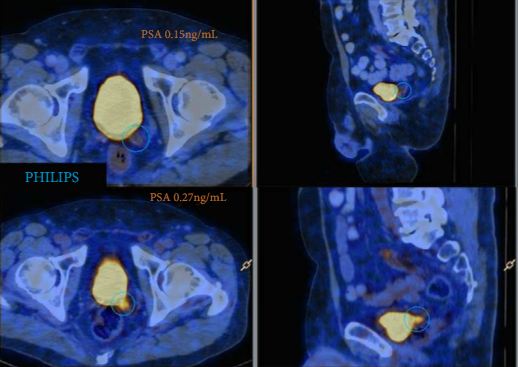
A total of 70 patients were included, and 53 positive 68Ga-PSMA lesions were detected in 38 (54%) patients. Among patients with PSA levels 0.05–0.09 ng/mL, 8% were definitely positive; the corresponding percentages for the other PSA ranges were as follows: PSA 0.1–0.19 ng/mL, 23%; PSA 0.2–0.29 ng/mL, 58%; PSA 0.3–0.49 ng/mL, 36%; and PSA 0.5–0.99 ng/mL, 57%. Eighteen of 70 patients (27%) had pathological 68Ga-PSMA uptake in the prostatic fossa, 11 (14.3%) in the pelvic nodes, and five (4.3%) in both the fossa and pelvic lymph nodes. Finally, there was uptake outside the pelvis with or without a lesion in the fossa or pelvic lymph nodes in four cases (8.6%). As a result of the 68Ga-PSMA findings there was a major management change in 20 (28.6%) patients.

Conclusions

68Ga-PSMA appears to be useful for re-staging of PCa in patients with rising PSA levels who are being considered for salvage RT even at PSA levels <0.5 ng/mL. These results underline the need for further prospective trials to evaluate the changes in RT volume or management attributable to 68Ga-PSMA findings.

Editorial: PSMA-targeted imaging of PCa – the best is yet to come

In recent years there has been increasing interest in imaging recurrent or metastatic prostate cancer with positron-emission tomography (PET) radiotracers targeting prostate-specific membrane antigen (PSMA [1]). The majority of this work has been performed using urea-based small molecules labelled with gallium-68 (68Ga). Within this class of radiotracers, 68Ga-PSMA-11 (also known as 68Ga-PSMA-HBED-CC) has been the most widely studied. In this month’s edition of BJUI, van Leeuwen et al. [2] report on the clinical utility of 68Ga-PSMA-11 PET/CT in men with rising PSA levels after radical prostatectomy being considered for salvage radiation therapy. In their study, 70 patients with negative conventional imaging findings and a median PSA of 0.2 ng/mL (all <1 ng/mL) were imaged with 68Ga-PSMA-11 PET/CT prior to initiating treatment. On PSMA-targeted PET/CT, 53 lesions were detected in 38 (54%) patients. Perhaps most significant among their findings was that 28.6% of men had radiotracer uptake outside of the prostatic fossa leading to a major change in clinical management. In total, these data demonstrate the great potential of PSMA-targeted imaging, particularly in men with biochemically recurrent prostate cancer.

While a great deal of encouraging data with 68Ga-PSMA-11 has appeared in the medical literature, it is worth noting that several other small molecules that offer potential advantages over this agent have seen early clinical development. For example, PSMA-617 makes use of the DOTA chelation moiety in place of HBED-CC, allowing for a scaffold that can accommodate both diagnostic 68Ga and therapeutic lutetium-177 (177Lu) [3]. Additionally, our group has focused on fluorine-18 (18F)-labelled urea-based small molecules targeting PSMA, most recently 18F-DCFPyL [4]. 18F-labelled small molecules offer several potential advantages over those labelled with 68Ga. These include more favourable dosimetry allowing for higher injected radiotracer doses and lower-energy emitted positrons that have shorter path lengths to annihilation and therefore higher intrinsic spatial resolution [5]. Notably, a recent direct comparison of 68Ga-PSMA-11 and 18F-DCFPyL performed by Dietlein et al. [6] seems to confirm these advantages, having observed a higher rate of lesion detection as well as superior mean tumour-to-background ratios with the radiofluorinated compound. An additional advantage of 18F-labelled compounds is related to their longer half-life for radionuclide decay (109 vs 68 min for 68Ga). Given this difference, agents incorporating 68Ga typically require an on-site generator for radiotracer production, whereas 18F-based radiotracers can be produced en masse at a central site with a cyclotron and then delivered to remote locations via pre-existing distribution infrastructure (e.g. PETNET in the USA). Table 1 summarizes several relevant differences in the physical properties of 68Ga and 18F.

Table 1. Comparison of gallium-68 and fluorine-18
Radionuclide 68Ga 18F
Half-life, min 68 109
Method of production Generator Cyclotron
Average positron energy, keV 836.0 249.3
Average path length in soft tissue, mm 8.1 2.4
Positron yield per 100 disintegrations 89.14 96.86


In summary, these are exceptionally exciting times for the study of PSMA-targeted imaging of prostate cancer. With continued radiotracer development and accompanying well-designed clinical trials, there is no doubt we can drastically improve the care of men with prostate cancer.

Michael A. Gorin*, Martin G. Pomper† and Steven P. Rowe


*The James Buchanan Brady Urological Institute and Department of Urology, and Russell H. Morgan Department of Radiology and Radiological Science, Johns Hopkins University School of Medicine, Baltimore, MD, USA


References


© 2025 BJU International. All Rights Reserved.