Tag Archive for: Article of the Week

Posts

Video: Use of mpMRI and fusion‐guided biopsies to properly select and follow African‐American men on active surveillance

Use of multiparametric magnetic resonance imaging and fusion‐guided biopsies to properly select and follow African‐American men on active surveillance

Abstract

Objectives

To determine the rate of Gleason Grade Group (GGG) upgrading in African‐American (AA) men with a prior diagnosis of low‐grade prostate cancer (GGG 1 or GGG 2) on 12‐core systematic biopsy (SB) after multiparametric magnetic resonance imaging (mpMRI) and fusion biopsy (FB); and whether AA men who continued active surveillance (AS) after mpMRI and FB fared differently than a predominantly Caucasian (non‐AA) population.

Patients and methods

A database of men who had undergone mpMRI and FB was queried to determine rates of upgrading by FB amongst men deemed to be AS candidates based on SB prior to referral. After FB, Kaplan–Meier curves were generated for AA men and non‐AA men who then elected AS. The time to GGG upgrading and time continuing AS were compared using the log‐rank test.

Results

AA men referred with GGG 1 disease on previous SB were upgraded to GGG ≥3 by FB more often than non‐AA men, 22.2% vs 12.7% (P = 0.01). A total of 32 AA men and 258 non‐AA men then continued AS, with a median (interquartile range) follow‐up of 39.19 (24.24–56.41) months. The median time to progression was 59.7 and 60.5 months, respectively (P = 0.26). The median time continuing AS was 61.9 months and not reached, respectively (P = 0.80).

Conclusions

AA men were more likely to be upgraded from GGG 1 on SB to GGG ≥3 on initial FB; however, AA and non‐AA men on AS subsequently progressed at similar rates following mpMRI and FB. A greater tendency for SB to underestimate tumour grade in AA men may explain prior studies that have shown AA men to be at higher risk of progression during AS.


Article of the week: mpMRI and follow‐up to avoid prostate biopsy in 4259 men

Every week, the Editor-in-Chief selects an Article of the Week from the current issue of BJUI. The abstract is reproduced below and you can click on the button to read the full article, which is freely available to all readers for at least 30 days from the time of this post.

In addition to the article itself, there is an editorial written by a prominent member of the urological community and a video prepared by the authors. These are intended to provoke comment and discussion and we invite you to use the comment tools at the bottom of each post to join the conversation. 

If you only have time to read one article this week, it should be this one.

Multiparametric magnetic resonance imaging and follow‐up to avoid prostate biopsy in 4259 men

Wulphert Venderink*, Annemarijke van Luijtelaar*, Marloes van der Leest*, Jelle O. Barentsz*, Sjoerd F.M. Jenniskens*, Michiel J.P. Sedelaar,Christina Hulsbergen-van de Kaa, Christiaan G. Overduin* and Jurgen J. Fütterer*

*Department of Radiology and Nuclear Medicine, Department of Urology, and Department of Pathology, Radboud University Medical Center, Nijmegen, the Netherlands

Read the full article

Abstract

Objective

To determine the proportion of men avoiding biopsy because of negative multiparametric magnetic resonance imaging (mpMRI) findings in a prostate MRI expert centre, and to assess the number of clinically significant prostate cancers (csPCa) detected during follow‐up.

Patients and method

Retrospective study of 4259 consecutive men having mpMRI of the prostate between January 2012 and December 2017, with either a history of previous negative transrectal ultrasonography‐guided biopsy or biopsy naïve. Patients underwent mpMRI in a referral centre. Lesions were classified according to Prostate Imaging Reporting And Data System (PI‐RADS) versions 1 and 2. Negative mpMRI was defined as an index lesion PI‐RADS ≤2. Follow‐up until 13 October 2018 was collected by searching the Dutch Pathology Registry (PALGA). Gleason score ≥3 + 4 was considered csPCa. Kaplan–Meier analysis and univariable logistic regression models were used in the cohort of patients with negative mpMRI and follow‐up.

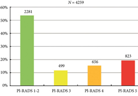
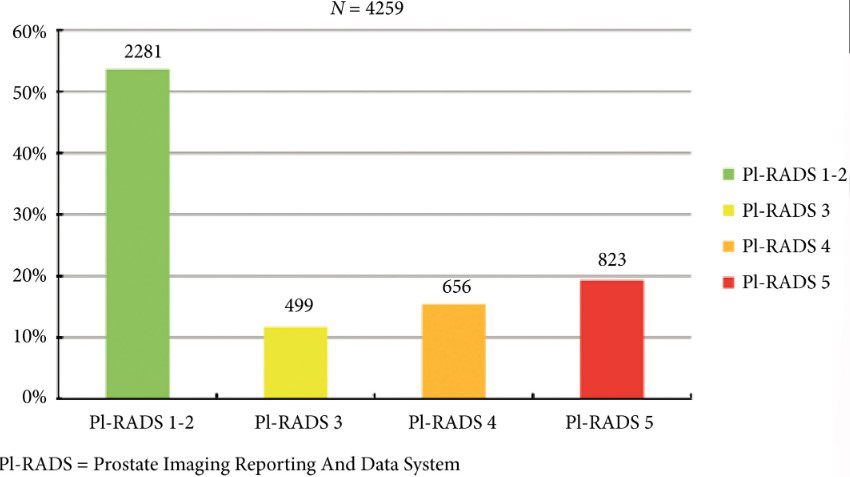
Fig. 2. Distribution of PI‐RADS scored in the entire cohort.

Results

Overall, in 53.6% (2281/4259) of patients had a lesion classified as PI‐RADS ≤2. In 320 patients with PI‐RADS 1 or 2, follow‐up mpMRI was obtained after a median (interquartile range) of 57 (41–63) months. In those patients, csPCa diagnosis‐free survival (DFS) was 99.6% after 3 years. Univariable logistic regression analysis revealed age as a predictor for csPCa during follow‐up (P < 0.05). In biopsied patients, csPCa was detected in 15.8% (19/120), 43.2% (228/528) and 74.5% (483/648) with PI‐RADS 3, 4 and 5, respectively.

Conclusion

More than half of patients having mpMRI of the prostate avoided biopsy. In those patients, csPCa DFS was 99.6% after 3 years.

Read more Articles of the week

Editorial: Avoiding biopsy in men with PI‐RADS scores 1 and 2 on mpMRI of the prostate, ready for prime time?

In 2019 is it safe to avoid prostate biopsy in men with Prostate Imaging Reporting and Data System (PI‐RADS) score 1 and 2 lesions reported on their multiparametric MRI (mpMRI)? In this journal, Venderink et al. [1] suggest that more than half the men being investigated for suspected prostate cancer could indeed safely avoid an initial biopsy. However, like other investigators in this field, the authors make an assumption in their study that there is such a paucity of clinically significant cancer in men with PI‐RADS 1 and 2 lesions, that biopsy is not deemed necessary, as in the PRECISION study [2]. In this study [1] from the Netherlands, of the 2281 men with an initial diagnosis of PI‐RADS 1 or 2 lesions, only 320 men had follow‐up mpMRI, and biopsies were only performed in a small number of men with PI‐RADS scores ≥ 3. Whilst one could conclude that 84% of men did not progress, based on serial imaging, one cannot prove what may have been missed.

Comparing mpMRI of the prostate to the reference standard of radical prostatectomy whole‐mount specimens, a study from the University of California, Los Angeles showed that mpMRI can potentially miss up to 35% of clinically significant cancers, and up to 20% of high grade cancers. It found that 74% of missed solitary tumours were clinically significant, including 23% with Gleason ≥4 + 3 = 7, and that 38.7% were >1 cm in diameter [3]. As such, these missed cancers were not all small, low grade and clinically insignificant. An Italian study confirmed these findings with a detection rate of clinically significant prostate cancer outside the index lesion seen on mpMRI in 30% of patients [4]. All urologists are aware that biopsy by any means can never detect all the cancers seen on formal whole‐mount histopathology, but we do have evidence using transperineal prostate mapping biopsies as the reference standard as to what may be missed. The PROMIS study [5] provides the best evidence using several definitions of clinically significant cancer. Using Gleason ≥4 + 3 or cancer core length >6 mm the negative predictive value (NPV) of a negative mpMRI was 89%. However, if the criteria were altered to any Gleason 7 cancer, the NPV falls to 76%. This is also supported by a multicentre study by Hansen et al. [6], which demonstrated that the NPV of a negative mpMRI for excluding Gleason 7–10 cancer was 80%, but improved to 91% with a PSA density of <0.1 ng/mL/mL, and to 89% with a PSA density of <0.15 ng/mL/mL. It is important to note that these studies used transperineal biopsies rather than 12‐core transrectal biopsies, suggesting the latter to be a more unreliable reference test with a greater probability of missing clinically significant cancer on systematic sampling.

Are all Gleason 3 + 4 = 7 cancers < 6 mm in core length, for example, 5 mm Gleason 3 + 4 (40%) = 7 cancer, truly clinically insignificant? If that were the case, favourable intermediate‐risk prostate cancer would have to be an accepted indication for active surveillance (AS) in men, and in most cases this is not the case. National Comprehensive Cancer Network guidelines recommend that men with favourable intermediate‐risk prostate cancer should only be offered AS if the PSA is <10 ng/mL, the lesion is cT1 and the percentage of positive cores is <50%. Prostate Cancer Research International Active Surveillance (PRIAS) criteria only accept men with favourable intermediate‐risk prostate cancer if there is a maximum of two cores involved, PSA density is <0.2 ng/mL/mL, and if it represents <10% of the core. Both European Association of Urology and AUA guidelines caution that if men are offered AS with favourable intermediate‐risk disease, they should be warned of the greater risk of developing metastatic spread. It is therefore clear that major international guidelines do not fully support AS for intermediate‐risk prostate cancers and therefore it may not be acceptable to be missing Gleason 3 + 4 cancers in up to 10–20% of men with normal prostate mpMRI results.

Multiparametric MRI of the prostate has been a huge advance in prostate cancer diagnostics and is now widely used internationally, but does have limitations. Based on the available data, men who choose not to be biopsied with a normal prostate mpMRI should be warned, as part of informed consent, that a clinically significant cancer could be missed in up to 10–20% of cases (depending on PSA density) and close follow‐up should be recommended. One could easily argue that men with normal prostate mpMRI but with PSA density >0.15 ng/mL/mL should still be offered a systematic biopsy. Perhaps the future lies in the genomics of mpMRI‐visible vs ‐invisible lesions, with a recent study showing that there is a confluence of aggressive molecular and pathological features in lesions visible on MRI. Future research may be able to determine if indeed it is safe to leave some Gleason 3 + 4 = 7 cancers undetected if invisible on mpMRI because of their lack of genomic and metabolic aggression rather than based on their Gleason pattern [7].

by Mark Frydenberg

References

  1. Verderink WVan Luijtelaar AVan der Leest M et al. Multiparametric MRI and follow up to avoid prostate biopsy in 4259 men. BJU Int 2019124775– 84
  2. Kasivisvanathan ASRannikko MBorghi V et al. MRI targeted or standard biopsy for prostate cancer diagnosis. N Engl J Med 20183781767– 77
  3. Johnson DCRaman SSMirak SA et al. Detection of individual prostate cancer foci via multiparametric magnetic resonance imaging. Eur Urol 201975712– 20
  4. Stabile Adell’Oglio Pde Cobelli F et al. Association between prostate Imaging Reporting and data system (PIRADS) score for the index lesion and multifocal clinically significant prostate cancer. Eur Urol Oncol 2018129– 3336
  5. Ahmed HUBasally ABrown LC et al. Diagnostic accuracy of multiparametric MRI and TRUS biopsy in prostate cancer (PROMIS): a paired validating confirmatory study. Lancet 2017389815– 22
  6. Hansen NLBarrett TKesch C et al. Multicentre evaluation of magnetic resonance imaging supported transperineal prostate biopsy in biopsy naïve men with suspicion of prostate cancer. BJU Int 201812240– 9
  7. Houlahan KESalmasi ASadun TY et al. Molecular hallmarks of multiparametric magnetic resonance imaging visibility in prostate cancer. Eur Urol 20197618– 23


Video: mpMRI and follow-up to avoid prostate biopsy in 4259 men

Multiparametric magnetic resonance imaging and follow-up to avoid prostate biopsy in 4259 men

Read the full article

Abstract

Objective

To determine the proportion of men avoiding biopsy because of negative multiparametric magnetic resonance imaging (mpMRI) findings in a prostate MRI expert centre, and to assess the number of clinically significant prostate cancers (csPCa) detected during follow‐up.

Patients and methods

Retrospective study of 4259 consecutive men having mpMRI of the prostate between January 2012 and December 2017, with either a history of previous negative transrectal ultrasonography‐guided biopsy or biopsy naïve. Patients underwent mpMRI in a referral centre. Lesions were classified according to Prostate Imaging Reporting And Data System (PI‐RADS) versions 1 and 2. Negative mpMRI was defined as an index lesion PI‐RADS ≤2. Follow‐up until 13 October 2018 was collected by searching the Dutch Pathology Registry (PALGA). Gleason score ≥3 + 4 was considered csPCa. Kaplan–Meier analysis and univariable logistic regression models were used in the cohort of patients with negative mpMRI and follow‐up.

Results

Overall, in 53.6% (2281/4259) of patients had a lesion classified as PI‐RADS ≤2. In 320 patients with PI‐RADS 1 or 2, follow‐up mpMRI was obtained after a median (interquartile range) of 57 (41–63) months. In those patients, csPCa diagnosis‐free survival (DFS) was 99.6% after 3 years. Univariable logistic regression analysis revealed age as a predictor for csPCa during follow‐up (P < 0.05). In biopsied patients, csPCa was detected in 15.8% (19/120), 43.2% (228/528) and 74.5% (483/648) with PI‐RADS 3, 4 and 5, respectively.

Conclusion

More than half of patients having mpMRI of the prostate avoided biopsy. In those patients, csPCa DFS was 99.6% after 3 years.

Article of the week: Androgen receptor (AR) splice variant 7 and full‐length AR expression is associated with clinical outcome: a translational study in patients with castrate‐resistant prostate cancer

Every week, the Editor-in-Chief selects an Article of the Week from the current issue of BJUI. The abstract is reproduced below and you can click on the button to read the full article, which is freely available to all readers for at least 30 days from the time of this post.

In addition to the article itself, there is an editorial written by a prominent member of the urological community. These are intended to provoke comment and discussion and we invite you to use the comment tools at the bottom of each post to join the conversation. 

If you only have time to read one article this week, it should be this one.

Androgen receptor (AR) splice variant 7 and full‐length AR expression is associated with clinical outcome: a translational study in patients with castrate‐resistant prostate cancer

Marzia Del Re*, Stefania Crucitta*, Andrea Sbrana, Eleonora Rofi*, Federico Paolieri, Giulia Gianfilippo*, Luca Galli, Alfredo Falcone, Riccardo Morganti, Camillo Porta§¶, Eleni Efstathiou**, Ron van Schaik††, Guido Jenster‡‡ and Romano Danesi*

*Unit of Clinical Pharmacology and Pharmacogenetics, Department of Clinical and Experimental Medicine, Medical Oncology Unit, Department of Translational Research and New Technologies in Medicine, Section of Statistics, Department of Clinical and Experimental Medicine, University of Pisa, Pisa, §Department of Internal Medicine, University of Pavia, Division of Translational Oncology, I.R.C.C.S. Istituti Clinici Scientifici Maugeri, Pavia, Italy, **Division of Cancer Medicine, Department of Genitourinary Medical Oncology, University of Texas MD Anderson Cancer Centre, Houston, TX, USA, ††Department of Clinical Chemistry, and ‡‡Department of Urology, Erasmus University Medical Centre, Rotterdam, The Netherlands

Read the full article

Abstract

Objectives

To investigate if full‐length androgen receptor (AR‐FL) is associated with resistance to androgen receptor (AR)‐directed therapy independently and/or combined with AR splice variant 7 (AR‐V7).

Patients and Methods

Plasma samples were prospectively collected from 73 patients with castrate‐resistant prostate cancer before first‐ or second‐line AR‐directed therapy. mRNA was isolated from exosomes and AR‐FL and AR‐V7 were analysed by droplet digital PCR.

Results

AR‐FL was detected in all patients and 22% of them were AR‐V7‐positive at baseline. AR‐FL expression was significantly higher in AR‐V7‐positive vs AR‐V7‐negative patients (P < 0.0001). After stratifying patients by tertile for AR‐FL expression, progression‐free survival (PFS) was 22 vs 18 vs 4 months for lower vs intermediate vs higher tertile, respectively (P = 0.0003). The median PFS and overall survival were significantly longer in AR‐V7‐negative vs AR‐V7‐positive patients (20 vs 4 months, P < 0.0001; not reached vs 9 months, P < 0.0001, respectively).

Conclusions

Resistance to AR‐directed therapy was associated with the presence of AR‐V7; however, AR‐FL expression may help better refine response and survival of patients to AR‐directed therapy. Both biomarkers, if validated in prospective trials, could be used to select the best treatment strategy.

Read more Articles of the week

Editorial: Androgen receptor splice variant 7 (AR‐V7) and AR full‐length (AR‐FL) as predictive biomarkers of therapeutic resistance: partners in crime?

The prostate cancer treatment armamentarium has expanded over the last decade to include taxane‐based chemotherapies (docetaxel, cabazitaxel), sipulecel‐T, radium‐233, and newer androgen receptor (AR) signalling (ARS) inhibitors (abiraterone, enzalutamide, apalutamide). Despite these improvements, persistent ARS remains a key driver of prostate cancer progression after androgen‐deprivation therapy (ADT), transition to castrate‐resistant prostate cancer (CRPC), and even after resistance to ARS inhibitors. Cross‐resistance between ARS inhibitors is common. Predictive biomarkers are therefore needed to optimise treatment selection. Mechanisms of resistance have been attributed to genomic heterogeneity; molecular alterations to the AR and/or upregulation of bypass mechanisms that drive AR activation, including expression of AR splice variants lacking the ligand‐binding domain. AR splice variant 7 (AR‐V7), the most abundant AR splice variant, has been implicated in abiraterone and enzalutamide resistance and poor patient outcomes. Whilst knowledge of AR‐V7 status may guide treatment decisions, AR‐V7 alone cannot sufficiently predict response; detection of other variants (ARv567es) or partners, such as AR full‐length (AR‐FL), might improve prediction.

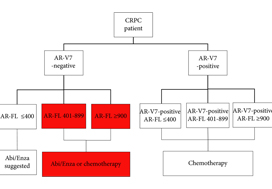
In this issue of BJUI, Del Re et al. [1] evaluated the expression of AR‐V7 and AR‐FL in exosomal RNA as combined predictive biomarkers of resistance to ARS therapy. AR‐FL was detected in all 73 patients (22% were AR‐V7 positive), and AR‐FL expression was significantly higher in AR‐V7‐positive vs AR‐V7‐negative patients (P < 0.001). These findings that AR‐V7 detection has the higher impact on response to therapy confirmed several previous studies; however, the authors took a novel approach to refine the predictive value by stratifying the patient pool into AR‐V7‐positive and ‐negative populations, and then into tertiles based on AR‐FL expression. Analysis of patient outcomes, both in terms of overall (OS) and progression‐free survival (PFS), in these six groups reveals a more nuanced potential treatment strategy. Although AR‐V7 expression better predicts OS and PFS to ARS therapy than does AR‐FL expression, patients with discordant AR‐FL expression relative to their AR‐V7 expression may also benefit from treatment different from that which their AR‐V7 status would suggest. For example, patients positive for AR‐V7 but in the bottom tertile of AR‐FL expression may be effectively treated with anti‐androgen therapies; a breakthrough for patients ineligible for chemotherapy. Additionally, patients negative for AR‐V7 but in the top tertile of AR‐FL expression may respond better to front‐line taxane chemotherapy. Thus, the addition of AR‐FL to AR‐V7 may aid in better treatment selection.

Recently, the Development of Circulating Molecular Predictors of Chemotherapy and Novel Hormonal Therapy Benefit in Men With Metastatic Castration‐Resistant Prostate Cancer (PROPHECY) trial (NCT02269982) prospectively validated the clinical utility of AR‐V7 by demonstrating that detection of AR‐V7 in circulating tumour cells (by two blood‐based assays) is predictive of whether patients with CRPC have become resistant to ARS inhibitors, thereby reducing future benefit from further ARS inhibitor therapy. Although the PROPHECY study found a strong association between positive AR‐V7 and anti‐androgen therapy resistance, some AR‐V7‐negative men did still exhibit resistance to anti‐androgen therapy, showing that a second predictive marker (like AR‐FL) would be helpful to further guide patient selection [2]. It would be interesting to see whether the approach described in Del Re et al. [1] could be replicated using the PROPHECY trial data. Moreover, a recent study established that AR‐V7 is very rarely expressed in primary tissue, with expression emerging in response to primary ADT (and in CRPC progression) and further enhanced in resistance to ARS inhibitors [3]. AR‐V7 was shown to associate with AR‐FL expression and copy number in CRPC, with many cases of high AR‐FL expression having undetectable/low AR‐V7 expression, indicating that mRNA splicing remains crucial for AR‐V7 generation. Although AR‐V7‐negative tumours responded to ARS therapy as expected, some AR‐V7‐positive tumours also responded, suggesting that AR‐V7 detection does not preclude response to ARS therapy and providing further evidence that a second marker could be useful as a predictive tool. Finally, AR‐V7 status in determining taxane response/resistance remains in conflict with studies either showing that taxanes retain activity in patients with positive AR‐V7 or that the absence of AR splice variants (AR‐V7 and ARv567es) may be associated with superior response to taxane treatment, leading to the hypothesis that AR‐FL would be most sensitive to taxane treatment, followed by ARv567es and AR‐V7 [4,5].

While several studies have shown that the AR‐V7/AR‐FL ratio tends to be elevated in CRPC tissues, the role of AR‐FL as a predictive biomarker for AR‐targeted therapy remains controversial. One study found that positive AR‐V7, but not higher AR‐FL, was associated with worse prognosis [6]. As detection methodologies in liquid biopsies and AR data analysis improve over time, the interplay between AR‐FL and AR‐V7, as well other AR variants, warrants further study, which should shed light on whether AR‐V7 and/or AR‐FL (either as homodimers or possibly heterodimers with AR‐V7) are driving resistance to ARS inhibitors. Are they equal partners or is one the dominant driver of the crime?

by Roberto H. Barbier, Cindy H. Chau and William D. Figg

References

  1. Del Re MCrucitta SSbrana A et al. AR‐V7 and AR‐FL expression is associated with clinical outcome: a translational study in patients with castrate-resistant prostate cancer. BJU Int 2019124693– 700
  2. Armstrong AJHalabi SLuo J et al. Prospective multicenter validation of androgen receptor splice variant 7 and hormone therapy resistance in high‐risk castration‐resistant prostate cancer: the PROPHECY study. J Clin Oncol 2019371120– 9
  3. Sharp AColeman IYuan W et al. Androgen receptor splice variant‐7 expression emerges with castration resistance in prostate cancer. J Clin Invest 2019129192– 208
  4. Scher HIGraf RPSchreiber NA et al. Assessment of the validity of nuclear‐localized androgen receptor splice variant 7 in circulating tumor cells as a predictive biomarker for castration‐resistant prostate cancer. JAMA Oncol 201841179– 86
  5. Tagawa STAntonarakis ESGjyrezi A et al. Expression of AR‐V7 and ARv(567es) in circulating tumor cells correlates with outcomes to taxane therapy in men with metastatic prostate cancer treated in TAXYNERGY. Clin Cancer Res 2019251880– 8
  6. Zhu YSharp AAnderson CM et al. Novel junction‐specific and quantifiable in situ detection of AR‐V7 and its clinical correlates in metastatic castration‐resistant prostate cancer. Eur Urol 201873727– 35


Article of the week: The global prevalence of erectile dysfunction: a review

Every week, the Editor-in-Chief selects an Article of the Week from the current issue of BJUI. The abstract is reproduced below and you can click on the button to read the full article, which is freely available to all readers for at least 30 days from the time of this post.

In addition to the article itself, there is a video prepared by the authors. Please use the comment tools at the bottom of each post to join the conversation. 

If you only have time to read one article this week, it should be this one.

The global prevalence of erectile dysfunction: a review

Anna Kessler*, Sam Sollie*, Ben Challacombe, Karen Briggs and Mieke Van Hemelrijck*

*School of Cancer and Pharmaceutical Sciences, King’s College London, Translational Oncology and Urology Research (TOUR) and Urology Centre, Guy’s and St Thomas’ NHS Foundation Trust, London, UK

Read the full article

Abstract

Objective

To evaluate the global prevalence of erectile dysfunction (ED); as well as its association with physiological and pathological ageing by examining the relationship between ED and cardiovascular disease (CVD), benign prostatic hyperplasia (BPH), and dementia. We also aimed to explain the treatment for erectile dysfunction and characterize discrepancies caused by the use of different ED screening tools.

Methods

The Excerpta Medica dataBASE (EMBASE) and Medical Literature Analysis and Retrieval System Online (MEDLINE) were searched to find population‐based studies investigating the prevalence of ED and the association between ED and CVD, BPH and dementia in the general population.

Results

The global prevalence of ED was 3–76.5%. ED was associated with increasing age. Use of the International Index of Erectile Function (IIEF) and Massachusetts Male Aging Study (MMAS)‐derived questionnaire identified a high prevalence of ED in young men. ED was positively associated with CVD. Men with ED have an increased risk of all‐cause mortality odds ratio (OR) 1.26 (95% confidence interval [CI] 1.01–1.57), as well as CVD mortality OR 1.43 (95% CI 1.00–2.05). Men with ED are 1.33–6.24‐times more likely to have BPH then men without ED, and 1.68‐times more likely to develop dementia than men without ED.

Conclusion

ED screening tools in population‐based studies are a major source of discrepancy. Non‐validated questionnaires may be less sensitive than the IIEF and MMAS‐derived questionnaires. ED constitutes a large burden on society given its high prevalence and impact on quality of life, and is also a risk factor for CVD, dementia and all‐cause mortality.

Read more Articles of the week

Video: The global prevalence of erectile dysfunction

The global prevalence of erectile dysfunction: a review

Read the full article

Abstract

Objective

To evaluate the global prevalence of erectile dysfunction (ED); as well as its association with physiological and pathological ageing by examining the relationship between ED and cardiovascular disease (CVD), benign prostatic hyperplasia (BPH), and dementia. We also aimed to characterise discrepancies caused by the use of different ED screening tools.

Methods

The Excerpta Medica dataBASE (EMBASE) and Medical Literature Analysis and Retrieval System Online (MEDLINE) were searched to find population‐based studies investigating the prevalence of ED and the association between ED and CVD, BPH, and dementia in the general population.

Results

The global prevalence of ED was 3–76.5%. ED was associated with increasing age. Use of the International Index of Erectile Function (IIEF) and Massachusetts Male Aging Study (MMAS)‐derived questionnaire identified a high prevalence of ED in young men. ED was positively associated with CVD. Men with ED have an increased risk of all‐cause mortality odds ratio (OR) 1.26 (95% confidence interval [CI] 1.01–1.57), as well as CVD mortality OR 1.43 (95% CI 1.00–2.05). Men with ED are 1.33–6.24‐times more likely to have BPH then men without ED, and 1.68‐times more likely to develop dementia than men without ED.

Conclusion

ED screening tools in population‐based studies are a major source of discrepancy. Non‐validated questionnaires may be less sensitive than the IIEF and MMAS‐derived questionnaire. ED constitutes a large burden on society given its high prevalence and impact on quality of life, and is also a risk factor for CVD, dementia, and all‐cause mortality.


View more videos

Article of the week: A four‐group urine risk classifier for predicting outcomes in patients with prostate cancer

Every week, the Editor-in-Chief selects an Article of the Week from the current issue of BJUI. The abstract is reproduced below and you can click on the button to read the full article, which is freely available to all readers for at least 30 days from the time of this post.

In addition to the article itself, there is an editorial written by a prominent member of the urological community. These are intended to provoke comment and discussion and we invite you to use the comment tools at the bottom of each post to join the conversation. 

If you only have time to read one article this week, it should be this one.

A four‐group urine risk classifier for predicting outcomes in patients with prostate cancer

Shea P. Connell*, Marcelino Yazbek-Hanna*, Frank McCarthy, Rachel Hurst*, MartynWebb*, Helen Curley*, Helen Walker, Rob Mills, Richard Y. Ball, Martin G. Sanda§, Kathryn L. Pellegrini§, Dattatraya Patil§, Antoinette S. Perry, Jack Schalken**, Hardev Pandha††, Hayley Whitaker‡‡, Nening Dennis, Christine Stuttle, Ian G. Mills§§¶¶***, Ingrid Guldvik¶¶, Movember GAP1 Urine Biomarker Consortium1, Chris Parker†††, Daniel S. Brewer*‡‡‡, Colin S. Cooper* and Jeremy Clark*

*Norwich Medical School, University of East Anglia, Norwich, Institute of Cancer Research, Sutton, Norfolk and Norwich University Hospitals NHS Foundation Trust, Norwich, UK, §Department of Urology, Winship Cancer Institute, Emory University School of Medicine, Atlanta, GA, USA , School of Biology and Environmental Science, Science West, University College Dublin, Dublin 4, Ireland, **Nijmegen Medical Centre, Radboud University Medical Centre, Nijmegen, The Netherlands, ††Faculty of Health and Medical Sciences, The University of Surrey, Guildford, ‡‡Molecular Diagnostics and Therapeutics Group, University College London, London, §§School of Medicine, Dentistry and Biomedical Sciences, Institute for Health Sciences, Centre for Cancer Research and Cell Biology, Queen’s University Belfast, Belfast, UK, ¶¶Centre for Molecular Medicine, University of Oslo, Oslo, Norway, ***Nuffield Department of Surgical Sciences, University of Oxford, Oxford, †††Royal Marsden Hospital, Sutton and ‡‡‡Earlham Institute, Norwich, UK

Read the full article

Abstract

Objectives

To develop a risk classifier using urine‐derived extracellular vesicle (EV)‐RNA capable of providing diagnostic information on disease status prior to biopsy, and prognostic information for men on active surveillance (AS).

Patients and Methods

Post‐digital rectal examination urine‐derived EV‐RNA expression profiles (n = 535, multiple centres) were interrogated with a curated NanoString panel. A LASSO‐based continuation ratio model was built to generate four prostate urine risk (PUR) signatures for predicting the probability of normal tissue (PUR‐1), D’Amico low‐risk (PUR‐2), intermediate‐risk (PUR‐3), and high‐risk (PUR‐4) prostate cancer. This model was applied to a test cohort (n = 177) for diagnostic evaluation, and to an AS sub‐cohort (n = 87) for prognostic evaluation.
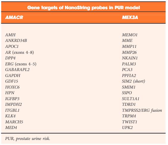
Table 2. NanoString gene probes incorporated by LASSO regularization in the final optimal model used to produce the prostate urine risk signatures

Results

Each PUR signature was significantly associated with its corresponding clinical category (P < 0.001). PUR‐4 status predicted the presence of clinically significant intermediate‐ or high‐risk disease (area under the curve = 0.77, 95% confidence interval [CI] 0.70–0.84). Application of PUR provided a net benefit over current clinical practice. In an AS sub‐cohort (n = 87), groups defined by PUR status and proportion of PUR‐4 had a significant association with time to progression (interquartile range hazard ratio [HR] 2.86, 95% CI 1.83–4.47; P < 0.001). PUR‐4, when used continuously, dichotomized patient groups with differential progression rates of 10% and 60% 5 years after urine collection (HR 8.23, 95% CI 3.26–20.81; P < 0.001).

Conclusion

Urine‐derived EV‐RNA can provide diagnostic information on aggressive prostate cancer prior to biopsy, and prognostic information for men on AS. PUR represents a new and versatile biomarker that could result in substantial alterations to current treatment of patients with prostate cancer.

Read more Articles of the week
Read the blog article


Editorial: Do you need further assistance in diagnosing and risk stratifying prostate cancer?

I would hope the answer to the question posed in the title is a universal ‘yes’; at least that is my experience with this complex and common disease. The concept that in 2019, we have unmet needs in prostate cancer diagnostics is somewhat remarkable, given that we have access to: (i) one of the most widely used biomarkers in oncology (PSA), (ii) a readily accessible organ to examine (DRE), (iii) state of the art imaging (MRI, positron emission tomography), (iv) specialty biopsy systems (fusion/transperineal template), (v) enhanced risk stratification systems (National Comprehensive Cancer Network [NCCN], Cancer of the Prostate Risk Assessment [CAPRA], etc.), (vi) numerous nomograms, (vii) secondary urine/serum biomarkers (Prostate Health Index [PHI], prostate cancer antigen 3 [PCA3], SelectMDx, ExoDx, four‐kallikrein panel [4K]), and (viii) commercially available genomic platforms (Prolaris, OncotypeDx, Decipher).

The paper by Connell et al. [1] in this issue of BJUI asks you to consider adding another diagnostic test to your list. You might correctly assume from the title that the test is in discovery/validation stages, and lacks a fancy commercialised name. Many steps await any promising biomarker to make it to your clinic. So why pay attention to this one? Let me reiterate a few points made by the authors and suggest where new paradigms might emerge if the test delivers on its promises.

First, the test crosses over the current barriers between screening patients and active surveillance (AS). In both populations we care about Gleason Grade Group ≥2. Yet a SelectMDx or similar tests are validated for diagnosis but not for monitoring Grade Group 1 on AS. Genomic profiling tests have strong validation and prognostic value for AS, but require tissue and external laboratory work flows. This marker is being tested for both settings, with potentially meaningful distinctions for both patient groups.

Second, this test is in the urine and does not need imaging or needles to obtain samples. It may have serial use (if cost‐effective) for monitoring AS.

Third, for AS cohorts, the test seems to be able to identify progression well in advance. This would potentially allow for early intervention in the correct patients, and less intense monitoring in the remaining.

Fourth, the test metrics looked favourable in PSA screened and unscreened populations; will we ever see a novel biomarker bold enough to move to primary/independent screening status?

Fifth, some of the secondary biomarkers you may be using now are included in this model: PCA3, transmembrane protease serine 2:v‑ets erythroblastosis virus E26 oncogene homolog (TMPRSS2‐ERG), Homeobox C6 (HOXC6).

To be critical, this biomarker will need significant validation in other cohorts, and we can always hope for head‐to‐head data with existing strategies. I will remain optimistic these authors can move this biomarker strategy along and help bridge some of the gaps that remain in disease detection and risk stratification. I may even attempt to insert some of those lovely new equations in the methods section into future lectures.

Reference

  1. Connell SPYazbek‐Hanna MMcCarthy F et al. A four‐group urine risk classifier for predicting outcomes in patients with prostate cancer. BJU Int 2019124609– 20
© 2024 BJU International. All Rights Reserved.