Tag Archive for: Article of the Week

Posts

Editorial: Prediction and Predicament – Complications after ILND for Penile cancer

In the current issue of BJUI, Gopman et al. [1] report the findings of an international multicentre study examining postoperative complications after inguinal lymph node dissection (ILND) for penile cancer. Their study is the largest to date, and despite its retrospective nature, provides detailed insight into this complex and morbid procedure.

ILND is a critical step in penile cancer treatment, and according to the guidelines of the European Association of Urology, is warranted when the clinical suspicion of lymph node invasion arises [2]. ILND helps to refine pathological staging and has been incorporated into prognostic tools estimating cancer-specific survival after treatment [3]. Despite clinical necessity, ILND is associated with exceptionally high complication rates, as reflected by the current studies’ 55.4% postoperative complication rate. As expected, most of the complications were due to wound complications. Although the authors recognised a decrease in major wound infections after 2008, the overall rate of morbidity after ILND for penile cancer has not changed substantially when compared with historical series [4].

The process of care for these patients can be long and tedious; it affects the personal well-being of the patient and is also responsible for a heavy societal financial burden [5]. The results of the current retrospective analysis are particularly sobering, given that the current data are exclusively from centres specialising in the care of patients with penile cancer. The number of unreported complications at lower volume centres may well be much higher than those evidenced by Gopman et al. [1].

So what can we do to improve our surgical results? The study by Gopman et al. [1] provides us with some tools for advancement. They found that the numbers of removed lymph nodes was a predictor for overall complications in their cohort. Specifically, higher pathological stages were accountable for all wound infections, while age and sartorius flap transposition affected major wound infections significantly. Unfortunately, the study could not provide granular information on preoperative comorbidities, e.g. diabetes mellitus, chronic steroid use and smoking status among others, which could have offered a deeper understanding of the determinants of complication.

Nonetheless, the authors are to be commended for their efforts to provide the urological community with the best available evidence, collected thus far, about complications of ILND for penile cancer. The rarity of penile cancer may limit a clinician’s ability to perceive the early warning signs of a deviation from the routine postoperative course. As such, the current study will not only help us to better counsel our patients but may also help raise our postoperative awareness of complications, thereby achieving improvements in operative outcomes.

Christian P. Meyer*, Julian Hanske*‡ and Jesse D. Sammon*§


*Center for Surgery and Public Health, Brigham and WomenHospital, Harvard Medical School, Boston, MA, USA, Department of Urology, University Hospital Hamburg- Eppendorf, Hamburg, Department of Urology, Marien Hospital, Ruhr-University Bochum, Herne, Germany and §VUI Center for Outcomes Research Ana lytics and Evaluation, Henry Ford Hospital, Detroit, MI, USA


References


2 Hakenberg OW, Comperat EM, Minhas S, Necchi A, Protzel C, Watkin N. EAU guidelines on penile cancer: 2014 update. Eur Urol 2014; 67: 142– 50


4 Ravi R. Morbidity following groin dissection for penile carcinoma. Br Urol 1993; 72: 9415


5 Drew P, Posnett J, Rusling L, Wound Care Audit Team. The cost of wound care for a local population in England. Int Wound J 2007; 4: 14955


Video: Predicting postoperative complications of ILND for penile cancer

Predicting postoperative complications of inguinal lymph node dissection for penile cancer in an international multicentre cohort

Jared M. Gopman, Rosa S. Djajadiningrat*, Adam S. Baumgarten, Patrick N. EspirituSimon Horenblas*, Yao Zhu, Chris Protzel , Julio M. Pow-Sang*, Timothy Kim, Wade J. Sexton, Michael A. Poch and Philippe E. Spiess


Department of Genitourinary Oncology, Moftt Cancer Center, Tampa, FL, USA, *Department of Urological Oncology, The Netherlands Cancer Institute, Amsterdam, The Netherlands, Department of Urological Oncology, Fudan University Shanghai Cancer Center, Shanghai, China, and Department of Urology, University of Rostock, Rostock, Germany


OBJECTIVES

To assess the potential complications associated with inguinal lymph node dissection (ILND) across international tertiary care referral centres, and to determine the prognostic factors that best predict the development of these complications.

MATERIALS AND METHODS

A retrospective chart review was conducted across four international cancer centres. The study population of 327 patients underwent diagnostic/therapeutic ILND. The endpoint was the overall incidence of complications and their respective severity (major/minor). The Clavien–Dindo classification system was used to standardize the reporting of complications.

RESULTS

A total of 181 patients (55.4%) had a postoperative complication, with minor complications in 119 cases (65.7%) and major in 62 (34.3%). The total number of lymph nodes removed was an independent predictor of experiencing any complication, while the median number of lymph nodes removed was an independent predictor of major complications. The American Joint Committee on Cancer stage was an independent predictor of all wound infections, while the patient’s age, ILND with Sartorius flap transposition, and surgery performed before the year 2008 were independent predictors of major wound infections.

CONCLUSIONS

This is the largest report of complication rates after ILND for squamous cell carcinoma of the penis and it shows that the majority of complications associated with ILND are minor and resolve without prolonged morbidity. Variables pertaining to the extent of disease burden have been found to be prognostic of increased postoperative morbidity.

Read more articles of the week

Article of the Month: Perineal repair of PFUI – In pursuit of a successful outcome

Every Month the Editor-in-Chief selects the Article of the Month from the current issue of BJUI. The abstract is reproduced below and you can click on the button to read the full article, which is freely available to all readers for at least 30 days from the time of this post.

In addition to the article itself, there is an accompanying editorial written by a prominent member of the urological community. This blog is intended to provoke comment and discussion and we invite you to use the comment tools at the bottom of each post to join the conversation.

If you only have time to read one article this week, it should be this one.

Perineal repair of pelvic fracture urethral injury – In pursuit of a successful outcome


Mamdouh M. Koraitim and Mohamed I. Kamel*


Department of Urology and *Occupational and Enviromental Medicine, College of Medicine, University of Alexandria, Alexandria, Egypt


Read the full article
OBJECTIVE

To determine perioperative factors that may optimize the outcome after delayed perineal repair of a pelvic fracture urethral injury (PFUI).

PATIENTS AND METHODS

In all, 86 consecutive patients who underwent perineal repair of a PFUI between 2004 and 2011 were prospectively enrolled in this study. The mean (range) patient age was 23 (5–50) years. The mean (range) follow-up was 5.5 (2–8) years. We examined seven perioperative variables that might influence the outcome including: prior failed treatment, condition of the bulbar urethra, displacement of the prostate, excision of scarred tissues, fixation of the mucosae of the two urethral ends, and the number and size of sutures used for urethral anastomosis. Univariate and multivariate analyses were used to identify factors that influence postoperative outcome.

RESULTS

Of the patients, 76 (88%) had successful outcomes and 10 (12%) were considered treatment failures. On univariate analysis, four variables were significant factors influencing the outcome: excision of scarred tissues, prostatic displacement, condition of the bulbar urethra and fixation of the mucosae. On multivariate analysis only two remained strong and independent factors namely complete excision of scarred tissues and prostatic displacement in a lateral direction.

CONCLUSIONS

Meticulous and complete excision of scar tissue is critically important to optimise the outcome after perineal urethroplasty. This is particularly emphasised in cases associated with lateral prostatic displacement. Six sutures of 3/0 or 4/0 polyglactin 910 are usually sufficient to create a sound urethral anastomosis. Prior treatment and scarring of the anterior urethra do not affect the outcome.

Read more articles of the week

Editorial: Specialty within a specialty – posterior urethroplasty

Posterior urethral distractions occur in up to 25% of cases of blunt force pelvic fractures. Proper repair of these pelvic fracture urethral injuries (PFUI) is an art that requires exquisite attention to technique and tissue handling. Koraitim and Kamel [1] recently reported their single-surgeon series of PFUI repairs on 86 patients, with the specific aim of characterizing risk factors for treatment failure. Success was defined subjectively as absence of urinary symptoms and normal postoperative urethrography. Requirement for repeat procedures constituted failure. At a mean 5.5 years of direct follow-up, 88% of patients were considered to have had successful treatment. Multivariate logistic regression showed that incomplete scar excision and lateral prostatic displacement (as opposed to superior or no displacement) were predictive of treatment failure (odds ratios 122 and 34, respectively). All other factors analysed, including previous treatment, relative bulbar urethral scarring, mucosal fixation, suture size and number of sutures, were not significant predictors of urethral outcomes.

Large patient series of posterior urethroplasty report treatment success rates of 86–97%, although follow-up has been short in general [2-4]. The present report by Koraitim and Kamel compares favourably with these series, despite longer patient follow-up. This suggests that late failures after posterior urethral repair are rare. The authors should be commended for their desire to ascertain risk factors for failure after repair of these urethral injuries; however, several factors that probably affect outcomes were not evaluated and may at least partially explain some of their treatment failures.

Erectile dysfunction (ED) is known to occur in ~5% of men after pelvic fracture, and to increase to a mean of 42% in those with a concomitant urethral injury [5]. A portion of these men with ED will have arterial insufficiency and will be at increased risk of bulbar necrosis and ischaemic stenosis. Before urethral reconstruction, men with ED should be evaluated with penile duplex ultrasonography and, if arteriogenic ED is suggested, pelvic angiogram. In those with bilateral complete obstruction of the deep internal pudendal or common penile arteries, revascularization should be offered before urethral reconstruction. In this patient population, penile revascularization has been shown to reverse arterial insufficiency, leading to both improved erections and enhanced tissue perfusion for optimum outcomes after posterior urethral reconstruction [6].

A progressive perineal approach has been popularized by Webster and Ramon [4] and generally accepted by those regularly performing posterior urethral reconstruction. While the present authors report extensively on relative excision of fibrosis and number, type and location of suture utilization, they do not provide insight into the number of ancillary measures necessary for a tension-free repair. While some argue that the importance of crural separation and infrapubectomy are overstated [3], these techniques are essential in some patients in order to achieve a tension-free anastomosis. Given that fibrosis was incompletely excised in 15% of patients in this cohort, some of these same patients may also have had some degree of tension of the urethral anastomosis. Alternatively, these adjunctive procedures may be independent predictors of treatment success or failure and their role in this series would be interesting to note.

It is our experience, and surely that of others, that direct long-term follow-up after urethroplasty at a tertiary referral centre is often difficult or non-existent. These authors should be applauded for their ability to follow their patients for a mean 5.5 years in this series. They have provided much needed extended outcome data after posterior urethral reconstruction. The challenge going forward will be for high-volume centres of reconstruction to design studies prospectively that answer specific questions using standardized instruments and objective results.

Read the full article
Jack M. Zuckerman, Kurt A. McCammon and Gerald H. Jordan


Department of Urology, Eastern Virginia Medical School, Norfolk, VA, USA


References


2 Cooperberg MR, McAninch JW, Alsika NF, Elliott SP. Urethral reconstruction for traumatic posterior urethral disruption: outcomes of 25-year experience. J Urol 2007;178:200610; discussion 10


3 Kizer WS, Armenakas NA, Brandes SB, Cavalcanti AG, Santucci RAMorey AF. Simplied reconstruction of posterior urethral disruption defects: limited role of supracrural rerouting. J Urol 2007;177:137881; discussion 812


Article of the Week: Robotic Surgery – Development Of A Standardised Training Curriculum

Every Week the Editor-in-Chief selects the Article of the Week from the current issue of BJUI. The abstract is reproduced below and you can click on the button to read the full article, which is freely available to all readers for at least 30 days from the time of this post.

In addition to the article itself, there is an accompanying editorial written by a prominent member of the urological community. This blog is intended to provoke comment and discussion and we invite you to use the comment tools at the bottom of each post to join the conversation.

If you only have time to read one article this week, it should be this one.

Development Of A Standardised Training Curriculum For Robotic Surgery: A Consensus Statement From An International Multidisciplinary Group Of Experts

Kamran Ahmed, Reenam Khan, Alexandre Mottrie1, Catherine Lovegrove, Ronny Abaza2, Rajesh Ahlawat3, Thomas Ahlering4, Goran Ahlgren5, Walter Artibani6, Eric Barret7, Xavier Cathelineau7, Ben Challacombe, Patrick Coloby8, Muhammad S. KhanJacques Hubert9, Maurice Stephan Michel10, Francesco Montorsi11, Declan Murphy12Joan Palou13, Vipul Patel14, Pierre-Thierry Piechaud15, Hendrik Van Poppel16, PascalRischmann17, Rafael Sanchez-Salas7, Stefan Siemer18, Michael Stoeckle18, Jens-Uwe Stolzenburg19, Jean-Etienne Terrier20, Joachim W. Thuroff21, Christophe Vaessen22, Henk G. Van Der Poel23, Ben Van Cleynenbreugel16, Alessandro Volpe 1,24, Christian Wagner25Peter Wiklund26, Timothy Wilson27, Manfred Wirth28, Jorn Witt26 and Prokar Dasgupta


Department of Urology, Medical Research Council (MRC) Centre for Transplantation, Kings College London, GuyHospital, London, UK, 1Department of Urology, OLV Vattikuti Robotic Surgery Institute, OLV Hospital, Aalst, Belgium, 2Department of Urology, The Ohio State University Comprehensive Cancer Center, Arthur G James Cancer Hospital Richard J Solove Research Institute, Columbus, OH, USA, 3Medanta The Medicity, Gurgaon, Haryana, India, 4Department of Urology, University of California, Irvine, Orange, CA, USA, 5Department of Urology, Lund University Hospital, Lund, Sweden, 6Urology Clinic, A.O.U.I. Verona, Verona, Italy, 7Department of Urology, Institut Mutualiste Montsouris, Paris, France, 8Service dUrologie, Centre Hospitalier Rene-Dubos, Cergy-Pontoise, France, 9Service dUrologie, CHRU Nancy, Vandoeeuvre-les-Nancy, France, 10University Hospital, Mannheim, Germany, 11Department of Urology, San Raffaele Scientic Institute, Milan, Italy, 12Peter MacCallum Cancer Centre and the Royal Melbourne Hospital, Melbourne, Vic., Australia, 13Department of Urology, Fundacio Puigvert, Universitat Autonoma de Barcelona, Barcelona, Spain, 14Global Robotics Institute, Florida Hospital Celebration Health, Celebration, FL, USA, 15Clinique Saint-Augustin, Bordeaux, France, 16Department of Urology, University Hospital, KU Leuven, Leuven, Belgium, 17Service de Chirurgie Urologique, CHU Purpan, Toulouse, France, 18Klinik fur Urologie und Kinderurologie, Universitatsklinikum des Saarlandes, Homburg/Saar, Germany, 19Department of Urology, University of Leipzig, Leipzig, Germany, 20Department of Urology, Foch Hospital, Suresnes, France, 21Department of Urology, Ulm University Medical Center, Ulm, Germany, 22Service D’Urologie et de Transplantation Réno-Pancréatique, Hôpital Pitié-Salpêtrière, Paris, France, 23Department Urology, Netherlands Cancer Institute, Amsterdam, The Netherlands, 24University of Eastern Piedmont, Novara, Italy, 25St. Antonius-Hospital Gronau, Gronau, Germany, 26Department of Oncology and Pathology, Karolinska Institute, Stockholm, Sweden, 27Division of Urology, City of Hope, Duarte, CA, USA, and 28Department of Urology, University Hospital Carl Gustav Carus, Technical University of Dresden, Dresden, Germany


Read the full article
OBJECTIVES

To explore the views of experts about the development and validation of a robotic surgery training curriculum, and how this should be implemented.

MATERIALS AND METHODS

An international expert panel was invited to a structured session for discussion. The study was of a mixed design, including qualitative and quantitative components based on focus group interviews during the European Association of Urology (EAU) Robotic Urology Section (ERUS) (2012), EAU (2013) and ERUS (2013) meetings. After introduction to the aims, principles and current status of the curriculum development, group responses were elicited. After content analysis of recorded interviews generated themes were discussed at the second meeting, where consensus was achieved on each theme. This discussion also underwent content analysis, and was used to draft a curriculum proposal. At the third meeting, a quantitative questionnaire about this curriculum was disseminated to attendees to assess the level of agreement with the key points.

RESULTS

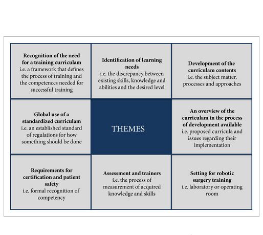
In all, 150 min (19 pages) of the focus group discussion was transcribed (21 316 words). Themes were agreed by two raters (median agreement κ 0.89) and they included: need for a training curriculum (inter-rater agreement κ 0.85); identification of learning needs (κ 0.83); development of the curriculum contents (κ 0.81); an overview of available curricula (κ 0.79); settings for robotic surgery training ((κ 0.89); assessment and training of trainers (κ 0.92); requirements for certification and patient safety (κ 0.83); and need for a universally standardised curriculum (κ 0.78). A training curriculum was proposed based on the above discussions.

CONCLUSION

This group proposes a multi-step curriculum for robotic training. Studies are in process to validate the effectiveness of the curriculum and to assess transfer of skills to the operating room.

Read more articles of the week

Editorial: Towards a Standardized Training Curriculum For Robotic Surgery

The work of the authors [1] towards robotic training and credentialing is much needed and should be applauded as increased scrutiny is being placed on complications associated with robotic surgery [2]. The authors held three separate meetings in 2012 and 2013 in which they identified themes, developed a training curriculum, and assessed expert agreement with their proposed curriculum. The authors’ [1]quantitative survey of 24 experts revealed that all ‘agreed’ or ‘agreed strongly’ with the proposed curriculum. The curriculum includes three areas, cognitive, psychomotor, and teamwork/communication skills, which we feel are vital for good outcomes [3]. As was noted, there are available ‘E-learning’ tools online from organisations such as the AUA and from Intuitive Surgical, and these can be further expanded and validated [4, 5]. The AUA also has recommendations for credentialing requirements that are available online.

We agree with the authors [1] that simulation should include inanimate models, which provide a good cost to benefit ratio. There are increasing numbers of inanimate models for the simulation of procedures, e.g. partial nephrectomy and pyeloplasty. One limitation of inanimate training is that the entire robotic surgical system is used and it may only be free for training on nights and weekends when the robotic systems are not being used clinically. Virtual reality simulators offer a more convenient way to become familiar with the robotic environment, but at a cost of ≈$100 000 (American dollars). Virtual reality simulation is predominantly used to develop skills for a junior trainee or a novice surgeon. However, procedure-specific and augmented-reality simulation is being developed and will greatly enhance robotic training.

The authors [1] should be applauded for offering a specific curriculum consisting of online training, an 8-day ‘discovery’ course for simulation and observation, and a 6-month fellowship for step-wise progression to ‘live’ surgical console time. As the authors note, credentialing should be based on competency and not on the number of cases logged or the duration of training alone. The duration of the fellowship should be based on the learning objectives and research/academic requirements.

In the USA, robotic surgical training is included during residency in urology and a fellowship may not be required if a graduating resident is proficient according to the programme directors’ assessment. For surgeons who have not been trained during residency, proctoring by an experienced surgeon is recommended by the AUA [5], after completing a structured robotic surgical curriculum as described in this article [1]. However, a validated curriculum and benchmarks for competency have not been established. The Fundamentals of Robotic Surgery (FRS) curriculum will be validated during the next year for a multidisciplinary curriculum with skills testing [6].

We also agree with the authors [1] that non-technical skills such as trouble-shooting, teamwork, leadership, and communication are critically important for preventing adverse events. Many if not most complications occur due to failures in patient selection, trocar positioning, and bedside assisting. Also, many complications can be traced to ‘system’ problems rather than console performance. Robotic surgery requires a proficient team to ensure good outcomes.

Currently, there are no uniform credentialing requirements to practice robotic surgery in the USA or many other countries. A validated robotic training curriculum with competency-based assessments is essential and can be integrated into residency programmes where robotic technology is readily available. Where robotic surgical volume is inadequate, fellowship programmes can provide the needed training. A validated competency-based approach offers the hope of better patient outcomes and the continued acceptance of new technologies such as robotic surgery.

Read the full article
Clinton D. Bahler and Chandru P. Sundaram
Department of Urology, Indiana University, Indianapolis, IN, USA


References


2 Alemzadeh H, Iyer RK, Raman J. Safety Implications of Robotic Surgery: Analysis of Recalls and Adverse Event Reports of da Vinci Surgical Systems. The Society of Thoracic Surgeons Annual Meeting2014; Orlando, Florida. Available at: https://www.sts.org/sites/default/les/documents/pdf/annmtg/2014AM/50AM_MonJan27.pdf. Accessed February 2015.


3 Bahler CD, Sundaram CP. Training in Robotic surgery: simulatorssurgery, and credentialing. Urol Clin North Am 2014; 41: 5819.


4 The American Urological Association. E-Learning: Urologic Robotic Surgery Course. The American Urological Association Education and Research, Inc, 2012. Available at: https://www.auanet.org/education/modules/robotic-surgery/. Accessed April 2014.


5 The American Urological Association. Standard Operating Practices (SOPS) for Urologic Robotic Surgery. The American Urological Association, 2013. Available at: https://www.auanet.org/common/pdf/about/SOP-Urologic-Robotic-Surgery.pdf. Accessed April 2014.


Article of the Week: HAL fluorescence cystoscopy in the diagnosis of NMIBC

Every Week the Editor-in-Chief selects the Article of the Week from the current issue of BJUI. The abstract is reproduced below and you can click on the button to read the full article, which is freely available to all readers for at least 30 days from the time of this post.

In addition to the article itself, there is an accompanying editorial written by a prominent member of the urological community. This blog is intended to provoke comment and discussion and we invite you to use the comment tools at the bottom of each post to join the conversation.

If you only have time to read one article this week, it should be this one.

Effectiveness of hexaminolevulinate fluorescence cystoscopy for the diagnosis of non-muscle-invasive bladder cancer in daily clinical practice: a Spanish multicentre observational study

Juan Palou, Carlos Hernandez*, Eduardo Solsona, Ramon Abascal, Juan P. Burgues§, Carlos Rioja, Jose A. Cabrera**, Carlos Gutierrez††, Oscar Rodrıguez, Inmaculada Iborra†, Felipe Herranz**, Jose M. Abascal, Guillermo Conde†† and Jose Oliva


Fundacio Puigvert, Universitat Autonoma de Barcelona, Barcelona,*Hospital Gregorio Maranon, Madrid†Instituto Valenciano de Oncologıa, ValenciaHospital Central de Asturias, Oviedo§Hospital Universitari Son Dureta, Palma de Mallorca Hospital Royo Vilanova, Zaragoza , **Hospital Central de Defensa Gomez Ulla, Madrid, and ††Hospital Son Llatzer, Palma de Mallorca, Spain


Read the full article
OBJECTIVE

To assess the sensitivity and specificity of blue-light cystoscopy (BLC) with hexaminolevulinate as an adjunct to white-light cystoscopy (WLC) vs WLC alone for the detection of non-muscle-invasive bladder cancer (NMIBC), in routine clinical practice in Spain.

PATIENTS AND METHODS

An intra-patient comparative, multicentre, prospective, observational study. Adults with suspected or documented primary or recurrent NMIBC at eight Spanish centres were included in the study. All patients were examined with WLC followed by BLC with hexaminolevulinate. We evaluated the detection rate of bladder cancer lesions by WLC and BLC with hexaminolevulinate, overall and by tumour stage and compared with histological examination of the biopsied lesions. Sensitivity and specificity was calculated.

RESULTS

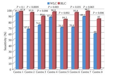
In all, 1 569 lesions were identified from 283 patients: 621 were tumour lesions according to histology and 948 were false-positives. Of the 621 tumour lesions, 475 were detected by WLC (sensitivity 76.5%, 95% confidence interval [CI] 73.2–79.8) and 579 were detected by BLC (sensitivity 93.2%, 95% CI 91.0–95.1; P < 0.001). There was a significant improvement in the sensitivity in the detection of all types of NMIBC lesions with BLC compared with WLC. Of 219 patients with tumours, 188 had NMIBC [highest grade: carcinoma in situ (CIS), n = 36; Ta, n = 87; T1, n = 65). CIS lesions were identified more with BLC (n = 27) than with WLC [n = 19; sensitivity: BLC 75.0% (95% CI 57.8–87.9) vs WLC 52.8% (95% CI 35.5–69.6); P = 0.021]. Results varied across centres.

CONCLUSIONS

This study shows that improvement in diagnosis of NMIBC, mainly CIS and Ta tumours, obtained with BLC with hexaminolevulinate as an adjunct to WLC vs WLC alone can be shown in routine clinical practice.

Read more articles of the week


Editorial: Fluorescence cystoscopy – the end of biopsies for CIS detection?

The present prospective study by Palou et al. [1], conducted in eight Spanish centres, documents the use of hexaminolevulinate fluorescence cystoscopy (FC)-guided bladder tumour resection and biopsies in 283 patients with non-muscle-invasive bladder cancer (NMIBC). It is an inpatient comparison between white-light cystoscopy and FC. The study presents data from routine practice in Spain and the results show an improvement in diagnosis of NMIBC, especially Ta tumours and carcinoma in situ (CIS) with FC-guided resections. These results are confirmation of reports in the literature, including a number of randomized controlled trials and a recent large meta analysis [2]. Although the magnitude of the difference between FC and white-light cystoscopy was somewhat lower in the present study, apparently even in normal daily practice the difference was significant. Moreover, as the rate of CIS in Spain is very high, up to 19% in a large Spanish series [3], I can imagine that the use of FC is of specific interest in this country.

Apart from the confirmation of the better detection rate (75.5 and 93.2% for white-light cystoscopy and FC, respectively, figures similar to those in the recent literature) and confirmation of safety of hexaminolevulinate FC, there are two particular points regarding the present study that I would like to highlight.

The first item that deserves some discussion is mucosal biopsies. In this study the number of false-positive results (948/1569; 60.4%) was very high. This was predominantly explained by the inclusion or mucosal biopsies from ‘normal appearing urothelium’ in these calculations. Only 36 lesions were detected with biopsies, which suggests a very low detection rate. Assuming that >800 random biopsies were taken (apparently six biopsies were taken per patient, and biopsies were taken in 49.1% of the 283 patients), the detection rate was <4%, and one might ask whether it was still worthwhile to take these biopsies. Even though 26.7% of patients with CIS were only diagnosed by biopsies in this study, the number was small. The authors also indicate that it was surprising that CIS was not found more often with FC, but they blame it on the learning curve for FC. The value of mucosal biopsies was also questioned by some reviewers, and in fact by the present authors too. In their introduction they explain the biopsy policy by the high rate of CIS in Spain; however, they also indicate that this incidence seems to be decreasing. Taking together the disappointing detection rate of mucosal biopsies and the high detection rate of CIS with FC, the message should be clear: stop taking mucosal biopsies from normal-looking urothelium. As a matter of fact, this had already been suggested before the era of FC by the authors of other large studies, such as an analysis by the European Organisation for the Research and Treatment of Cancer [4] and a large Dutch study [5]. The false-positive rate for FC was not very high (15.7%, similar to the more recent studies with hexaminolevulinate FC). And I assume that these figures would even be much lower if biopsies of normal urothelium were to be excluded from these calculations.

The second point that deserves attention is the learning curve for FC and its impact on the results. The authors indeed mention as an important limitation of this study ‘the investigators’ lack of experience’ with FC. They point out that this might be the reason that the advantage of FC for the detection of CIS seemed to be less pronounced than in published series, although still significantly better than with white light. In their cohort, 27 of 36 patients with CIS (75%) were detected with FC. The impact of the learning curve and the limited experience of some of the centres is also illustrated by the wide range between centres in, for example, the false-positive rates. Indeed, some training with this FC technique is mandatory. Unfortunately, however, the authors were not able to provide details of the relationship between experience and detection of CIS or false-positive rates.

In conclusion, even in routine practice, FC significantly improves the detection of NMIBC. The advantage is seen especially in Ta tumours and CIS, similarly to recent publications. The use of FC can, in my view, replace the use of random mucosal biopsies of normal-looking urothelium with white light because the detection rate of these biopsies is only a few percent. Finally, the present study also shows that a learning curve significantly improves the detection rate of NMIBC with FC and decreases the rate of false-positives. This should probably be somewhere between 5 (the number used in some of the registration studies for hexaminolevulinate) and 20 as suggested by a recent Canadian study [6].

Read the full article
J. Alfred Witjes
Department of Urology, Radboud University Nijmegen Medicalm Centre, Nijmegen, The Netherlands


References


3 Millan-Rodriguez F, Chechile-Toniolo G, Salvador-Bayarri J, Palou JAlgaba F, Vicente-Rodriguez J. Primary supercial bladder cancer risk groups according to progression, mortality and recurrence. J Urol 2000; 164: 6804


5 Kiemeney LA, Witjes JA, Heijbroek RP, Koper NP, Verbeek ALDebruyne FM. Should random urothelial biopsies be taken from patients with primary supercial bladder cancer? A decision analysis Br J Urol 1994; 73: 16471


6 Gravas S, Efstathiou K, Zachos I, Melekos MD, Tzortzis V.Ithere a learning curve for photodynamic diagnosis of bladder cancer with hexaminolevulinate hydrochloride? Can J Urol 2012; 19: 6269– 73


Video: Full immersion simulation – validation of a distributed simulation environment for technical and non-technical skills training in Urology


Full immersion simulation: validation of a distributed simulation environment for technical and non-technical skills training in Urology

James Brewin, Jessica Tang*, Prokar Dasgup ta, Muhammad S. Khan, Kamran AhmedFernando Bello*, Roger Kneebone* and Peter Jaye


Kings Health Partners, Guys and St Thomas NHS Foundation Trust, and *Imperial College, London,UK


Read the full article
OBJECTIVE

To evaluate the face, content and construct validity of the distributed simulation (DS) environment for technical and non-technical skills training in endourology. To evaluate the educational impact of DS for urology training.

SUBJECTS AND METHODS

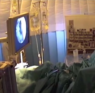
DS offers a portable, low-cost simulated operating room environment that can be set up in any open space. A prospective mixed methods design using established validation methodology was conducted in this simulated environment with 10 experienced and 10 trainee urologists. All participants performed a simulated prostate resection in the DS environment. Outcome measures included surveys to evaluate the DS, as well as comparative analyses of experienced and trainee urologist’s performance using real-time and ‘blinded’ video analysis and validated performance metrics. Non-parametric statistical methods were used to compare differences between groups.

RESULTS

The DS environment demonstrated face, content and construct validity for both non-technical and technical skills. Kirkpatrick level 1 evidence for the educational impact of the DS environment was shown. Further studies are needed to evaluate the effect of simulated operating room training on real operating room performance.

CONCLUSIONS

This study has shown the validity of the DS environment for non-technical, as well as technical skills training. DS-based simulation appears to be a valuable addition to traditional classroom-based simulation training.


Read more articles of the week

Article of the Week: Significance of LVI in organ-confined, node-negative UCB – the p53-MVAC trial

Every Week the Editor-in-Chief selects the Article of the Week from the current issue of BJUI. The abstract is reproduced below and you can click on the button to read the full article, which is freely available to all readers for at least 30 days from the time of this post.

In addition to the article itself, there is an accompanying editorial written by a prominent member of the urological community. This blog is intended to provoke comment and discussion and we invite you to use the comment tools at the bottom of each post to join the conversation.

If you only have time to read one article this week, it should be this one.

Significance of lymphovascular invasion in organ-confined, node-negative urothelial cancer of the bladder: data from the prospective p53-MVAC trial

Friedrich-Carl von Rundstedt, Douglas A. Mata*, Susan Groshen , John P. Stein1Donald G. Skinner, Walter M. Stadler§, Richard J. Cote¶**, Oleksandr N. Kryvenko††Guilherme Godoy and Seth P. Lerner Scott


Department of Urology, Baylor College of Medicine, Houston, TX, *Department of Pathology, Brigham and Womens Hospital, Harvard Medical School, Boston, MA,USC Norris Comprehensive Cancer Center, ‡Aresty Department of Urology, Keck School of Medicine, University of Southern California, Los Angeles, CA, §Section of Hematology/Oncology, Department of Medicine, University of Chicago, Chicago, IL, Departments of Pathology, **Biochemistry and Molecular Biology, and ††Urology, Miller School of Medicine, University of Miami, Miami, FL, USA


Read the full article

OBJECTIVES

To investigate the association between lymphovascular invasion (LVI) and clinical outcome in organ-confined, node-negative urothelial cancer of the bladder (UCB) in a post hoc analysis of a prospective clinical trial. To explore the effect of adjuvant chemotherapy with methotrexate, vinblastine, doxorubicin, and cisplatin (MVAC) on outcome in the subset of patients whose tumours exhibited LVI.

PATIENTS AND METHODS

Surgical and tumour factors were extracted from the operative and pathology reports of 499 patients who had undergone radical cystectomy (RC) for pT1–T2 N0 UCB in the p53-MVAC trial (Southwest Oncology Group 4B951/NCT00005047). The presence or absence of LVI was determined by pathological examination of transurethral resection or RC specimens. Variables were examined in univariate and multivariate Cox proportional hazards models for associations with time to recurrence (TTR) and overall survival (OS).

RESULTS

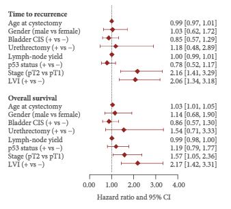
Among 499 patients with a median follow-up of 4.9 years, a subset of 102 (20%) had LVI-positive tumours. Of these, 34 patients had pT1 and 68 had pT2 disease. LVI was significantly associated with TTR with a hazard ratio (HR) of 1.78 [95% confidence interval (CI) 1.15–2.77; number of events (EV) 95; P = 0.01) and with OS with a HR of 2.02 (95% CI 1.31–3.11; EV 98; P = 0.001) after adjustment for pathological stage. Among 27 patients with LVI-positive tumours who were randomised to receive adjuvant chemotherapy, receiving MVAC was not significantly associated with TTR (HR 0.70, 95% CI 0.16–3.17; EV 7; P = 0.65) or with OS (HR 0.45, 95% CI 0.11–1.83; EV 9; P = 0.26).

CONCLUSIONS

Our post hoc analysis of the p53-MVAC trial revealed an association between LVI and shorter TTR and OS in patients with pT1–T2N0 disease. The analysis did not show a statistically significant benefit of adjuvant MVAC chemotherapy in patients with LVI, although a possible benefit was not excluded.

Read more articles of the week


© 2024 BJU International. All Rights Reserved.