Tag Archive for: Article of the Week

Posts

Editorial: Responsiveness to Medical BPH Therapy – Is There a Genetic Factor?

In this issue, Lee et al. [1] from Taiwan demonstrate that the endothelial nitric oxide synthase (eNOS) G894T gene polymorphism predicts responsiveness to α1-blocker therapy in men with BPH/LUTS.

There is a long-standing interest in the establishment of a genetic marker for BPH/LUTS predicting clinical status, the natural history and – ideally – also responsiveness to for example medical therapy. The high prevalence of disease, the socioeconomic impact of diagnosis, medical and surgical treatment, and the availability of drugs (5α-reductase inhibitors) that alter the natural course of the disease justify the intensive search for a genetic marker for BPH/LUTS.

The few familial and twin studies suggest a (moderate) genetic background for this disease to an extent similar to other chronic diseases such as hypertension or diabetes mellitus type II [2, 3]. However, BPH/LUTS is a complex disorder and it is very unlikely that the pathogenesis can be reduced to a single gene or gene defect. Most likely, genetic alterations – besides inflammation, endocrine, myogenic, neurogenic, and morphological factors – act as co-factors.

Within the past decade numerous polymorphisms in the steroid-metabolism pathway, in cytokine genes, in the vitamin D receptor gene, the α-adrenoceptor gene, in homeobox genes, in the angiotensin converting enzyme, the glutathione S-transferase gene, and in the nitric oxide system (just to mention the most frequently studied ones) have been correlated to several clinical parameters of BPH/LUTS, such as symptom status, prostate volume, maximum urinary flow rate and the natural history of the disease [4, 5]. None of these studies provided compelling evidence that one of these polymorphisms (or combinations thereof) could serve as a clinically relevant marker [4, 5]. As indicated by Cartwright et al. [5], many of these genetic studies are hampered by a small sample size, lack of genotyping quality control, inadequate adjustment for populations from heterogeneous descent groups, and poorly defined/inhomogeneous study endpoints.

There is increasing evidence that the nitric oxide (NO)/cGMP pathway plays an important role in controlling the smooth muscle tone of the lower urinary tract. Decreases in the NO/cGMP pathway with age would result in decreased levels of intracellular cGMP and calcium, leading to less smooth muscle relaxation of the bladder and the prostate, thus worsening LUTS. NO is synthesised by at least three isoenzymes of NOS, inducible NOS (iNOS), neuronal NOS (nNOS) and eNOS [1]. The close relationship between NOS/NO pathway and the pathophysiology of BPH/LUTS was the rationale for the study by Lee et al. [1]. Using multiple logistic regression analysis adjusted for age and IPSS, the data showed that the eNOS 894T allele carrier was an independent factor for drug non-responders [1]. However, responsiveness to α1-blocker therapy was also strongly dependent on diabetes mellitus and hypertension, suggesting that the metabolic syndrome plays an important role in the pathogenesis of BPH/LUTS [1]. This is indeed the first study showing that a genetic factor is predictive of the responsiveness to medical BPH/LUTS therapy, therefore this study is significant.

Further studies in populations with other genetic backgrounds (e.g. Caucasian) are required to confirm and to generalise these data. Phosphodiesterase type 5 inhibitors have been proposed to act in BPH/LUTS via the NO systems; therefore, it would be interesting to test this genetic marker also in men treated with tadalafil 5 mg/day. Finally, one has to be aware of the fact that the authors have tested α-blocker monotherapy. All major guidelines recommend a combination of α-blocker and 5α-reductase inhibitor for men with larger prostates (e.g. prostate volume >30–40 mL) [6]. The mean prostate volume in this cohort was 35 mL suggesting that, according to guideline recommendations, these men would have required combined therapy [6].

Stephan Madersbacher, Professor and Chairman
Department of Urology, Kaiser-Franz-Josef Spital, Vienna, Austria


References


2 Partin AW, Page WF, Lee BR, Sanda MG, Miller RN, Walsh PCConcordance rates for benign prostatic disease among twins suggest hereditary inuence. Urology 1994; 44: 64650


3 Rohrmann S, Fallin MD, Page WF et al. Concordance rates and modiable risk factors for lower urinary tract symptoms in twins. Epidemiology 2006; 17: 41927


4 Konwar R, Chattopadhyay N, Bid HK. Genetic polymorphism and pathogenesis of benign prostatic hyperplasia. BJU Int 2008; 102: 53643


6 Gravas S, Bach T, Bachmann A et al. Treatment of Non-Neurogenic Male LUTS. Available at: www.uroweb.org. Accessed March 2016.


Article of the Month: Combined mpMRI Fusion and Systematic Biopsies Predict the Final Tumour Grading after RP

Every Month the Editor-in-Chief selects an Article of the Month from the current issue of BJUI. The abstract is reproduced below and you can click on the button to read the full article, which is freely available to all readers for at least 30 days from the time of this post.

In addition to the article itself, there is an accompanying editorial written by a prominent member of the urological community. This blog is intended to provoke comment and discussion and we invite you to use the comment tools at the bottom of each post to join the conversation.

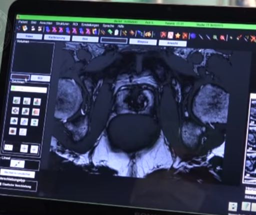
Finally, the third post under the Article of the Week heading on the homepage will consist of additional material or media. This week we feature a video from Angelika Borkowetz, discussing her paper.

If you only have time to read one article this week, it should be this one.

Direct comparison of multiparametric magnetic resonance imaging (MRI) results with final histopathology in patients with proven prostate cancer in MRI/ultrasonography-fusion biopsy

Angelika Borkowetz*, Ivan Platzek, Marieta Toma, Theresa Renner*, Roman Herout*, Martin Baunacke*, Michael Laniado, Gustavo Baretton, Michael Froehner*, Stefan Zastrow* and Manfred Wirth*


*Department of Urology, Department of Radiology and Interventional Radiology, and
Department of Pathology, Technische Universitat Dresden, Dresden, Germany


Read the full article

Objective

To compare multiparametric magnetic resonance imaging (mpMRI) of the prostate and histological findings of both targeted MRI/ultrasonography-fusion prostate biopsy (PBx) and systematic PBx with final histology of the radical prostatectomy (RP) specimen.

Patients and Methods

A total of 105 patients with prostate cancer (PCa) histopathologically proven using a combination of fusion Pbx and systematic PBx, who underwent RP, were investigated. All patients had been examined using mpMRI, applying the European Society of Urogenital Radiology criteria. Histological findings from the RP specimen were compared with those from the PBx. Whole-mount RP specimen and mpMRI results were directly compared by a uro-pathologist and a uro-radiologist in step-section analysis.

AugAOTM1

Results

In the 105 patients with histopathologically proven PCa by combination of fusion PBx and systematic PBx, the detection rate of PCa was 90% (94/105) in fusion PBx alone and 68% (72/105) in systematic PBx alone (P = 0.001). The combination PBx detected 23 (22%) Gleason score (GS) 6, 69 (66%) GS 7 and 13 (12%) GS ≥8 tumours. Fusion PBx alone detected 25 (26%) GS 6, 57 (61%) GS 7 and 12 (13%) GS ≥8 tumours. Systematic PBx alone detected 17 (24%) GS 6, 49 (68%) GS 7 and 6 (8%) GS ≥8 tumours. Fusion PBx alone would have missed 11 tumours (4% [4/105] of GS 6, 6% [6/105] of GS 7 and 1% [1/105] of GS ≥8 tumours). Systematic PBx alone would have missed 33 tumours (10% [10/105] of GS 6, 20% [21/105] of GS 7 and 2% [2/105] of GS ≥8 tumours). The rates of concordance with regard to GS between the PBx and RP specimen were 63% (n = 65), 54% (n = 56) and 75% (n = 78) in fusion, systematic and combination PBx (fusion and systematic PBx combined), respectively. Upgrading of the GS between PBx and RP specimen occurred in 33% (n = 34), 44% (n = 46) and 18% (n = 19) in fusion, systematic and combination PBx, respectively. γ-correlation for detection of any cancer was 0.76 for combination PBx, 0.68 for fusion PBx alone and 0.23 for systematic PBx alone. In all, 84% (n = 88) of index tumours were identified by mpMRI; 86% (n = 91) of index lesions on the mpMRI were proven in the RP specimen.

Conclusions

Fusion PBx of tumour-suspicious lesions on mpMRI was associated with a higher detection rate of more aggressive PCa and a better tumour prediction in final histopathology than systematic PBx alone; however, combination PBx had the best concordance for the prediction of GS. Furthermore, the additional findings of systematic PBx reflect the multifocality of PCa, therefore, the combination of both biopsy methods would still represent the best approach for the prediction of the final tumour grading in PCa.

Read more articles of the week

Editorial: Role of systematic biopsy in the era of mpMRI and US fusion guidance

The success of multiparametric MRI (mpMRI) and MRI/ultrasound (US) fusion-guided biopsies in improving the detection of prostate cancer in patients with occult disease (elevated PSA level with prior negative biopsies) and optimising the detection of clinically significant cancer has been reported by centres that have served as early adopters of these techniques [1, 2]. Technological advances in MRI and associated imaging protocols, as well as increased clinical experience with MRI interpretation have led to increased prospective detection and characterisation of clinically significant prostate cancer. This, in conjunction with increasing experience with MRI/US fusion-guided prostate biopsy techniques, has led to the re-evaluation of the contributory role and utility of systematic template US-guided prostate biopsies in the diagnosis of prostate cancer. It is an attractive proposition to forego the systematic biopsy when performing MRI-directed fusion biopsy, as this would minimise the duration, morbidity, and overall cost of the biopsy procedure and post-biopsy pathology processing. However, before adopting this approach, it is important to first consider the potential possibility of missing clinically significant cancer diagnoses when relying on the targeted biopsy cores in isolation.

In this issue of BJUI, Borkowetz et al. [3] report their results of biopsy histological yields on systematic biopsies compared with MRI/US fusion biopsies in their series of patients who underwent radical prostatectomy (RP). These results corroborate previously reported comparisons of fusion biopsy of suspicious lesions on MRI performed concurrently with systematic biopsy, consistently showing an improved detection of both overall prostate cancer foci and, more importantly, an improved detection of clinically significant higher grade cancer foci [1, 2]. It is important to note that the overall detection rate and detection rate for clinically significant prostate cancer was highest when fusion and systematic biopsies were evaluated in conjunction with each other. Another important factor to consider when evaluating the utility and value of these biopsy techniques is the concordance of the pathology of the biopsy specimen with the final pathology of the RP specimen, the ‘gold standard’. The concordance of Gleason grade assigned on targeted fusion-biopsy cores and RP outperformed that of systematic biopsy cores and RP. This, in essence, suggests that targeted biopsy can perform as well, and likely better, than the systematic biopsy approach of sampling the prostate with a systematic-sextant approach, which has been the long standing standard of care for the diagnosis of prostate cancer. Again, it is important to note that the greatest concordance in this study was achieved when the results of the fusion and systematic biopsy cores were combined.

The question now arises regarding the ‘cost’ for the incremental improvement in cancer detection provided by the combination of both MRI-directed fusion biopsy and the systematic biopsy approach. The improved negative predictive value parallels the increased sensitivity for cancer detection by having a larger sampling of the prostate by augmenting the number of biopsy cores sampled and submitted for histopathological evaluation. The area under the curve for detection of clinically significant cancer reported by Borkowetz et al. [3] was not improved by adding systematic biopsies to the targeted biopsies. However, this experience described a mixed population of patients, most of whom had undergone prior prostate biopsy with benign pathology. This creates an enriched population who likely harbours prostate cancers that are more occult to the systematic biopsy approach, thus improving the diagnostic yield of MRI-directed biopsies even further. This is concordant with the work presented by Mendhiratta et al. [4], where systematic biopsies added little to the diagnosis of clinically significant prostate cancer in a population of men undergoing MRI/US fusion-guided biopsy after prior cancer-negative biopsy sessions.

Alternatively, current datasets for biopsy naïve patients have not shown the same degree of convincingly improved detection with targeted biopsies over systematic biopsies. In fact, Delongchamps et al. [5] recently reported a slightly lower rate of overall cancer detection with fusion-guided targeted biopsies vs systematic biopsy cores; however, the difference in detection of clinically significant prostate cancer was not statistically significant. Further study of the role of targeted biopsy in the biopsy naïve patient population is warranted, as there is suggestion that cancer detection efficiency per needle core is significantly improved with MRI-directed biopsies over systematic biopsies [6]. Alternatively, in patients with prior negative systematic biopsies and continued clinical suspicion for prostate cancer, a repeat biopsy session with targeted cores alone may be appropriate, particularly as these patients have previously undergone standard-of-care, extended sextant biopsy.

Read the full article
Jason A. Pietryga* and Soroush Rais-Bahrami*,
*Department of Radiology, and Department of Urology, University of Alabama at Birmingham, Birmingham, AL, USA


References


Video: Combined mpMRI Fusion and Systematic Biopsies Predict the Final Tumour Grading after RP

Direct comparison of multiparametric magnetic resonance imaging (MRI) results with final histopathology in patients with proven prostate cancer in MRI/ultrasonography-fusion biopsy

Angelika Borkowetz*, Ivan Platzek, Marieta Toma, Theresa Renner*, Roman Herout*, Martin Baunacke*, Michael Laniado, Gustavo Baretton, Michael Froehner*, Stefan Zastrow* and Manfred Wirth*


*Department of Urology, Department of Radiology and Interventional Radiology, and
Department of Pathology, Technische Universitat Dresden, Dresden, Germany


Read the full article

Objective

To compare multiparametric magnetic resonance imaging (mpMRI) of the prostate and histological findings of both targeted MRI/ultrasonography-fusion prostate biopsy (PBx) and systematic PBx with final histology of the radical prostatectomy (RP) specimen.

Patients and Methods

A total of 105 patients with prostate cancer (PCa) histopathologically proven using a combination of fusion Pbx and systematic PBx, who underwent RP, were investigated. All patients had been examined using mpMRI, applying the European Society of Urogenital Radiology criteria. Histological findings from the RP specimen were compared with those from the PBx. Whole-mount RP specimen and mpMRI results were directly compared by a uro-pathologist and a uro-radiologist in step-section analysis.

AugAOTM1

Results

In the 105 patients with histopathologically proven PCa by combination of fusion PBx and systematic PBx, the detection rate of PCa was 90% (94/105) in fusion PBx alone and 68% (72/105) in systematic PBx alone (P = 0.001). The combination PBx detected 23 (22%) Gleason score (GS) 6, 69 (66%) GS 7 and 13 (12%) GS ≥8 tumours. Fusion PBx alone detected 25 (26%) GS 6, 57 (61%) GS 7 and 12 (13%) GS ≥8 tumours. Systematic PBx alone detected 17 (24%) GS 6, 49 (68%) GS 7 and 6 (8%) GS ≥8 tumours. Fusion PBx alone would have missed 11 tumours (4% [4/105] of GS 6, 6% [6/105] of GS 7 and 1% [1/105] of GS ≥8 tumours). Systematic PBx alone would have missed 33 tumours (10% [10/105] of GS 6, 20% [21/105] of GS 7 and 2% [2/105] of GS ≥8 tumours). The rates of concordance with regard to GS between the PBx and RP specimen were 63% (n = 65), 54% (n = 56) and 75% (n = 78) in fusion, systematic and combination PBx (fusion and systematic PBx combined), respectively. Upgrading of the GS between PBx and RP specimen occurred in 33% (n = 34), 44% (n = 46) and 18% (n = 19) in fusion, systematic and combination PBx, respectively. γ-correlation for detection of any cancer was 0.76 for combination PBx, 0.68 for fusion PBx alone and 0.23 for systematic PBx alone. In all, 84% (n = 88) of index tumours were identified by mpMRI; 86% (n = 91) of index lesions on the mpMRI were proven in the RP specimen.

Conclusions

Fusion PBx of tumour-suspicious lesions on mpMRI was associated with a higher detection rate of more aggressive PCa and a better tumour prediction in final histopathology than systematic PBx alone; however, combination PBx had the best concordance for the prediction of GS. Furthermore, the additional findings of systematic PBx reflect the multifocality of PCa, therefore, the combination of both biopsy methods would still represent the best approach for the prediction of the final tumour grading in PCa.

Read more articles of the week

Article of the Week: Impact of Re-TUR on BCG-Treated T1 HG/G3 Bladder Cancer

Every Week the Editor-in-Chief selects an Article of the Week from the current issue of BJUI. The abstract is reproduced below and you can click on the button to read the full article, which is freely available to all readers for at least 30 days from the time of this post.

In addition to the article itself, there is an accompanying editorial written by a prominent member of the urological community. This blog is intended to provoke comment and discussion and we invite you to use the comment tools at the bottom of each post to join the conversation.

Finally, the third post under the Article of the Week heading on the homepage will consist of additional material or media. This week we feature a video from Francesca Pisano and Paolo Gontero, discussing their paper.

If you only have time to read one article this week, it should be this one.

The impact of re-transurethral resection on clinical outcomes in a large multicentre cohort of patients with T1 high-grade/Grade 3 bladder cancer treated with bacille Calmette–Guerin

Paolo Gontero1, Richard Sylvester2, Francesca Pisano1, Steven Joniau3, Marco Oderda1, Vincenzo Serretta4,Stephane Larre5, Savino Di Stasi6, Bas Van Rhijn7, Alfred J.Witjes8, Anne J. Grotenhuis8, Renzo Colombo9, Alberto Briganti9, Marek Babjuk10, Viktor Soukup10, Per-Uno Malmstrom11, Jacques Irani12, Nuria Malats13, Jack Baniel14, RoyMano14, Tommaso Cai15, Eugene K. Cha16, Peter Ardelt17, John Vakarakis18, Riccardo Bartoletti19, Guido Dalbagni20, Shahrokh F. Shariat16, Evanguelos Xylinas16, Robert J.Karnes21 and Joan Palou22


1Urology Clinic, Citta della Salute e della Scienza di Torino, University of Studies of Turin, Turin ,4Department of Surgical, Oncological and Stomatological Sciences, University of Palermo, Palermo, 6Policlinico Tor Vergata-University of Rome, Rome, 9Dipartimento di Urologia, Universita Vita-Salute. Ospedale S. Raffaele, Milan, 15Department of Urology, SantaChiara Hospital, Trento, 19Department of Experimental and Clinical Medicine, University of Florence, Florence, Italy, 2Formerly Department of Biostatistics, EORTC Headquarters, Brussels, 3Oncologic and Reconstructive Urology, Department of Urology, University Hospitals Leuven, Leuven, Belgium, 5Department of Surgical Science, John Radcliffe Hospital, University of Oxford, Oxford, UK, 7Department of Urology, Netherlands Cancer Institute – Antoni van Leeuwenhoek Hospital, Amsterdam, 8Department of Urology, Radboud University Nijmegen Medical Centre, Nijmegen, The Netherlands, 10Department of Urology, Motol Hospital, University of Praha, Praha, Czech Republic, 11Department of Urology, Academic Hospital, Uppsala University, Uppsala, Sweden, 12Department of Urology, Centre Hospitalier Universitaire La Miletrie, University of Poitiers, Poitiers, France, 13Genetic and Molecular Epidemiology Group, Spanish National Cancer Research Centre (CNIO), Madrid, 22Department of Urology, Fundacio Puigvert, University of Barcelona, Barcelona, Spain, 14Department of Urology, Rabin Medical Centre, Tel Aviv, Israel, 16Department of Urology, Weill Medical College of Cornell University in New York City, 20Department of Urology, Memorial Sloan Kettering Cancer Center, New York, NY, 21Department of Urology, Mayo Clinic, Rochester, MN, USA, 17Facharzt fur Urologie, Abteilung fur Urologie. Chirurgische Universitats klinik, Freiburg, Germany, and 18Department of Urology, Sismanoglio Hospital, University of Athens, Athens, Greece


Read the full article

Objectives

To determine if a re-transurethral resection (TUR), in the presence or absence of muscle at the first TUR in patients with T1-high grade (HG)/Grade 3 (G3) bladder cancer, makes a difference in recurrence, progression, cancer specific (CSS) and overall survival (OS).

Patients and methods

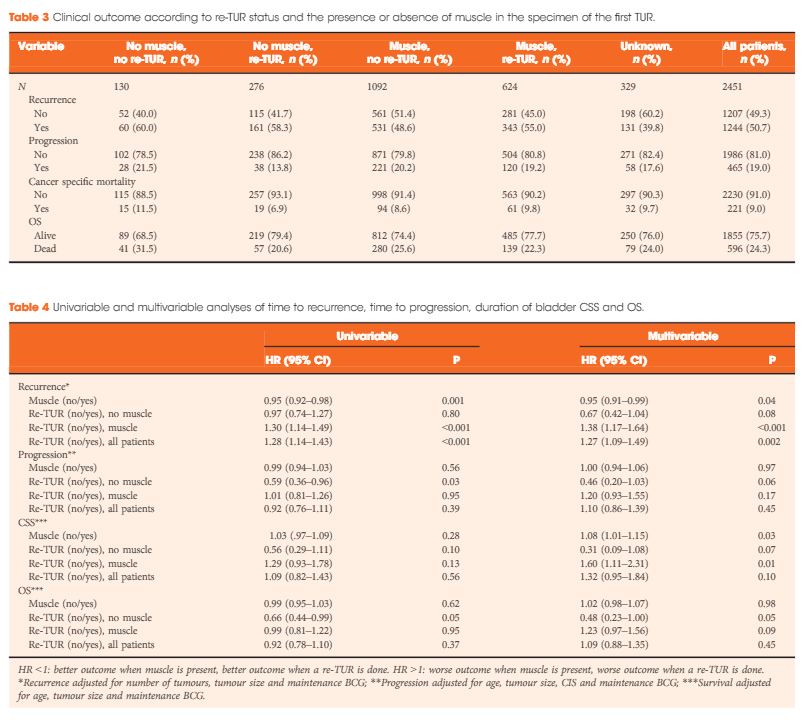
In a large retrospective multicentre cohort of 2451 patients with T1-HG/G3 initially treated with bacille Calmette–Guérin, 935 (38%) had a re-TUR. According to the presence or absence of muscle in the specimen of the primary TUR, patients were divided in four groups: group 1 (no muscle, no re-TUR), group 2 (no muscle, re-TUR), group 3 (muscle, no re-TUR) and group 4 (muscle, re-TUR). Clinical outcomes were compared across the four groups.

JUlAOTW4Results

Results

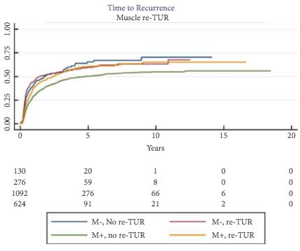
Re-TUR had a positive impact on recurrence, progression, CSS and OS only if muscle was not present in the primary TUR specimen. Adjusting for the most important prognostic factors, re-TUR in the absence of muscle had a borderline significant effect on time to recurrence [hazard ratio (HR) 0.67, P = 0.08], progression (HR 0.46, P = 0.06), CSS (HR 0.31, P = 0.07) and OS (HR 0.48, P = 0.05). Re-TUR in the presence of muscle in the primary TUR specimen did not improve the outcome for any of the endpoints.

Conclusions

Our retrospective analysis suggests that re-TUR may not be necessary in patients with T1-HG/G3, if muscle is present in the specimen of the primary TUR.

Read more articles of the week

Editorial: Time to re-evaluate and refine re-TUR in bladder cancer?

In this issue of BJUI, Gontero et al. [1] present data from a large multi-centre study that should allow us to re-evaluate and refine the indications for re-transurethral resection (TUR) in bladder cancer.

Herr [2] first described this procedure in 1999 and for the past 16 years the indications have remained largely unchanged and are summarised in the latest European Association of Urology guidelines on non-muscle-invasive bladder cancer (NMIBC) [3]:

  • After incomplete initial TUR of bladder tumour (TURBT).
  • If there is no muscle in the specimen after initial resection.
  • In all T1 tumours.
  • In all Grade 3 tumours except primary carcinoma in situ.

In a multi-centre retrospective study of 2 451 patients with high-grade (HG)/Grade 3 (G3) T1 NMIBC treated with BCG, Gontero et al. [1]examined 935 patients who had re-TUR (38% of the total, itself a low figure). Patients were divided into four groups according to the presence or absence of detrusor muscle in the first TURBT specimen:

  • No muscle, no re-TUR
  • No muscle, re-TUR
  • Muscle, no re-TUR
  • Muscle, re-TUR

The authors found that re-TUR only had a positive impact on recurrence, progression, cancer-specific and overall survival, if detrusor muscle was not present in the original specimen. Importantly, in the presence of detrusor muscle in the original specimen, re-TUR did not improve outcomes. The authors conclude that re-TUR may be unnecessary in HG/G3 T1 patients if detrusor muscle is present at the first TURBT.

These findings are important for two reasons: firstly, Herr’s [2] paper was the first to draw attention to the finding that TURBT, a routine urological procedure, was often carried out inadequately. In recent years, the importance of carrying out a high-quality TURBT has been increasingly recognised [4], whilst the presence of detrusor muscle in the TURBT specimen has been shown to be a good measure of the technical quality of a TURBT [5]. This paper [1] further reinforces the importance of obtaining detrusor muscle in the first TURBT. Indeed, as failure to do so results in the patient having to have a second operation and delays their treatment, perhaps we should start to think of a failure to obtain detrusor muscle at the first TURBT in much the same way as positive margin rates are used as a measure of the quality of radical prostatectomy and by inference, the skill of the surgeon.

Secondly, re-TUR arguably serves one overarching purpose: to identify patients with muscle-invasive bladder cancer (MIBC) who have been under-staged by an inadequate first TURBT and who without a re-TUR would be inadequately treated.

Although a secondary role of re-TUR is to identify patients with residual NMIBC, which has some prognostic value, in practice it rarely changes the patient’s management in this setting, which is intravesical therapy usually with BCG. However, in many healthcare systems the timely organisation of a re-TUR within the recommended 6 weeks is challenging and there is usually a further delay of at least 2 weeks until the pathology is reviewed and a patient with NMIBC can finally commence treatment. In this context, it is not surprising that a recent paper in BJUI showed that the interval to re-TUR was a predictor of recurrence and progression and that a re-TUR after 7 weeks was associated with a much worse outcome [6]. It therefore seems logical to reserve re-TUR only for those patients who truly need it, so that limited resources are focused on ensuring that they receive their operation in a timely manner, ideally within 2–4 weeks. If adopted into day-to-day urological practice, the findings by Gontero et al. [1] will allow many patients with HG/G3 T1 and detrusor muscle in the first TURBT specimen to avoid a re-TUR and start intravesical therapy without further delay. Pragmatically, the same should apply to patients with HG/G3 Ta with detrusor muscle in the specimen. On the other hand, HG/G3 T1 patients without detrusor muscle should be fast-tracked for re-TUR as soon as is practicable and certainly no later than 6 weeks.

The article [1] does have some shortcomings. The study design excludes patients with MIBC, so we do not know by comparison how many patients with MIBC were under-staged at the initial TUR based on subsequent re-TUR but as the authors point out, their conclusions would hold true even in this group, as it is very unlikely that one would miss MIBC if there was adequate detrusor muscle in the pathology specimen.

In conclusion, we should consider refining the indications for re-TUR to improve the utilisation of healthcare resources and ensure that for those that need it, a re-TUR is carried promptly whilst for those that do not, essential intravesical treatment is not delayed.

Read the full article
A. Hugh Mostad, Consultant Urologist and Honorary
Senior Lecturer The Royal Surrey County Hospital, Guildford, Surrey, UK


References


Video: Impact of Re-TUR on BCG-Treated T1 HG/G3 Bladder Cancer

The impact of re-transurethral resection on clinical outcomes in a large multicentre cohort of patients with T1 high-grade/Grade 3 bladder cancer treated with bacille Calmette–Guerin

Paolo Gontero1, Richard Sylvester2, Francesca Pisano1, Steven Joniau3, Marco Oderda1, Vincenzo Serretta4,Stephane Larre5, Savino Di Stasi6, Bas Van Rhijn7, Alfred J.Witjes8, Anne J. Grotenhuis8, Renzo Colombo9, Alberto Briganti9, Marek Babjuk10, Viktor Soukup10, Per-Uno Malmstrom11, Jacques Irani12, Nuria Malats13, Jack Baniel14, RoyMano14, Tommaso Cai15, Eugene K. Cha16, Peter Ardelt17, John Vakarakis18, Riccardo Bartoletti19, Guido Dalbagni20, Shahrokh F. Shariat16, Evanguelos Xylinas16, Robert J.Karnes21 and Joan Palou22


1Urology Clinic, Citta della Salute e della Scienza di Torino, University of Studies of Turin, Turin ,4Department of Surgical, Oncological and Stomatological Sciences, University of Palermo, Palermo, 6Policlinico Tor Vergata-University of Rome, Rome, 9Dipartimento di Urologia, Universita Vita-Salute. Ospedale S. Raffaele, Milan, 15Department of Urology, SantaChiara Hospital, Trento, 19Department of Experimental and Clinical Medicine, University of Florence, Florence, Italy, 2Formerly Department of Biostatistics, EORTC Headquarters, Brussels, 3Oncologic and Reconstructive Urology, Department of Urology, University Hospitals Leuven, Leuven, Belgium, 5Department of Surgical Science, John Radcliffe Hospital, University of Oxford, Oxford, UK, 7Department of Urology, Netherlands Cancer Institute – Antoni van Leeuwenhoek Hospital, Amsterdam, 8Department of Urology, Radboud University Nijmegen Medical Centre, Nijmegen, The Netherlands, 10Department of Urology, Motol Hospital, University of Praha, Praha, Czech Republic, 11Department of Urology, Academic Hospital, Uppsala University, Uppsala, Sweden, 12Department of Urology, Centre Hospitalier Universitaire La Miletrie, University of Poitiers, Poitiers, France, 13Genetic and Molecular Epidemiology Group, Spanish National Cancer Research Centre (CNIO), Madrid, 22Department of Urology, Fundacio Puigvert, University of Barcelona, Barcelona, Spain, 14Department of Urology, Rabin Medical Centre, Tel Aviv, Israel, 16Department of Urology, Weill Medical College of Cornell University in New York City, 20Department of Urology, Memorial Sloan Kettering Cancer Center, New York, NY, 21Department of Urology, Mayo Clinic, Rochester, MN, USA, 17Facharzt fur Urologie, Abteilung fur Urologie. Chirurgische Universitats klinik, Freiburg, Germany, and 18Department of Urology, Sismanoglio Hospital, University of Athens, Athens, Greece


Read the full article

Objectives

To determine if a re-transurethral resection (TUR), in the presence or absence of muscle at the first TUR in patients with T1-high grade (HG)/Grade 3 (G3) bladder cancer, makes a difference in recurrence, progression, cancer specific (CSS) and overall survival (OS).

Patients and methods

In a large retrospective multicentre cohort of 2451 patients with T1-HG/G3 initially treated with bacille Calmette–Guérin, 935 (38%) had a re-TUR. According to the presence or absence of muscle in the specimen of the primary TUR, patients were divided in four groups: group 1 (no muscle, no re-TUR), group 2 (no muscle, re-TUR), group 3 (muscle, no re-TUR) and group 4 (muscle, re-TUR). Clinical outcomes were compared across the four groups.

JUlAOTW4Results

Results

Re-TUR had a positive impact on recurrence, progression, CSS and OS only if muscle was not present in the primary TUR specimen. Adjusting for the most important prognostic factors, re-TUR in the absence of muscle had a borderline significant effect on time to recurrence [hazard ratio (HR) 0.67, P = 0.08], progression (HR 0.46, P = 0.06), CSS (HR 0.31, P = 0.07) and OS (HR 0.48, P = 0.05). Re-TUR in the presence of muscle in the primary TUR specimen did not improve the outcome for any of the endpoints.

Conclusions

Our retrospective analysis suggests that re-TUR may not be necessary in patients with T1-HG/G3, if muscle is present in the specimen of the primary TUR.

Read more articles of the week

Article of the Week: TRUS-Guided RB PCa Detection – Reasons for Targeted Biopsy Failure

Every Week the Editor-in-Chief selects an Article of the Week from the current issue of BJUI. The abstract is reproduced below and you can click on the button to read the full article, which is freely available to all readers for at least 30 days from the time of this post.

In addition to the article itself, there is an accompanying editorial written by a prominent member of the urological community. This blog is intended to provoke comment and discussion and we invite you to use the comment tools at the bottom of each post to join the conversation.

Finally, the third post under the Article of the Week heading on the homepage will consist of additional material or media. This week we feature a video from Hannes Cash and Patrick Asbach, discussing their paper.

If you only have time to read one article this week, it should be this one.

Prostate cancer detection on transrectal ultrasonography-guided random biopsy despite negative real-time magnetic resonance imaging/ultrasonography fusion-guided targeted biopsy: reasons for targeted biopsy failure

Hannes Cash*, Karsten Gunzel*, Andreas Maxeiner*, Carsten Stephan*, Thomas Fischer, Tahir Durmus, Kurt Miller*, Patrick Asbach, Matthias Haas† and Carsten Kempkensteffen*


*Department of Urology, and Department of Radiology, ChariteUniversity of Medicine Berlin, Berlin, Germany M. H. and C.K. contributed equally to the study.


Read the full article

Objective

To examine the value of additional transrectal ultrasonography (TRUS)-guided random biopsy (RB) in patients with negative magnetic resonance imaging (MRI)/ultrasonography (US) fusion-guided targeted biopsy (TB) and to identify possible reasons for TB failure.

Patients and Methods

We conducted a subgroup analysis of 61 men with prostate cancer (PCa) detected by 10-core RB but with a negative TB, from a cohort of 408 men with suspicious multiparametric magnetic resonance imaging (mpMRI) between January 2012 and January 2015. A consensus re-reading of mpMRI results (using Prostate Imaging Reporting and Data System [PI-RADS] versions 1 and 2) for each suspicious lesion was performed, with the image reader blinded to the biopsy results, followed by an unblinded anatomical correlation of the lesion on mpMRI to the biopsy result. The potential reasons for TB failure were estimated for each lesion. We defined clinically significant PCa according to the Epstein criteria and stratified patients into risk groups according to the European Association of Urology guidelines.

JulAOTW3Results

Results

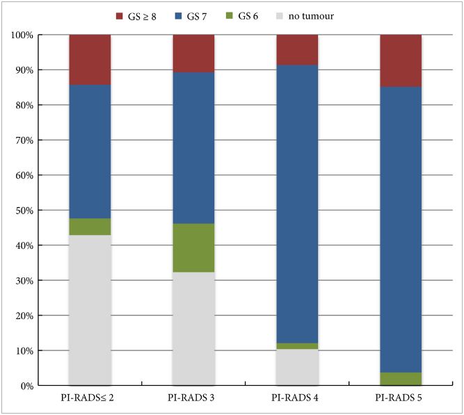
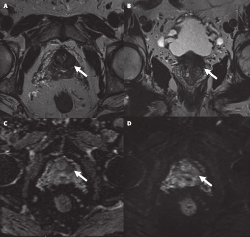
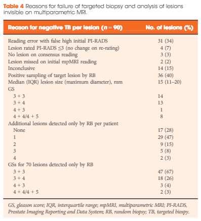
Our analysis showed that RB detected significant PCa in 64% of patients (39/61) and intermediate-/high-risk PCa in 57% of patients (35/61). The initial mpMRI reading identified 90 suspicious lesions in the cohort. Blinded consensus re-reading of the mpMRI led to PI-RADS score downgrading of 45 lesions (50%) and upgrading of 13 lesions (14%); thus, negative TB could be explained by falsely high initial PI-RADS scores for 32 lesions (34%) and sampling of the target lesion by RB in the corresponding anatomical site for 36 out of 90 lesions (40%) in 35 of 61 patients (57%). Sampling of the target lesion by RB was most likely for lesions with PI-RADS scores of 4/5 and Gleason scores (GS) of ≥7. A total of 70 PCa lesions (67% with GS 6) in 44 patients (72%) were sampled from prostatic sites with no abnormalities on mpMRI.

Conclusion

In cases of TB failure, RB still detected a high rate of significant PCa. The main reason for a negative TB was a TB error, compensated for by positive sampling of the target lesion by the additional RB, and the second reason for TB failure was a falsely high initial PI-RADS score. The challenges that arise for both MRI diagnostics and prostate lesion sampling are evident in our data and support the integration of RB into the TB workflow.

Read more articles of the week

Editorial: MRI-Fusion Biopsy – Behind the Scenes

MRI information of the prostate is increasingly used for improving the diagnostic yield of prostate biopsies [1]. However, increasing complexity of a procedure makes it prone to errors at multiple technical and human levels. Incorporating MRI information and ultrasonography (US) images for MRI-fusion biopsies is a technically challenging task. It involves various steps such as the acquisition and fusion of MRI and US images, the needle guidance during biopsy, and the diligence of the pathological evaluation of biopsy specimens. These different steps and interfaces between different medical professions influence the diagnostic performance of MRI-fusion biopsies.

For example, in daily clinical practice, MRIs from different institutions still harbour a great variance of sequences and reporting, despite the European Society of Urogenital Urology (ESUR) recently introducing acquisition and imaging protocols and a new and advanced version of the Prostate Imaging Reporting and Data System (PIRADS) version 2.0 [2]. The usefulness of such reporting schemes is evidenced by a moderate-to-good interobserver agreement between uro-radiologists for tumour lesion interpretation and corresponding κ values ranging from 0.55 to 0.80 [3]. Important pitfalls of image interpretation are benign lesions such as prostatitis, BPH and fibrosis, which might score similarly to prostate cancer lesions. This problem is further aggravated by a high proportion of patients that receive their first multiparametric MRI (mpMRI) of the prostate in the repeat-biopsy setting with a high burden of post-biopsy artefacts (haemorrhage, capsular irregularity) and lower overall cancer detection rate. Also, during MRI-fusion biopsy patient movement, prostate deformation by the US probe, and mismatch of image planes can lead to a biopsy error exceeding 4 mm. Moreover, targeting error might be aggravated by MRI underestimation of the tumour volume compared with final pathology [4]. After various authors reported the advantages and accuracy of MRI/US-fusion biopsy approaches, Cash et al. [5] address potential reasons for targeted biopsy failure to detect prostate cancer compared with random biopsy. Within their analyses the authors address potential limitations and technical considerations. Based on different technical biopsy strategies (with the patient placed within the MRI scanner (‘in-bore’) vs outside) and different technical approaches, these considerations are very important.

In contrast to cognitive fusion, most MRI/US platforms allow needle tracking by archiving the needle orientation, either by an electromagnetic, image-based or stepper-based mechanism [1]. However, lesion targeting by needle guidance is highly dependent on the dimensions of the primary lesion, numbers of relevant lesions, localisation, and overall prostate volume, making MRI-US fusion and cognitive fusion more error prone (i.e. aiming off the mark with the needle) than in-bore biopsies. Moreover, different technical fusion approaches provide different degrees of manual/automated adjustment tools, with for example either rigid or elastic image transformation to facilitate MRI/US image alignment.

In their analyses, Cash et al. [5] found that 34% of negative targeted biopsies could be explained by initially too high estimated PIRADS scores that were downgraded at re-reading. Interestingly, the remaining lesions were without an mpMRI correlate but within this group 92.9% showed a primary Gleason 3 pattern in biopsy pathology, suggesting a high degree of invisibility on mpMRI. Subanalyses did not show an association of targeted biopsy failures in the ventral location. Therefore, the study by Cash et al. [5] is an important precursor for further analyses to address other underlying reasons for targeted biopsy failure. Moreover, it reveals the need for a tight collaboration of radiologists, urologists, and pathologists as interdisciplinary partners involved in MRI-fusion biopsy. Consequently, the optimal diagnostic performance of MRI-fusion biopsies can only be achieved through standardised MRI performance, reading and reporting of MRI findings, as well as final correlation of MRI findings with histopathological work up.

Read the full article
Lars Budaus and Sami-Ramzi Leyh-Bannurah
Martini-Clinic University Hospital Hamburg-Eppendorf, Hamburg, Germany


References


Article of the Week: Muscolofascial Reconstruction after RP

Every Week the Editor-in-Chief selects an Article of the Week from the current issue of BJUI. The abstract is reproduced below and you can click on the button to read the full article, which is freely available to all readers for at least 30 days from the time of this post.

In addition to the article itself, there is an accompanying editorial written by a prominent member of the urological community. This blog is intended to provoke comment and discussion and we invite you to use the comment tools at the bottom of each post to join the conversation.

Finally, the third post under the Article of the Week heading on the homepage will consist of additional material or media. This week we feature a video from Dr. Francesco Alessandro Mistretta, discussing his paper.

If you only have time to read one article this week, it should be this one.

Posterior musculofascial reconstruction after radical prostatectomy: an updated systematic review and a meta-analysis


Angelica A.C. Grasso*, Francesco A. Mistretta*, Marco Sandri, Gabriele Cozzi*, Elisa De Lorenzis*, Marco Rosso*, Giancarlo Albo*, Franco Palmisano*, Alex MottrieAlexander Haese§, Markus Graefen§, Rafael Coelho, Vipul R. Patel¶ and Bernardo Rocco*


*Department of Urology, Fondazione IRCCS Ca Granda-Ospedale Maggiore Policlinico, University of Milan, Milan, Italy, DMS StatLab, Data Methods and Systems Statistical Laboratory, University of Brescia, Brescia, Italy, OLV Robotic Surgery Institute, Aalst, Belgium, §Martini Clinic Prostate Cancer Center, University Clinic Eppendorf, Hamburg, Germany, and Global Robotics Institute, Florida Hospital-Celebration Health Celebration, University of Central Florida School of Medicine, Orlando, FL, USA


Read the full article
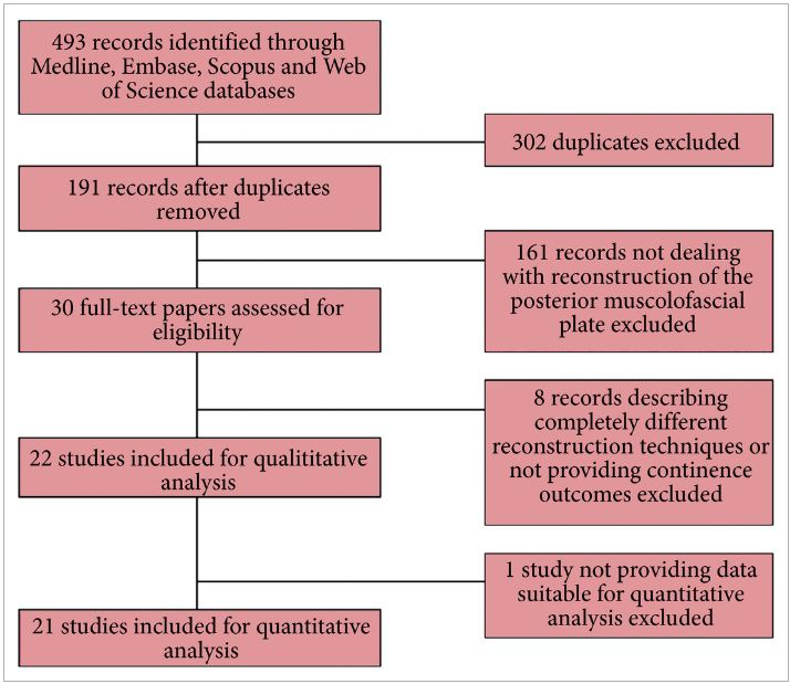
To evaluate the influence of posterior musculofascial plate reconstruction (PR) on early return of continence after radical prostatectomy (RP); an updated systematic review of the literature. A systematic review of the literature was performed in June 2015, following the Preferred Reporting Items for Systematic Reviews and Meta-Analyses (PRISMA) statement and searching Medline, Embase, Scopus and Web of Science databases. We searched the terms posterior reconstruction prostatectomy, double layer anastomosis prostatectomy across the ‘Title’ and ‘Abstract’ fields of the records, with the following limits: humans, gender (male), and language (English). The authors reviewed the records to identify studies comparing cohorts of patients who underwent RP with or without restoration of the posterior aspect of the rhabdosphincter. A meta-analysis of the risk ratios estimated using data from the selected studies was performed. In all, 21 studies were identified, including three randomised controlled trials. The overall analysis of comparative studies showed that PR improved early continence recovery at 3–7, 30, and 90 days after catheter removal, while the continence rate at 180 days was statistically but not clinically affected. Statistically significantly lower anastomotic leakage rates were described after PR. There were no significant differences for positive surgical margins rates or for complications such as acute urinary retention and bladder neck stricture. The analysis confirms the benefits at 30 days after catheter removal already discussed in the review published in 2012, but also shows a significant advantage in terms of urinary continence recovery in the first 90 days. A multicentre prospective randomised controlled trial is currently being conducted in several institutions around the world to better assess the effectiveness of PR in facilitating an earlier recovery of postoperative urinary continence.


Read more articles of the week


© 2024 BJU International. All Rights Reserved.