Tag Archive for: Article of the Week

Posts

Article of the Month: Comparing VEILND with OILND for penile cancer

Every Month the Editor-in-Chief selects an Article of the Month from the current issue of BJUI. The abstract is reproduced below and you can click on the button to read the full article, which is freely available to all readers for at least 30 days from the time of this post.

In addition to the article itself, there are accompanying editorials written by prominent members of the urological community. These blogs are intended to provoke comment and discussion and we invite you to use the comment tools at the bottom of each post to join the conversation.

Finally, the third post under the Article of the Week heading on the homepage will consist of additional material or media. This week we feature a video discussing the paper.

If you only have time to read one article this week, it should be this one.

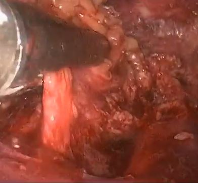
Prospective study comparing video-endoscopic radical inguinal lymph node dissection (VEILND) with open radical ILND (OILND) for penile cancer over an 8-year period

Vivekanandan Kumar and K. Krishna Sethia
Norfolk and Norwich University Hospital, Norwich, UK


How to Cite this article:

Kumar, V. and Sethia, K. K. (2017), Prospective study comparing video-endoscopic radical inguinal lymph node dissection (VEILND) with open radical ILND (OILND) for penile cancer over an 8-year period. BJU International, 119: 530–534. doi: 10.1111/bju.13660

Abstract

Objective

To compare the complications and oncological outcomes between video-endoscopic inguinal lymph node dissection (VEILND) and open ILND (OILND) in men with carcinoma of the penis.

Patients and methods

A prospectively collected institutional database was used to determine the outcomes in 42 consecutive patients undergoing ILND between 2008 and 2015 in a centre for treating penile cancer. Before 2013 all procedures were OILNDs. Since 2013 we have performed VEILND on all patients in need of ILND. The wound-related and non-wound-related complications, length of stay, and oncological safety between OILND and VEILND groups were compared. The mean duration of follow-up was 71 months for OILND and 16 months for the VEILND groups.

ergerger

Results

In the study period 42 patients underwent 68 ILNDs (OILND 35, VEILND 33). The patients’ demographics, primary stage and grade, and indications were comparable in both groups. There were no intraoperative complications in either group. The wound complication rate was significantly lower in the VEILND group at 6% compared to 68% in the OILND group. Lymphocoele rates were similar in both the groups (27% and 20%). The VEILND group had a better or the same lymph node yield, mean number of positive lymph nodes, and lymph node density confirming oncological safety. There were no groin recurrences in either group of patients. VEILND significantly reduced the mean length of stay by 4.8 days (P < 0.001).

Conclusion

VEILND is an oncologically safe procedure with considerably low morbidity and reduced length of stay, at a mean (range) follow-up of 16 (4–35) months.

Editorial: VEIL – is a new standard ready to be accepted?

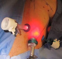
In this interesting prospective study, Kumar and Sethia [1] provide further evidence that video endoscopic inguinal lymphadenectomy (VEIL) could be considered the procedure of choice for most patients with an indication for ILND. In their comparison with historical patients submitted to open ILND (OILND) in a reference centre in the UK, VEIL achieved reduced morbidity, shorter hospital stay, and equivalent oncological control. Also, we must consider that some cases of the VEIL learning curve were included in this study, suggesting that good results can be obtained with the endoscopic technique from its inception.

The idea for an endoscopic approach to ILND was conceived and developed in a cadaveric model by Bishoff et al. [2] in 2003. In 2005, our group reported the first successful experience in humans [3]. In 2006, in collaboration with the Brazilian National Institute of Cancer, we published a landmark study in the Journal of Urology comparing OILND in one limb and VEIL in the contralateral limb in the same patient to minimise comparative bias [4]. In all, 10 patients with impalpable nodes underwent bilateral ILND. We found that morbidity was reduced with VEIL (20% vs 70%) with the removal of the same number of nodes as with OILND [4]. In 2008, we reported on a series of bilateral VEIL obtaining reduced postoperative morbidity and a mean hospital stay of 1 day [5]. Several other small series have reported essentially the same results that we initially described.

Robot-assisted VEIL duplicates the procedure but is more ergonomic, with excellent freedom of movement and amplification of lymphatic visualisation. Matin et al. [6] reported that robot-assisted VEIL could remove the same number of nodes as the open procedure.

The present study [1] reports a large experience comparing bilateral procedures (VEIL and OILND). The follow-up data for OILND are longer but oncological indicators suggest equivalence. This is also the first report of VEIL after node cytology and dynamic sentinel node biopsy with no related adverse events.

To date, a few series have reported long-term oncological results [1, 7] and future studies will be important to evaluate and reproduce the oncological efficacy of VEIL.

We congratulate Kumar and Sethia [1] for these significant data to establish VEIL in the modern urological armamentarium. Indeed, I really have some doubt as whether a randomised controlled study would be necessary considering the accumulated worldwide data on VEIL in the last 10 years.

This year we present at the University of California meeting the promising results of our Latin America collaborative group with 150 VEIL in 110 patients. The mean (range) follow up was 6 (2–10) years, with an overall morbidity of 30% (Clavien–Dindo grade III–IV, 2%), mean lymph node removal of 8 nodes/groin, node positive disease rate of 25%, inguinal recurrence rate of 2%, cancer-specific survival of 90%, and overall survival of 85% (Tobias-Machado et al., 2016, unpublished data). Our data suggest that we can obtain a dramatic reduction in severe complications and also that VEIL is surviving the test of time.

In my opinion, as the reduction in morbidity is already confirmed, we only need larger series with long-term reports of oncological equivalence with OILND to designate VEIL as a ‘gold standard’ in centres with appropriately trained surgeons.

I speculate that in the near future the utilisation of robotic surgery associated with improvements in imaging methods and contrasts, which will improve the identification of lymph nodes to be removed and lymphatic channels to be clipped, will achieve further reductions in morbidity and optimisation of oncological control.

How to Cite this article:

Tobias-Machado, M. (2017), Video endoscopic inguinal lymphadenectomy (VEIL): is a new standard ready to be accepted?. BJU International, 119: 504–505. doi: 10.1111/bju.13723

Marcos Tobias-Machado
Department of Urology, ABC Medical School, Sao PauloBrazil


References


2 Bishoff JT, Basler JW, Teichman JM et al. Endoscopic subcutaneous modied inguinal lymph node dissection (ESMIL) for squamous cell carcinoma of the penis. J Urol (Suppl.) 2003; 169: 78 (abstract 301)


3 Tobias-Machado M, Tavares A, Molina WR Jr, Forseto PH Jr, Juliano RV, Wroclawski ER. Video endoscopic inguinal lymphadenectomy (VEIL): minimally invasive resection of inguinal lymph nodes. Int Braz Urol 2006; 32: 31621


4 Tobias-Machado M, Tavares A, Ornellas AA, Molina WR Jr, Juliano RVWroclawski ER. Video endoscopic inguinal lymphadenectomy: a new minimally invasive procedure for radical management of inguinal nodes in


7 SchwentneC, Todenhofer T, Seibold J e t al. Endoscopic inguinofemoral lymphadenectomy extended follow-up. JEndouro20 13; 27: 497503


Editorial: Is minimally invasive inguinal node dissection the way forward?

In this issue of BJUI, Kumar and Sethia report their experience of laparoscopic inguinal node dissection for patients with penile cancer [1]. The authors should be congratulated for what is a technically and feasibly difficult study to complete.

The step-wise spread of penile carcinoma first to the inguinal lymph nodes is well known. It is also recognized that accurate staging and treatment of inguinal metastases play a crucial part in a patient’s management pathway. Furthermore, evidence points to a survival advantage for those who undergo prophylactic inguinal lymph node dissection (ILND) and have positive nodes, compared with those treated with surveillance and delayed dissection when nodes become palpable or radiologically positive.

The overall postoperative complication rate after ILND has been reported to be nearly 50% or even up to 75%. Patients with comorbidities or locally advanced disease are at particular risk of postoperative morbidity [2]. High rates of wound dehiscence and skin necrosis after ILND have also been reported [3]. More recently, complication rates among men with no palpable adenopathy have been reported to be decreasing in comparison with historical experience, but overall complication rates still range from 10 to 40%.

Adoption of dynamic sentinel node biopsy has reduced the absolute number of prophylactic ILNDs, but in any high-volume unit (as is the case in the UK supra-network centres), a large number of inguinal node dissections will still be undertaken [4].

For these reasons it is desirable to minimize or modify the inguinal wound to reduce the complication rate, and the use of minimally invasive techniques has gradually progressed over the last 10 years.

The first case series reporting the use of laparoscopic node dissection (videoendoscopic inguinal node dissection [VEILND]) described 10 patients who underwent bilateral ILND using standard lymphadenectomy on one side and the VEILND technique on the contralateral side [5]. A more recent development has been the incorporation of robotic assistance as an enabling tool for performing ILND [6].

In these small series the technique appeared to be safe and oncologically comparable to open surgery, but the numbers were too small to give a meaningful assessment of complications. It is therefore welcome to read a paper on a high-volume unit’s experience of VEILND.

The authors describe their experience of VEILND on a sequential prospective group of patients and compared them with a historical control cohort who underwent open inguinal node dissection. The data appear to show a significant difference in length of stay and wound-related comorbidity in favour of the minimally invasive approach without compromising oncological outcome. Interestingly, the rate of lower limb lymphedema was also significantly reduced in the VEILND group.

The headline data are impressive but the comparison does have a number of potential weaknesses. Firstly, the rate of wound-related complications is quite high in the open group, with 23% of patients needing re-exploration or debridement and 37% some form of drainage of collection. The choice of incision position, length and preparation of skin flaps can influence the rate of wound breakdown and length of stay.

Secondly, the headline 6% complication rate for VEILND does not include the 27% lymphocoele rate which is still a troublesome complication in both the open and endoscopic groups.

Finally, there is no mention of the limitations of the technique with size and extent of the malignant node burden. The paper states that all patients were included, and yet in any high-volume centre there must be patients with grossly enlarged and sometimes fungating or fixed nodes and I would have thought that VEILND would not be appropriate in these instances.

In conclusion, there is much to commend this paper and the direction of travel may well be towards minimally invasive techniques; however, as with all comparisons between open and keyhole surgery, the focus should be on improving the open technique to its limit. Only then will the results be meaningful and adopted with confidence.

How to Cite this article:

Watkin, N. (2017), Is minimally invasive inguinal node dissection the way forward?. BJU International, 119: 505–506. doi: 10.1111/bju.13761

Nick Watkin
Department of Urology, St Geo rges Hospital, London, UK


References


Video: Comparing VEILND with OILND for Penile Cancer

Prospective study comparing video-endoscopic radical inguinal lymph node dissection (VEILND) with open radical ILND (OILND) for penile cancer over an 8-year period

Abstract

Objective

To compare the complications and oncological outcomes between video-endoscopic inguinal lymph node dissection (VEILND) and open ILND (OILND) in men with carcinoma of the penis.

Patients and methods

A prospectively collected institutional database was used to determine the outcomes in 42 consecutive patients undergoing ILND between 2008 and 2015 in a centre for treating penile cancer. Before 2013 all procedures were OILNDs. Since 2013 we have performed VEILND on all patients in need of ILND. The wound-related and non-wound-related complications, length of stay, and oncological safety between OILND and VEILND groups were compared. The mean duration of follow-up was 71 months for OILND and 16 months for the VEILND groups.

Results

In the study period 42 patients underwent 68 ILNDs (OILND 35, VEILND 33). The patients’ demographics, primary stage and grade, and indications were comparable in both groups. There were no intraoperative complications in either group. The wound complication rate was significantly lower in the VEILND group at 6% compared to 68% in the OILND group. Lymphocoele rates were similar in both the groups (27% and 20%). The VEILND group had a better or the same lymph node yield, mean number of positive lymph nodes, and lymph node density confirming oncological safety. There were no groin recurrences in either group of patients. VEILND significantly reduced the mean length of stay by 4.8 days (P < 0.001).

Conclusion

VEILND is an oncologically safe procedure with considerably low morbidity and reduced length of stay, at a mean (range) follow-up of 16 (4–35) months.

View more videos

Article of the Week: 3-Tesla mpMRI and TRUS-Bx in PCa patients on AS

Every week the Editor-in-Chief selects an Article of the Week from the current issue of BJUI. The abstract is reproduced below and you can click on the button to read the full article, which is freely available to all readers for at least 30 days from the time of this post.

In addition to the article itself, there is an accompanying editorial written by a prominent member of the urological community. This blog is intended to provoke comment and discussion and we invite you to use the comment tools at the bottom of each post to join the conversation.

If you only have time to read one article this week, it should be this one.

Value of 3-Tesla multiparametric magnetic resonance imaging and targeted biopsy for improved risk stratification in patients considered for active surveillance

Rodrigo R. Pessoa*, Publio C. Viana, Romulo L. Mattedi, Giuliano B. Guglielmetti*, Mauricio D. Cordeiro*, Rafael F. Coelho*, William C. Nahas* and Miguel Srougi*


Departments of *Urology, Diagnostic and Interventional Radiology, and Department of Pathology, Instituto do Cancer, Universidade de Sao Paulo Faculdade de Medicina Hospital das Clinicas, Sao Paulo, SP, Brazil
Read the full article

Abstract

Objective

To evaluate the role of multiparametric magnetic resonance imaging (mpMRI) of the prostate and transrectal ultrasonography guided biopsy (TRUS-Bx) with visual estimation in early risk stratification of patients with prostate cancer on active surveillance (AS).

Patients and Methods

Patients with low-risk, low-grade, localised prostate cancer were prospectively enrolled and submitted to a 3-T 16-channel cardiac surface coil mpMRI of the prostate and confirmatory biopsy (CBx), which included a standard biopsy (SBx) and visual estimation-guided TRUS-Bx. Cancer-suspicious regions were defined using Prostate Imaging Reporting and Data System (PI-RADS) scores. Reclassification occurred if CBx confirmed the presence of a Gleason score ≥7, greater than three positive fragments, or ≥50% involvement of any core. The performance of mpMRI for the prediction of CBx results was assessed. Univariate and multivariate logistic regressions were performed to study relationships between age, prostate-specific antigen (PSA) level, PSA density (PSAD), number of positive cores in the initial biopsy, and mpMRI grade on CBx reclassification. Our report is consistent with the Standards of Reporting for MRI-targeted Biopsy Studies (START) guidelines.

apr-aotw-1-results

Results

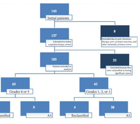
In all, 105 patients were available for analysis in the study. From this cohort, 42 (40%) had PI-RADS 1, 2, or 3 lesions and 63 (60%) had only grade 4 or 5 lesions. Overall, 87 patients underwent visual estimation TRUS-Bx. Reclassification among patients with PI-RADS 1, 2, 3, 4, and 5 was 0%, 23.1%, 9.1%, 74.5%, and 100%, respectively. Overall, mpMRI sensitivity, specificity, positive predictive value, and negative predictive value for disease reclassification were 92.5%, 76%, 81%, and 90.5%, respectively. In the multivariate analysis, only PSAD and mpMRI remained significant for reclassification (P < 0.05). In the cross-tabulation, SBx would have missed 15 significant cases detected by targeted biopsy, but SBx did detect five cases of significant cancer not detected by targeted biopsy alone.

Conclusion

Multiparametric magnetic resonance imaging is a significant tool for predicting cancer severity reclassification on CBx among AS candidates. The reclassification rate on CBx is particularly high in the group of patients who have PI-RADS grades 4 or 5 lesions. Despite the usefulness of visual-guided biopsy, it still remains highly recommended to retrieve standard fragments during CBx in order to avoid missing significant tumours.

Editorial: An end to the phenomenon of ‘upgrading’ in early prostate cancer?

The phenomenon of ‘upgrading’ in early prostate cancer is one of those unusual events that is both useful to us on the one hand and undesirable on the other; useful because the phenomenon gifts us a direct measure of the precision of our risk stratification methods for men recently diagnosed, and undesirable because the perfect pathway should, ideally, be free of any upgrading.

Upgrading occurs in a number of settings. We see it at play to some degree when an unreliable test is re-applied in the same subject. The REDUCE study [1] showed us that just under one fifth of men will convert from a status of ‘cancer-free’ to one of ‘cancer-present’ as a result of a second exposure to the same test; that is, TRUS-guided biopsy. We see it in full play when an unreliable test is followed by a more accurate test. Shaw et al. [2] have reminded us once again – as have a number of others – of our limited ability to risk-stratify patients with early prostate cancer. They reported a 50% upgrading when they compared the results of TRUS biopsy against the final pathology at radical prostatectomy. In other words, half the patients went on to their definitive therapy with an incorrect grade attribution [2].

It would be a great pity if, in the modern era, the only route available to patients who wanted to be sure of their risk status was to agree to surgical removal of the prostate. Surely, the value of accurate risk stratification is derived from using it to allocate appropriate and effective care. Risk stratification needs to be linked to or closely follow diagnosis if it is to be put to work for patients.

Nowhere is this need greater than in men whose treatment preference is tissue preservation. The study, in this issue of BJUI, by Pessoa et al. [3] adds to our knowledge on the subject and equips us with a strategy to mitigate some of the errors that are inherent to the standard diagnostic pathway.

In the present study, the authors evaluated the role of a single exposure to MRI (and the opportunity that resulted to undertake a targeted biopsy of an MRI-derived abnormality as well as systematic sampling) in 105 men who had been attributed a diagnosis of low-risk prostate cancer – and, as a result, were deemed to be suitable for active surveillance. The authors used prostate imaging reporting and data system (PIRADS) scoring to interpret and communicate MRI risk. In summary, men attributed a low PIRADS score (PIRADS 1–3) had a low probability of being re-classified to a higher risk. In contrast, men attributed PIRADS score 4 or 5 had a probability of 70–100% of being re-classified. The authors calculated a sensitivity of 93% for MRI to predict ‘re-classification’. This equates to a 93% sensitivity to predict the presence of clinically significant disease as re-classification occurred when there was a transition from low-risk to higher-risk disease.

These results concur with those of others who are working in this area [4] and are in line with current recommendations [5]. One observation that is worth highlighting – because it is a current controversy in the field – relates to the utility of the systematic (or semi-random) biopsies as a component of the confirmatory biopsy. Whilst targeted biopsy was superior to systematic biopsy at identifying clinically significant disease, omission of the systematic biopsies would have resulted in five significant cancers being overlooked. The less perfect the targeted biopsy, the greater the reliance on the systematic. In the present study, the lesion generation and the targeting may have been compromised by one or two issues. Using TRUS biopsy as the authors did (as opposed to transperineal biopsy) to access all areas of the prostate is always going to be a challenge. To do so without image registration makes it even harder. To use PIRADS – as opposed to a Likert scale – as a method of interpreting and communicating MRI outputs will, very likely, lead to an under-reporting of the smaller, high-grade lesions [6]. This is because PIRADS 2.0 is triggered by a volume threshold towards the upper end of the scale. Such lesions might be more prevalent in an apparently ‘low-risk’ population such as the one under scrutiny. If this is the case, they will not be identified as ‘targets’ by virtue of a high PIRADS score. As a consequence they cannot be identified by targeting but might be picked up by the random fall of the needles.

Mark Emberton
Division of Surgery and Interventional Science, University College London, London, UK
Read the full article
References

1 Andriole GL, Bostwick DG, Brawley OW et al. Rittmaster RS; REDUCE Study Group. Effect of dutasteride on the risk of prostate cancer. N Engl Med 2010; 362: 1192202


2 Shaw GL, Thomas BC, Dawson SN et al. Identication of pathologically insignicant prostate cancer is not accurate in unscreened men. Br Cancer 2014; 110: 240511


4 Nassiri N, Margolis DJ, Natarajan S et al. Targeted biopsy to detect Gleason score upgrading during active surveillance for men with low- vs. intermediate-risk prostate cancer. J Urol 2016; [Epub ahead of print]. doi: 10.1016/j.juro.2016.09.070.


5 Moore CM, Giganti F, Albertsen P et al. Reporting magnetic resonance imaging in men on active surveillance for prostate cancer: the PRECISE recommendations-a report of a European school of oncology task force. Eur Urol 2016; [Epub ahead of print]. doi: 10.1016/j.eururo.2016.06.011.


Article of the Week: PADUA and R.E.N.A.L. nephrometry scores correlate with perioperative outcomes of RAPN: analysis of the Vattikuti GQI-RUS database

Every week the Editor-in-Chief selects an Article of the Week from the current issue of BJUI. The abstract is reproduced below and you can click on the button to read the full article, which is freely available to all readers for at least 30 days from the time of this post.

In addition to the article itself, there is an accompanying editorial written by a prominent member of the urological community. This blog is intended to provoke comment and discussion and we invite you to use the comment tools at the bottom of each post to join the conversation.

If you only have time to read one article this week, it should be this one.

PADUA and R.E.N.A.L. nephrometry scores correlate with perioperative outcomes of robot-assisted partial nephrectomy: analysis of the Vattikuti Global Quality Initiative in Robotic Urologic Surgery (GQI-RUS) database


Riccardo Schiavina*, Giacomo Novara,, Marco Borghesi*, Vincenzo Ficarra§Rajesh Ahlawat, Daniel A. Moon**, Francesco Porpiglia††,BenjaminJ.Challacombe‡‡Prokar Dasgupta‡‡, Eugenio Brunocilla*, Gaetano La Manna§§, Alessandro Volpe¶¶Hema Verma***, Giuseppe Martorana* and Alexandre Mottrie,†††


*Department of Urology, University of Bologna, Bologna,† Department of Surgery, Oncology, and Gastroenterology – Urology Clinic, University of Padua, Padua, Italy, OLV Vattikuti Robotic Surgery Institute, Aalst, Belgium, §Department of
Experimental and Clinical Medical Sciences, University of Udine, Udine, Italy, Division of Urology and Renal Transplantation, Medanta Kidney and Urology Institute, Medanta-The Medicity, Gurgaon, India, **Department of Surgery, Peter MacCallum Cancer Centre, University of Melbourne, Melbourne, Vic., Australia, ††San Luigi Gonzaga Hospital, University of Turin, Orbassano, Italy, ‡‡Department of Urology, Guys and St Thomas NHS Foundation Trust and National Institute for Health Research (NIHR) Biomedical Research Centre, Kings College London, London, UK, §§Department Nephrology and Experimental, Diagnostic and Specialty Medicine, University of Bologna, Bologna, ¶¶University of Eastern Piedmont, Novara, Italy, ***Department of Radiology, Guys and St Thomas NHS Foundation Trust and National Institute for Health Research (NIHR) Biomedical Research Centre, Kings College London, London, UK, and †††Department of Urology, Onze-Lieve-Vrouw Hospital, Aalst, Belgium


Read the full article

Abstract

Objectives

To evaluate and compare the correlations between Preoperative Aspects and Dimensions Used for an Anatomical (PADUA) and R.E.N.A.L. [Radius (tumour size as maximal diameter), Exophytic/endophytic properties of the tumour, Nearness of tumour deepest portion to the collecting system or sinus, Anterior (a)/posterior (p) descriptor and the Location relative to the polar line] nephrometry scores and perioperative outcomes and postoperative complications in a multicentre, international series of patients undergoing robot-assisted partial nephrectomy (RAPN) for masses suspicious for renal cell carcinoma (RCC).

Patients and Methods

We retrospectively evaluated the clinical records of patients who underwent RAPN between 2010 and 2013 for clinical N0M0 renal tumours in four international centres that completed all the data required for the Vattikuti Global Quality Initiative in Robotic Urologic Surgery (GQI-RUS) database. All patients underwent preoperative computed tomography or magnetic resonance imaging to define the clinical stage and anatomical characteristics of the tumours. PADUA and R.E.N.A.L. scores were retrospectively assessed in each centre. Univariate and multivariate analyses were used to evaluate the correlations between age, gender, Charlson comorbidity index, clinical tumour size, PADUA and R.E.N.A.L. complexity group categories and warm ischaemia time (WIT) of >20 min, urinary calyceal system closure, and grade of postoperative complications.

aotw-mar-4-results

Results

Overall, 277 patients were evaluated. The median (interquartile range) tumour size was 33.0 (22.0–43.0) mm. The median PADUA and R.E.N.A.L. scores were eight and seven, respectively; 112 (40.4%), 86 (31.0%) and 79 (28.5%) patients were classified in the low-, intermediate- or high-complexity group according to PADUA score, while 118 (42.5%), 139 (50.1%) and 20 (7.2%) were classified in the low-, intermediate- or high-complexity group according to R.E.N.A.L. score, respectively. Both nephrometry tools significantly correlated with perioperative outcomes at univariate and multivariate analyses.

Conclusion

A precise stratification of patients before PN is recommended to consider both the potential threats and benefits of nephron-sparing surgery. In our present analysis, both PADUA and R.E.N.A.L. were significantly associated with predicting prolonged WIT and high-grade postoperative complications after RAPN.

Editorial: Nephrometry scoring systems: valuable research tools, but can they be applied in daily clinical practice?

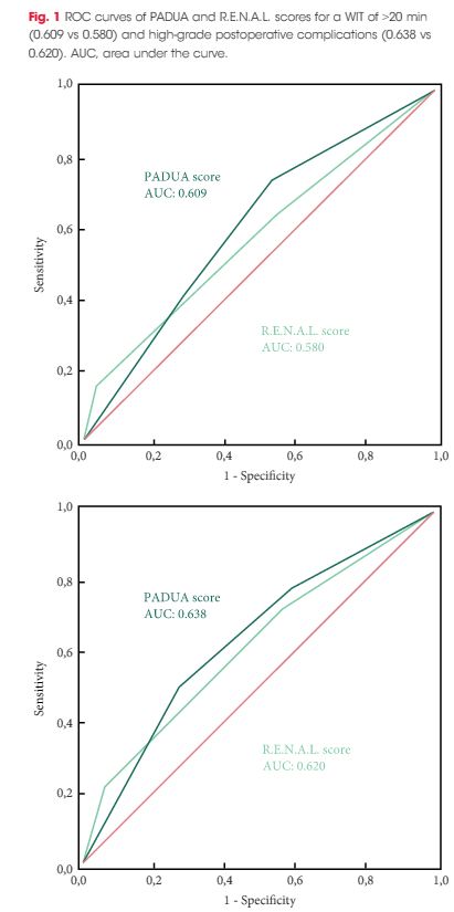
In this issue of BJUI Schiavina et al. [1] report on the RENAL and PADUA nephrometry scoring systems in predicting peri-operative outcomes, including warm ischaemia time and postoperative complications, in a multi-institutional cohort of patients undergoing robot-assisted partial nephrectomy. The authors showed that tumours classified as being of intermediate and high complexity on the PADUA score and high complexity on the RENAL score were associated with a nearly threefold higher risk of longer warm ischaemia times (>20 min). In addition, more complex tumours carried a higher risk of grade 3–4 postoperative complications (most commonly bleeding requiring angioembolization and urine leak requiring a ureteric stent). Notably, the two scoring systems were found to be similar predictors of these peri-operative outcomes on receiver-operating curve (ROC) analyses [1].

This represents the first large, multicentre study to evaluate the accuracy of these scoring systems in a cohort of patients who purely underwent robot-assisted surgery. A recent study by Borgmann et al. [2] found that, among the reported scoring systems, the RENAL nephrometry score correlated best with achieving negative surgical margins, shorter ischaemia times, and low postoperative complication rates; however, only 9% of patients underwent robot-assisted surgery. Another contemporary series showed concordance between the RENAL and PADUA scoring systems in predicting ischaemia times and complication rates, albeit in patients who only underwent open surgery [3].

Current guidelines recognize nephron-sparing approaches to small renal masses as the standard of care in well-selected patients, with the robot-assisted platform being predominantly adopted in clinical practice where available. Certainly, these nephrometry scores are valuable for urologists in counselling patients on the potential risk of complications specific to the surgical anatomy of the tumour. In addition, the RENAL and PADUA scores (and others) provide a quantitative, objective method for comparing data from different studies and different institutions.

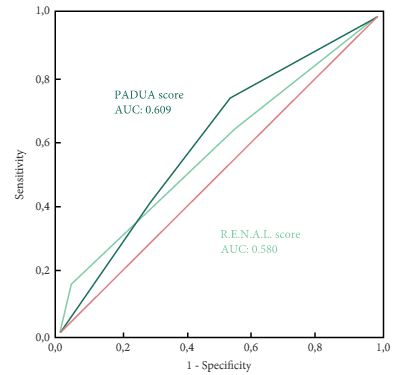
As nephrometry scoring systems continue to be critically evaluated in the robotic surgery era, the question that naturally arises is: which system is best? With regard to this question, the data in the present study do not necessarily favour one or the other for the prediction of clinically relevant peri-operative outcomes. One must recognize, however, that several other anatomy-based scoring systems exist and were not examined in this manuscript [4-6]. While these are very valuable research and patient counselling tools, one must caution against using these nephrometry tools to make clinical decisions; for example, attempting to predict benign vs malignant histology (without a biopsy), attempting to predict high vs low grade tumours, or deciding on whether to perform a radical vs partial nephrectomy, or an open vs minimally invasive approach. After all, one must keep in mind that the area under the curve for these tools is in the range of 0.58–0.63 (0.50 being equivalent to toss of a coin).

It would have been interesting to include clinical size only in the present multivariate analysis (as was done for RENAL and PADUA scoring) and ROC analysis to compare this simple variable with the studied nephrometry scores. Future research should examine additional confounders that could potentially affect postoperative complication rates, such as BMI, adherent perinephric fat, experience of the surgeon actually performing the partial nephrectomy, technique of resection used (e.g. enucleation or resection) among others. This may help to distinguish a single system as the optimum model for use in research and in patient counselling regarding potential postoperative complications.

Matthew A. Meissner and Jose A. Karam


Department of Urology, University of Texas MD Andersonn Cancer Center, Houston, TX, USA


Read the full article


References


3 Kriegmair MC, Mandel P, Moses A et al. Dening Renal Masses: comprehensive Comparison of RENAL, PADUA, NePhRO, and C-Index Score. Clin Genitourin Cancer 2016; [Epub ahead of print]. doi: 10.1016/ j.clgc.2016.07.029.


5 Hakky TS, Baumgarten AS, Allen B, Lin HY, Ercole CE, Sexton WJSpiess PE et al. Zonal NePhRO scoring system: a superior renal tumor complexity classication model. Clin Genitourin Cancer 2014; 12: e138


6 Simmons MN, Ching CB, Samplaski MK, Park CH, Gill IS et al. Kidney tumor location measurement using the C index method. J Urol 2010; 183: 170813


Article of the Week: Accuracy of ultrasonography for renal stone detection and size determination: is it good enough for management decisions?

Every week the Editor-in-Chief selects an Article of the Week from the current issue of BJUI. The abstract is reproduced below and you can click on the button to read the full article, which is freely available to all readers for at least 30 days from the time of this post.

In addition to the article itself, there is an accompanying editorial written by a prominent member of the urological community. This blog is intended to provoke comment and discussion and we invite you to use the comment tools at the bottom of each post to join the conversation.

Finally, the third post under the Article of the Week heading on the homepage will consist of additional material or media. This week we feature a video discussing the paper.

If you only have time to read one article this week, it should be this one.

Accuracy of ultrasonography for renal stone detection and size determination: is it good enough for management decisions?

Vishnu Ganesan*,, Shubha De*, Daniel Greene*, Fabio Cesar Miranda Torricelli* and Manoj Monga*


*Glickman Urological Kidney Institute, and Lerner College of Medicine, Cleveland Clinic, Cleveland, OH, USA


Read the full article

Abstract

Objectives

To determine the sensitivity and specificity of ultrasonography (US) for detecting renal calculi and to assess the accuracy of US for determining the size of calculi and how this can affect counselling decisions.

Materials and Methods

We retrospectively identified all patients at our institution with a diagnosis of nephrolithiasis who underwent US followed by non-contrast computed tomography (CT) within 60 days. Data on patient characteristics, stone size (maximum axial diameter) and stone location were collected. The sensitivity, specificity and size accuracy of US was determined using CT as the standard.

aotw-mar

Results

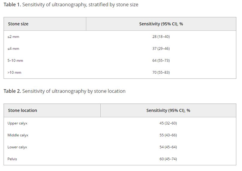
A total of 552 US and CT examinations met the inclusion criteria. Overall, the sensitivity and specificity of US was 54 and 91%, respectively. There was a significant association between sensitivity of US and stone size (P < 0.001), but not with stone location (P = 0.58). US significantly overestimated the size of stones in the 0–10 mm range (P < 0.001). Assuming patients with stones 0–4 mm in size will be selected for observation and those with stones ≥5 mm could be counselled on the alternative of intervention, we found that in 14% (54/384) of cases where CT would suggest observation, US would lead to a recommendation for intervention. By contrast, when CT results would suggest intervention as management, US would suggest observation in 39% (65/168) of cases. An average of 22% (119/552) of patients could be inappropriately counselled. Stones classified as 5–10 mm according to US had the highest probability (43% [41/96]) of having their management recommendation changed when CT was performed. The use of plain abdominal film of kidney, ureter and bladder and US increases sensitivity (78%), but 37% (13/35) of patients may still be counselled inappropriately to undergo observation.

Conclusions

Using US to guide clinical decision-making for residual or asymptomatic calculi is limited by low sensitivity and inability to size the stone accurately. As a result, one in five patients may be inappropriately counselled when using US alone.

Editorial: Ultrasonography vs computed tomography for stone size

In this edition of the BJUI Ganesan et al. [1] report a retrospective analysis of 552 ultrasonography (US) examinations that were followed by a non-contrast CT within 60 days in 486 patients collected over an 18-year period (1995–2012). The sensitivity of US for stone detection was 54% and its specificity was 91% when compared to CT, and sensitivity was positively associated with stone size (increasing from 73% for stones of 0–4 mm to 77% for 5–10 mm, and 89% for >10 mm; P < 0.001), but not with intra-renal location of stones (P = 0.58). US overestimated the size of stones that were <10 mm (P < 0.001), and had a tendency to underestimate size for those >10 mm (P = 0.05).

Stones were grouped into three size categories, based on clinical relevance to stone management: ≤4 mm (where observation would likely be recommended), 5–10 mm (where shockwave lithotripsy [SWL] would be chosen) or >10 mm where an endoscopic approach would be undertaken). Using these thresholds, 39% of cases would have been misassigned to observation and 14% of patients would have been inappropriately advised to undergo active treatment.

One may question the use of CT as the ‘gold standard’, as CT is also prone to sizing inaccuracy. Nevertheless, the headline findings that the inaccuracies inherent in US diagnosis and sizing may compromise clinical management are important. Other authors have made similar observations: in a literature review, Ray et al. [2] reported that US sensitivity was 45% for the detection of renal and ureteric calculi, with specificity up to 94% for ureteric stones and 88% for renal stones and that US overestimated stone size by a mean of 1.9 mm over CT, especially with stones of <5 mm. Similarly, Sternberg et al. [3] showed that the largest stone diameter was over-estimated by an average of 2.2 mm with US, and that errors increased with reducing stone size, rising from a 3% difference in stones >10 mm to 27% for those of 5–10 mm, and an 85% difference in stones ≤5 mm.

It is well established that, whilst having the advantage of no radiation dose, that US is a ‘user dependent’ study but there are also inherent limitations of US compared to CT for stone imaging. CT is capable of much finer spatial resolution, whilst US is prey to more diagnostic confounders. Reflectivity arising from sinus fat or the edges of the papillae may be mistaken for small calculi. For size, it can be difficult to delineate stone edges with the same precision as with CT. The sensitivity of US for stone detection can be improved by adjusting the imaging modalities between ray line (the conventional form of US), spatial compound and harmonic imaging (the most accurate stone size modality). Techniques such as increasing the gain and the transducer-to-stone depth and identifying ‘twinkle artefact’ using colour Doppler have also been used to improve stone detection [4].

However, manoeuvres to improve sensitivity of US may also compromise size measurement. An in vitro study has shown that each 2 cm increase in depth setting increases the size overestimation of stones by ~22% [5]. Using calcium oxalate monohydrate stones, the same group have shown that measuring the posterior acoustic shadow provided a more precise assessment of stone size than measurement of the stone itself [4]. Interestingly, the accuracy of stone width measurement was worse with greater transducer-to-stone depth, but measurement of the shadow width was independent of depth, and all US modalities (ray line, spatial compound, and harmonic imaging) performed similarly for shadow size. Shadow measurement was accurate to within 1 mm of the stone size [4], and similar findings have been shown in vivo, where 73% of the stone measurements and 85% of the shadow measurements were within 2 mm of the size on CT [6].

Unfortunately, not all stones cast an acoustic shadow, particularly the smaller ones, which are most likely to be over-sized. May et al. [6] showed that 89% of stones >5 mm, but only 53% of stones <5 mm demonstrated a posterior acoustic shadow. However, this may provide a further value for US-based clinical decision making, as stones that do not shadow are most likely <5 mm and are small enough to pass spontaneously, and therefore to be managed conservatively.

It is also important to be aware that CT stone measurements are also prone to error and inter-observer variability. Comparing in vitro CT measurements of stones in a ‘kidney sized potato model’, Eisner et al. [7] have shown that the most accurate measurements were obtained using magnified ‘bone window’ settings, which showed a mean 0.13 mm difference compared to a ‘gold standard’ measurement using callipers. This study also included a comparison of size estimate for spontaneously passed ureteric stones (thus a true reference standard) demonstrating that magnified ‘bone window’ measurements were equivalent to digital calliper measurements (the mean underestimation vs digital callipers was only 0.3 mm, P = 0.4), while measurements using magnified soft tissue windows were statistically different (mean underestimation 1.4 mm, P = 0.001) [7].

With its safety and accessibility, US should be the ideal modality for postoperative follow-up, both for assessment of stone recurrence, monitoring for enlargement of residual fragments, and for identifying the rare but important finding of ‘silent obstruction’, with the potential to lose renal function. However, given the ‘real-life’ data reported in this edition of the BJUI [1], and particularly the findings that 22% of patients might have been managed inappropriately when using US for decision making alone, increasing to 43% of patients who had stones between 5 and 10 mm on US, the authors have concluded that patients monitored by US might benefit from an additional CT if intervention is being considered, particularly for stones in the 5–10 mm range by US measurement.

Given the key importance of stone size to the outcome of interventions for stone disease, accurate imaging should translate into improved decision making and patient counselling and allow fairer inter-surgeon and departmental comparisons. Until the best US protocol and settings have been established, we recommend that, when US is used for diagnosis or follow-up, careful optimisation of the settings is crucial. Colour Doppler for ‘twinkle artefact’, and a high gain setting can be used to reduce the risk of missing stones, combined with removing all filtering and compressing the grey scale range to enhance the posterior shadowing. Harmonic imaging (which is now available on most commercial machines) is more accurate than cross beam or compound beams (that are used for standard renal US settings). When decisions need to be made, particularly those based on stone size, CT of the kidneys, ureters and bladder remains invaluable, from which the longest stone diameter should be measured, using magnified images and the ‘bone window’ setting. Current methods for accurate estimation of stone volume are impractical or imprecise. Manual segmentation can be accurate but is laborious, whilst standard semi-ellipsoid formulae cannot account for the wide variety of stone shapes seen in practice. Further studies devoted to simplifying stone volume estimation are necessary. There is also the wider challenge of how best to report stone imaging data. The key variables are stone size, density and location; and the morphology of the collecting system. Agreement between the various stakeholders – sonographers, radiologists and endourologists – over imaging standards and a minimal data set for stone imaging would improve management.

Daron Smith* and Uday Patel


*Institute of Urology, University College Hospital, and Department of Radiology, St Georges Hospital, London, UK
Read the full article


References


2 Ray AA, Ghiculete D, Pace KT, Honey RJ. Limitations to ultrasound in the detection and measurement of urinary tract calculi. Urology 2010; 76: 295300


3 Sternberg KM, Eisner B, Larson T, Hernandez N, Han J, Pais VMUltrasonography signicantly overestimates stone size when compared to low-dose, noncontrast computed tomography. Urology 2016; 95: 6771


4 Dunmire B, Harper JD, Cunitz BW et al. Use of the acoustic shadow width to determine kidney stone size with ultrasound. J Urol 2016; 195: 1717


5 Dunmire B, Lee FC, Hsi RS et al. Tools to improve the accuracy of kidney stone sizing with ultrasound. J Endourol 2015; 29: 14752


6 May PC, Haider Y, Dunmire B et al. Stone-mode ultrasound for determining renal stone size. J Endourol 2016; 30: 95862


© 2024 BJU International. All Rights Reserved.