Tag Archive for: Article of the Week

Posts

Editorial: Is there a role for pure clinical prediction models in prostate cancer in the contemporary era?

The identification of men with localised prostate cancer at higher risk of adverse pathological outcomes after radical prostatectomy (RP) would assist physicians in preoperative patient counselling and in tailoring the most appropriate treatment strategy. In this issue of the BJUI, Tosoian et al. [1] have updated the Partin Tables in contemporary patients with localised prostate cancer. The authors should be commended for undertaking a well-performed study evaluating a large cohort of patients treated at a high-volume centre. Notably, they were able to show that the Partin Tables still represent an accurate tool for identifying men at higher risk of adverse pathological features [1]. Having said this, the first question we should ask ourselves is whether preoperative models based on clinical variables only still play a role in contemporary patients. The Partin Tables were developed in 1993 and since then they have undergone a series of updates, all of which are based on virtually the same variables included in the original analyses [1]. However, recent implementations, including biomarkers and imaging, have been introduced to better stage prostate cancer. These novel approaches are usually added to clinical variables to improve patient risk stratification. Multi-parametric MRI (mp-MRI) represents the major game changer in this setting, being now recommended for prostate cancer staging in all men with high-risk disease and in those with less favourable intermediate-risk prostate cancer [2]. In the era of modern and sophisticated approaches, are models using clinical variables only still clinically valuable? To answer this question, we can consider two major settings, namely nodal and local staging.

When assessing the risk of lymph node invasion (LNI) at diagnosis, mp-MRI and positron emission tomography/CT scan are characterised by a low sensitivity and, therefore, are not recommended for the identification of patients who should receive a lymph node dissection (LND) [2, 3]. Conversely, the updated Partin Tables depicted a remarkably high accuracy (>90%) in predicting LNI. This supports what is currently recommended by virtually all guidelines, which indicate that candidates for extended LND (eLND) should still be identified according to a combination of clinical variables only. However, although the Partin Tables might assist clinicians in identifying patients more likely to harbour LNI, the lack of the uniform adoption of an eLND template might have resulted in a substantial under-estimation of the real LNI risk [4]. Other tools specifically developed to predict LNI among men treated with eLND could better assist clinicians in identifying men who should receive an eLND [2, 5].

Similarly, when considering local staging, mp-MRI is characterised by a high specificity but a relatively low sensitivity in detecting small, microscopic foci of extracapsular extension and seminal vesicle invasion (SVI) [6]. Conversely, the updated Partin Tables depicted a predictive accuracy of >80% in predicting SVI, despite the lack of individualised data on the extent and volume of extraprostatic extension. For all these reasons, clinical risk models still represent the cornerstone for the identification of men at higher risk of adverse pathological findings. Additional data coming from sophisticated imaging modalities may further improve individualised risk predictions [6] and better assist clinicians in tailoring the most appropriate treatment approach. However, imaging and biomarkers should complement, rather than substitute, currently available clinical risk models.

In conclusion, preoperative predictive tools based on clinical parameters still play an important role in the management of patients with clinically localised prostate cancer. Any staging model including additional approaches, such as imaging and/or biomarkers, is welcomed only when it is shown to improve prostate cancer staging in terms of both accuracy and cost-effectiveness.


How to Cite

Gandaglia, G., Fossati, N., Dell’Oglio, P., Montorsi, F. and Briganti, A. (2017), Is there a role for pure clinical prediction models in prostate cancer in the contemporary era?. BJU International, 119: 652–653. doi: 10.1111/bju.13833


Giorgio Gandaglia,*† Nicola Fossati,*Paolo DellOglio,*Francesco Montorsi,*† and Alberto Briganti*


*Division of Oncology/Unit of Urology, Urological Research Institute, LIstituto di Ricovero e Cura a Carattere Scientico (IRCCS), Ospedale San Raffaele, and Vita-Salute San Raffaele University, Milan, Italy


References


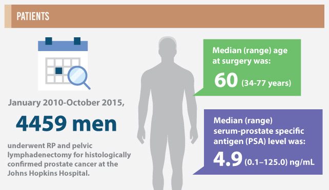
Infographic: Partin Tables in the Contemporary Era

The Partin Tables in the Contemporary Era: Infographic to accompany the May 2017 Article of the Month

Read the full article

Download Slides of this infographic:

PDF
PowerPoint

See more infographics


Article of the Week: Silicone renal models and complex tumour resections prior to RALPN

Every week the Editor-in-Chief selects an Article of the Week from the current issue of BJUI. The abstract is reproduced below and you can click on the button to read the full article, which is freely available to all readers for at least 30 days from the time of this post.

In addition to the article itself, there is an accompanying editorial written by a prominent member of the urological community. This blog is intended to provoke comment and discussion and we invite you to use the comment tools at the bottom of each post to join the conversation.

Finally, the third post under the Article of the Week heading on the homepage will consist of additional material or media. This week we feature a video discussing the paper.

If you only have time to read one article this week, it should be this one.

Utility of patient-specific silicone renal models for planning and rehearsal of complex tumour resections prior to robot-assisted laparoscopic partial nephrectomy

Friedrich-Carl von Rundstedt*,, Jason M. Scovell*, Smriti Agrawal, Jacques Zaneveld§ and Richard E. Link*,,**


*Scott Department of Urology, Baylor College of Medicine, Houston, TX, USA, Department of Urology, Jena University Hospital, Friedrich-Schiller University, Jena, Germany, Department of Molecular and Human Genetics, Baylor College of Medicine, §Lazarus 3D LLC, Dan L. Duncan Comprehensive Cancer Center, Baylor College of Medicine, and **Center for Reproductive Medicine, Baylor College of Medicine, Houston, TX, USA


Read the full article

How to Cite this Article

von Rundstedt, F.-C., Scovell, J. M., Agrawal, S., Zaneveld, J. and Link, R. E. (2017), Utility of patient-specific silicone renal models for planning and rehearsal of complex tumour resections prior to robot-assisted laparoscopic partial nephrectomy. BJU International, 119: 598–604. doi: 10.1111/bju.13712

Abstract

Objective

To describe our experience using patient-specific tissue-like kidney models created with advanced three-dimensional (3D)-printing technology for preoperative planning and surgical rehearsal prior to robot-assisted laparoscopic partial nephrectomy (RALPN).

Patients and Methods

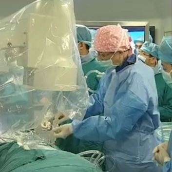
A feasibility study of 10 patients with solid renal masses who underwent RALPN after preoperative rehearsal using 3D-print kidney models. A single surgeon performed all surgical rehearsals and procedures. Using standard preoperative imaging and 3D reconstruction, we generated pre-surgical models using a silicone-based material. All surgical rehearsals were performed using the da Vinci® robotic system (Intuitive Surgical Inc., Sunnyvale, CA, USA) before the actual procedure. To determine construct validity, we compared resection times between the model and actual tumour in a patient-specific manner. Using 3D laser scanning in the operating room, we quantified and compared the shape and tumour volume resected for each model and patient tumour.

aotw-apr-results-5

Results

We generated patient-specific models for 10 patients with complex tumour anatomy. R.E.N.A.L. nephrometry scores were between 7 and 11, with a mean maximal tumour diameter of 40.6 mm. The mean resection times between model and patient (6:58 vs 8:22 min, P = 0.162) and tumour volumes between the computer model, excised model, and excised tumour (38.88 vs 38.50 vs 41.79 mm3, P = 0.98) were not significantly different.

Conclusions

We have developed a patient-specific pre-surgical simulation protocol for RALPN. We demonstrated construct validity and provided accurate representation of enucleation time and resected tissue volume. This simulation platform can assist in surgical decision-making, provide preoperative rehearsals, and improve surgical training.

Read more articles of the week

Editorial: Touching the future – 3D printing facilitates preoperative planning, realistic simulation and enhanced precision in RALPN

Practice taking that match-winning penalty knowing which way the keeper will dive, or take that last putt knowing the lie of the green; it would be very handy wouldn’t it?

Virtual reality (VR), augmented reality (AR), computer-generated images (CGI), and stereotactic overlay, have all been documented as adjuncts in enhancing operative patient care through planning, simulation and increased precision. But what if you could actually handle the specimen and practice operating on a model to refine operative technique before the definitive procedure? With three-dimensional (3D) printing this has now become a reality.

The work of von Rundstedt et al. [1] has the potential to transform surgical planning, operative accuracy, and training, with the development of a life-like kidney and tumour model. Their paper validates a patient-specific simulation protocol to assist in surgical decision-making through operative rehearsal. They assessed the benefits of 10 patient-specific 3D renal models for preoperative planning using tissue-like silicone, and performed model tumour excision with the robot before actual robot-assisted laparoscopic partial nephrectomy (RALPN). Nephrometry scores for tumours ranged from 7 to 11, with a relatively large mean maximal tumour diameter of 40.6 mm. In validating the model the investigators compared resection times between the model and patient (6.61 vs 7.93 min, P = 0.16) and tumour volumes between computer model, excised model, and excised tumour (38.88 vs 38.50 vs 41.79 mm3, P = 0.98), showing no significant differences.

The key principles in nephron-sparing surgery are adequate oncological excision, whilst preserving maximal renal parenchyma, with minimal ischaemia time, and avoiding complications. RALPN is challenging for complex tumours, with an extended learning curve, due partly to limitations in accurate surgical planning and surgical technique. Key anatomical considerations for planning including kidney orientation, tumour position and depth, and locality of adjacent anatomical structures (vessels, collecting system), are difficult to appreciate on conventional two-dimensional axial imaging platforms; with variance in imaging and model-planned approaches clearly noted in previous studies [2].

The development of nephrometry scores have been designed to predict surgical complexity along with various simulation and modelling reconstructions to aid excisional techniques. Several other surgical specialties (orthopaedics, maxillofacial and craniofacial surgery, neurosurgery, plastic surgery [3]) have used 3D-printing technology for organ/lesion modelling or to produce accurate imaging-based prostheses. In the era of minimally invasive surgery and personalised medicine, 3D printing can be a powerful tool for uro-oncologists to better understand individual tumour characteristics and anatomical variations.

Currently there is limited published data on 3D renal tumour printing. This paper [1] represents the first model and validation of its kind. Previous studies of 3D-printed kidneys with renal masses have been reported but limited to depictions of anatomical visualisation of arteries, collecting system and the tumour itself [4, 5]. This is the first time that a model not only provides a 3D representation of the tumour anatomy but also allows high-fidelity simulated excision. Construct validity of these reproductions has been assessed and demonstrate a striking similarity in tumour volume, morphology, and resection time, the main limitations of previous studies [4].

Preoperative planning has never achieved this accuracy before. Operative rehearsal significantly altered the ultimate approach to tumour excision in several cases as noted by the authors. In addition, 3D printing represents a breakthrough in surgical training as it offers a great opportunity, especially in facilities were wet laboratories are not available.

With the small sample assessed and the subjective nature of the surgical technique modification between model and tumour excision, early generalisation may not be appropriate. Other limitations include the models inability to replicate viable orthotopic anatomy such as adjacent organs, dissection planes, perinephric fat thickness/adherence, and bleeding; while also excluding the renorrhaphy component of the procedure. Additionally, production costs and 3D printer access may be an initial deterrent to widespread use of this technique; however, it does address the lack of tactile feeling in AR or VR, avoids the specialised facilities required by animal or cadaveric models, can readily be accurately reproduced, and most importantly provide an accurate anatomical representation of the individual patient.

This is an important and interesting paper as it presents and validates a novel model with extirpative technique in a prospective manner. It provides a life-like model useful for patient education, procedural practice with realistic simulation, an accurate training platform, and is the easiest to access given current technology. Clinical trials are needed to confirm how 3D modelling is ultimately useful in: i) improving patient education, ii) enhancing surgical training, and iii) conferring superior clinical outcomes. Evolution of 3D printers and shrinking production costs will eventually contribute to the widespread usage of this technology.

Further development will provide functional models that replicate not only macroscopic structures but elements such as the collecting system, segmental vessels, and bleeding parenchyma. However, it may be that high-fidelity VR simulators or CGI that can generate patient-specific graphics or even provide an intraoperative stereotactic 3D overlay to guide tumour excision may eventually supersede 3D modelling. Urology has entered the 3D printing era. This study [1] shows that 3D printing is both a feasible and useful technique that may enhance current practice, while providing an improved training platform. The future is here today.

Read the full article

How to Cite this Article

von Rundstedt, F.-C., Scovell, J. M., Agrawal, S., Zaneveld, J. and Link, R. E. (2017), Utility of patient-specific silicone renal models for planning and rehearsal of complex tumour resections prior to robot-assisted laparoscopic partial nephrectomy. BJU International, 119: 598–604. doi: 10.1111/bju.13712

Nicolo de Luyk, Benjamin Namdarian* and Benjamin Challacombe*
*Department of Urology, Guys and St Thomas Hospit als NHS Foundation Trust and Kings College, London, UK and Department of Urology, University Hospital of VeronaVerona, Italy


References


2 Wake N , Rude T, Kang SK et al. 3D printed renal cancer models derived from MRI data: application in pre-surgical planning. Abdom Radiol (NY) 2017; [Epub ahead of print]. doi: 10.1007/s00261-016-1022-2


3 LiJ, Chen M, Fan X, Zhou H. Recent advances in bioprinting techniques: approaches, applications and future prospects. J Transl Med 2016; 14: 271. doi:10.1186/s12967-016-1028-0


4 Silberstein JL, Maddox MM, Dorsey P, Feibus A, Thomas R, Lee BRPhysical models of renal malignancies using standard cross-sectional imaging and 3- dimensional printers: a pilot study. Urology 2014; 84: 26872


5 Bernhard JC, Isotani S, Matsugasumi T et al. Personalized 3D printed model of kidney and tumor anatomy: a useful tool for patient education. World J Urol 2016; 34: 33745


Video: Silicone renal models and complex tumour resections prior to RALPN

Utility of patient-specific silicone renal models for planning and rehearsal of complex tumour resections prior to robot-assisted laparoscopic partial nephrectomy

Read the full article

Abstract

Objective

To describe our experience using patient-specific tissue-like kidney models created with advanced three-dimensional (3D)-printing technology for preoperative planning and surgical rehearsal prior to robot-assisted laparoscopic partial nephrectomy (RALPN).

Patients and Methods

A feasibility study of 10 patients with solid renal masses who underwent RALPN after preoperative rehearsal using 3D-print kidney models. A single surgeon performed all surgical rehearsals and procedures. Using standard preoperative imaging and 3D reconstruction, we generated pre-surgical models using a silicone-based material. All surgical rehearsals were performed using the da Vinci® robotic system (Intuitive Surgical Inc., Sunnyvale, CA, USA) before the actual procedure. To determine construct validity, we compared resection times between the model and actual tumour in a patient-specific manner. Using 3D laser scanning in the operating room, we quantified and compared the shape and tumour volume resected for each model and patient tumour.

Results

We generated patient-specific models for 10 patients with complex tumour anatomy. R.E.N.A.L. nephrometry scores were between 7 and 11, with a mean maximal tumour diameter of 40.6 mm. The mean resection times between model and patient (6:58 vs 8:22 min, P = 0.162) and tumour volumes between the computer model, excised model, and excised tumour (38.88 vs 38.50 vs 41.79 mm3, P = 0.98) were not significantly different.

Conclusions

We have developed a patient-specific pre-surgical simulation protocol for RALPN. We demonstrated construct validity and provided accurate representation of enucleation time and resected tissue volume. This simulation platform can assist in surgical decision-making, provide preoperative rehearsals, and improve surgical training.

View more videos

Article of the Week: Comparing FG, USG and CG for renal access in mini-PCNL

Every week the Editor-in-Chief selects an Article of the Week from the current issue of BJUI. The abstract is reproduced below and you can click on the button to read the full article, which is freely available to all readers for at least 30 days from the time of this post.

In addition to the article itself, there is an accompanying editorial written by a prominent member of the urological community. This blog is intended to provoke comment and discussion and we invite you to use the comment tools at the bottom of each post to join the conversation.

Finally, the third post under the Article of the Week heading on the homepage will consist of additional material or media. This week we feature a video discussing the paper.

If you only have time to read one article this week, it should be this one.

A prospective and randomised trial comparing fluoroscopic, total ultrasonographic, and combined guidance for renal access in mini-percutaneous nephrolithotomy

Wei Zhu*, Jiasheng Li*, Jian Yuan*, Yongda Liu*, Shaw P.Wan*, Guanzhao Liu*† Wenzhong Chen*, Wenqi Wu*, Jintai Luo*, Dongliang Zhong*, Defeng Qi*, Ming
Lei*, Wen Zhong*, Ze Zhang*, Zhaohui He*, Zhijian Zhao*, Suilin Lu*, Yuji Wu*
and Guohua Zeng*


*Department of Urology, Minimally Invasive Surgery Center, The First Afliated Hospital of Guangzhou Medical University, and Guangdong Key Laboratory of Urology, Guangzhou, Guangdong, China


Read the full article

Abstract

Objective

To compare the safety and efficacy of fluoroscopic guidance (FG), total ultrasonographic guidance (USG), and combined ultrasonographic and fluoroscopic guidance (CG) for percutaneous renal access in mini-percutaneous nephrolithotomy (mini-PCNL).

Patients and methods

The present study was conducted between July 2014 and May 2015 as a prospective randomised trial at the First Affiliated Hospital of Guangzhou Medical University. In all, 450 consecutive patients with renal stones of >2 cm were randomised to undergo FG, USG, or CG mini-PCNL (150 patients for each group). The primary endpoints were the stone-free rate (SFR) and blood loss (haemoglobin decrease during the operation and transfusion rate). Secondary endpoints included access failure rate, operating time, and complications. S.T.O.N.E. score was used to document the complexity of the renal stones. The study was registered at https://clinicaltrials.gov/ (NCT02266381).

apr-saotw-4

Results

The three groups had similar baseline characteristics. With S.T.O.N.E. scores of 5–6 or 9–13, the SFRs were comparable between the three groups. For S.T.O.N.E. scores of 7–8, FG and CG achieved significantly better SFRs than USG (one-session SFR 85.1% vs 88.5% vs 66.7%, P = 0.006; overall SFR at 3 months postoperatively 89.4% vs 90.2% vs 69.8%, P = 0.002). Multiple-tracts mini-PCNL was used more frequently in the FG and CG groups than in the USG group (20.7% vs 17.1% vs 9.5%, P = 0.028). The mean total radiation exposure time was significantly greater for FG than for CG (47.5 vs 17.9 s, P < 0.001). The USG had zero radiation exposure. There was no significant difference in the haemoglobin decrease, transfusion rate, access failure rate, operating time, nephrostomy drainage time, and hospital stay among the groups. The overall operative complication rates using the Clavien–Dindo grading system were similar between the groups.

Conclusions

Mini-PCNL under USG is as safe and effective as FG or CG in the treatment of simple kidney stones (S.T.O.N.E. scores 5–6) but with no radiation exposure. FG or CG is more effective for patients with S.T.O.N.E. scores of 7–8, where multiple percutaneous tracts may be necessary.

Read more articles of the week

Editorial: Renal access during PCNL: increasing value of USG for a safer and successful procedure

Renal access to the pelvicalyceal system is the initial but highly important and crucial step of percutaneous nephrolithotomy (PCNL), which can significantly affect the final outcome of the procedure. Although the puncture of the kidney and subsequently dilatation of the tract has been commonly performed under fluoroscopic guidance [1]; renal access can also be established under ultrasonographic guidance (USG) with or without fluoroscopy.

To give a further insight into the role of both methods; in a prospective and randomised study published in this issue of the BJUI, Zhu et al. [2] have compared the safety and efficacy of fluoroscopic (FG), total ultrasonographic (USG), and combined ultrasonographic and fluoroscopic guidance (CG) for percutaneous renal access during mini-PCNL (mini-PCNL). In all, 450 consecutive patients with renal stones of >2 cm were randomised to undergo three different approaches during mini-PCNL. In addition to the stone-free rate (SFR) and blood loss as primary endpoints; access failure rate, operative time and complications were also evaluated. The S.T.O.N.E. [stone size (S), tract length (T), obstruction (O), number of involved calices (N), and essence or stone density (E)] scoring system was used for stone assessment [3] and the scores were further categorised into three grades (5–6, 7–8 and 9–13) for comparison.

While the overall operative complication rates, using the Clavien–Dindo grading system, were similar between the three groups; colonic injury treated with a temporary colostomy occurred in one case in the CG group. Although the SFRs were similar between the groups with S.T.O.N.E. scores of 5–6 and 9–13; the FG and CG approaches achieved significantly better SFRs than USG in patients with scores of 7–8, (P = 0.006). Multiple-tracts PCNL were used more frequently in the FG and CG group than USG group (P = 0.028). While the access failure rate was similar in the groups, the mean access time was longer in the CG group than in the FG and USG groups (P = 0.003). However, the mean total radiation exposure time was significantly greater for FG than for CG (47.5 vs 17.9 s, P < 0.001). The USG had zero radiation exposure. The operative time, hospital stay, nephrostomy drainage time, and the changes in the haemoglobin and creatinine levels were all similar in the three groups. The authors [1] concluded that mini-PCNL under total USG is as safe and effective as FG or CG in the treatment of simple kidney stones (S.T.O.N.E. scores 5–6) with no risk of radiation exposure. FG or CG is more effective for patients with S.T.O.N.E. scores of 7–8 where multiple percutaneous tracts may be necessary.

Percutaneous nephrolithotomy is now the preferred treatment method for larger stones (>2 cm) with successful outcomes. However, despite the high SFR obtained in a single session this approach can be associated with some severe complications such as bleeding, organ perforation, and sepsis. Such complications could be encountered during all steps of PCNL among which renal access seems to be the most critical one [4]. An appropriate puncture aiming a direct path from the skin through the papilla of the desired calyx of the kidney is of paramount importance to limit the above mentioned complications. Such an access to the renal collecting system can be established by either FG and/or USG. Although FG has been used commonly in the past; increasing experience in US applications has enabled endourologists to use this approach more often with some certain advantages in preventing renal puncture-related complications. When compared with FG, use of USG in establishing an access under vision allows the surgeon to identify the kidney pelvicalyceal system as well as the surrounding organs in a precise manner [5], with the benefit of minimising the risk of injury to such organs. Moreover, in addition to being free of ionising radiation; USG results in fewer punctures, has shorter operating times, and avoids contrast-related complications [1, 2]. Apart from helping to identify non-opaque residual stones at the end of the procedure; colour Doppler US can be used as a tool to localise the intrarenal arteries and avoid their puncture. However, the use of USG is an operator-dependent procedure requiring sufficient experience before routine performance and it may not be as efficient in the extremely obese patient and patients without hydronephrosis.

For the use of USG access in clinical practice, Agarwal et al. [5] reported a shorter mean time for successful puncture and significantly lower radiation exposure, yielding complete stone clearance with no substantial morbidity when compared with the FG technique. USG access was found also to increase puncture accuracy to a certain extent with a 96.5% SFR in another trial [6].

In conclusion, each of these techniques mentioned above have their own advantages and disadvantages. Despite its high success rate, radiation exposure and risk of multiple punctures are the main risks of the FG approach. USG renal access in experienced hands can produce high success rates following an appropriate puncture, lower risk of radiation exposure, and the ability to monitor all organs in the path of the puncture [7]. Depending on the surgeon’s experience, patient and stone-related factors, as well as the technical infrastructure, each approach may be used either alone or in combination for a complication-free and successful procedure. However, taking the above mentioned advantages of USG access into account, it is clear that all young urologist need to increase their experience in USG puncture to use it in appropriate cases (children, pregnant cases, dilated kidneys etc.) to lower the radiation risk and shorten the procedural duration.

Read the full article
Kemal Sarica, Professor of Urology, Chief


Department of Urology, Health Sciences University, Dr Lut Kirdar Kartal Research and Training Hospital, Istanbul, Turkey


References


1 Michel MS, Trojan L, Rassweiler JJ. Complications in percutaneous nephrolithotomy. Eur Urol 2007; 51: 899906


3 Okhunov Z, Friedlander JI, George AK et al. S.T.O.N.E. nephrolithometry: novel surgical classication system for kidney calculi. Urology 2013; 81: 115460


4 Aslam MZ, Thwaini A, Duggan B et al. Urologists versus radiologists made PCNL tracts: the UK experience. Urol Res 2011; 39: 21721


5 Agarwal M, Agrawal MS, Jaiswal A, Kumar D, Yadav H, Lavania PSafety and efcacy of ultrasonography as an adjunct to uoroscopy for renal access in percutaneous nephrolithotomy. BJU Int 2011; 108: 13469


6 BasiriA, Ziaee AM, Kianian HR, Mehrabi S, Ka rami H, Moghaddam SM. Ultrasonographic versus u oroscopic access for percutaneounephrolithotomy, a randomized clinical trial. J Enodourol 2008; 22: 28 14


7 Osman M, Wendt-Nordahl G, Heger K, Michel MS, Alken P, Knoll TPercutaneous nephrolithotomy with ultrasonography-guided renal access: experience from over 300 cases. BJU Int 2005; 96: 8758


Video: Comparing FG, USG and CG for renal access in mini-PCNL

A prospective and randomised trial comparing fluoroscopic, total ultrasonographic, and combined guidance for renal access in mini-percutaneous nephrolithotomy

Read the full article

Abstract

Objective

To compare the safety and efficacy of fluoroscopic guidance (FG), total ultrasonographic guidance (USG), and combined ultrasonographic and fluoroscopic guidance (CG) for percutaneous renal access in mini-percutaneous nephrolithotomy (mini-PCNL).

Patients and methods

The present study was conducted between July 2014 and May 2015 as a prospective randomised trial at the First Affiliated Hospital of Guangzhou Medical University. In all, 450 consecutive patients with renal stones of >2 cm were randomised to undergo FG, USG, or CG mini-PCNL (150 patients for each group). The primary endpoints were the stone-free rate (SFR) and blood loss (haemoglobin decrease during the operation and transfusion rate). Secondary endpoints included access failure rate, operating time, and complications. S.T.O.N.E. score was used to document the complexity of the renal stones. The study was registered at https://clinicaltrials.gov/ (NCT02266381).

Results

The three groups had similar baseline characteristics. With S.T.O.N.E. scores of 5–6 or 9–13, the SFRs were comparable between the three groups. For S.T.O.N.E. scores of 7–8, FG and CG achieved significantly better SFRs than USG (one-session SFR 85.1% vs 88.5% vs 66.7%, P = 0.006; overall SFR at 3 months postoperatively 89.4% vs 90.2% vs 69.8%, P = 0.002). Multiple-tracts mini-PCNL was used more frequently in the FG and CG groups than in the USG group (20.7% vs 17.1% vs 9.5%, P = 0.028). The mean total radiation exposure time was significantly greater for FG than for CG (47.5 vs 17.9 s, P < 0.001). The USG had zero radiation exposure. There was no significant difference in the haemoglobin decrease, transfusion rate, access failure rate, operating time, nephrostomy drainage time, and hospital stay among the groups. The overall operative complication rates using the Clavien–Dindo grading system were similar between the groups.

Conclusions

Mini-PCNL under USG is as safe and effective as FG or CG in the treatment of simple kidney stones (S.T.O.N.E. scores 5–6) but with no radiation exposure. FG or CG is more effective for patients with S.T.O.N.E. scores of 7–8, where multiple percutaneous tracts may be necessary.

View more videos

Article of the Week: Safety, reliability and accuracy of small renal tumour biopsies: results from a multi-institution registry

Every week the Editor-in-Chief selects an Article of the Week from the current issue of BJUI. The abstract is reproduced below and you can click on the button to read the full article, which is freely available to all readers for at least 30 days from the time of this post.

In addition to the article itself, there is an accompanying editorial written by a prominent member of the urological community. This blog is intended to provoke comment and discussion and we invite you to use the comment tools at the bottom of each post to join the conversation.

If you only have time to read one article this week, it should be this one.

Safety, reliability and accuracy of small renal tumour biopsies: results from a multi-institution registry

Patrick O. Richard*,, Michael A. S. Jewett*, Simon Tanguay, Olli Saarela§, Zhihui Amy Liu§, Frederic Pouliot, Anil Kapoor**, Ricardo Rendon†† and Antonio Finelli*


*Departments of Surgery and Surgical Oncology, Princess Margaret Cancer Centre, University Health Network and the University of Toronto, Toronto, ON, Canada, Centre Hospitalier Universitaire de Sherbrooke, Universite de Sherbrooke, Sherbrooke, QC, Canada, Department of Surgery, Division of Urology, McGill University Health Center, McGill University, Montreal§Dalla Lana School of Public Health, University of Toronto, Universite Laval, Centre de Recherche du Centre Hospitalier Universitaire de Quebec, Quebec **Department of Surgery, Division of Urology, McMaster University, Hamilton and ††QEII Health Sciences Centre, Department of Urology, Dalhousie University, Halifax, NS, Canada


Read the full article

Abstract

Objective

To validate, in a multi-institution review, the safety, accuracy and reliability of renal tumour biopsy (RTB) and its role in decreasing unnecessary treatment.

Materials and Methods

We conducted a multi-institution retrospective study of patients who underwent RTB to characterize a small renal mass (SRM) between 2011 and May 2015. Patients were identified using the prospectively maintained Canadian Kidney Cancer information system. Diagnostic and concordance rates were presented using proportions, whereas factors associated with a diagnostic RTB were identified using a logistic regression model.

scasca

Results

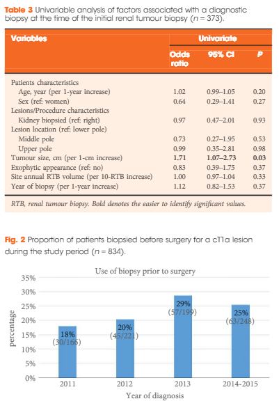
Of the 373 biopsied SRMs, the initial biopsy was diagnostic in 87% of cases. Of the 47 non-diagnostic biopsies, 15 had a repeat biopsy of which, 80% were diagnostic. When both were combined, therefore, a diagnosis was obtained in 91% of SRMs. Of these, 18% were benign. Size was the only factor found to be associated with achieving a diagnostic biopsy. RTB histology and nuclear grade (high or low) were found to be highly concordant with surgical pathology (86 and 81%, respectively). Of the discordant tumours (n = 16), all were upgraded from low to high grade on surgical pathology. Adverse events were rare (<1% of cases).

Conclusion

The present multi-institution study confirms that RTB of SRMs is safe, accurate and reliable across institutions, while decreasing unnecessary treatment. Given our findings, RTBs may be a helpful tool with which to triage SRMs and guide appropriate management.

Read more articles of the week

Editorial: Renal tumour biopsy: let’s talk about it

There has been a marked increase in the incidental diagnosis of small renal masses (SRMs), resulting in overtreatment of benign and indolent lesions. Renal tumour biopsy (RTB) has received increasing attention as a potential tool to help reduce this overtreatment, with single-institution studies reporting good safety, accuracy, and reliability. One of the purposes of paper by Richard et al. [1], appearing in this issue of BJUI, was to address whether these results of RTB were generalisable across multiple institutions. They evaluated 373 RTBs from 12 centres, reporting an initial diagnostic rate of 87%, with 32% of non-diagnostic RTBs undergoing repeat biopsy, for a combined diagnostic rate of 91%. They reported concordance rates between RTB and surgical pathology of >80% and a RTB complication rate of <1%. The generalisability of these impressive RTB results remains unclear because they were unable to report the numbers of RTB per centre (beyond ‘at least one’) and results were likely driven by a few high-volume centres.

The analysis is not without limitations. The negative predicative value (NPV) of RTB could not be assessed because there was no surgical specimen to confirm a benign RTB diagnosis. A meta-analysis by Patel et al. [2] raised concerns about a non-diagnostic or negative RTB. Of the 14% of patients with a non-diagnostic biopsy, 90% of those subsequently undergoing surgery were found to have cancer. Among patients having surgery, 37% with a negative biopsy who underwent surgical extirpation were found to have cancer on final pathology (NPV 63%). Another limitation of RTBs is that they tend to under grade tumours compared to surgical pathology. Even when using a simplified two-tiered grading system of low vs high grade tumours to improve concordance [3], 20% of patients with low-grade clear cell RCC (ccRCC) were upgraded to high-grade ccRCC at surgery. Concordance rates did not include non-diagnostic biopsies; nonetheless, their results support that a ‘good’ RTB can usually be trusted. Selection bias may have been a factor because patients who did not receive a RTB for a SRM were not included. It seems unlikely that reports of improved RTB outcomes would result in a change in guidelines to a ‘one size fits all’ policy recommending RTB in all patients with a SRM. RTB may not be feasible in some patients (anterior, hilar, cystic tumours) and may not always have potential to change clinical management, such as with a young healthy patient who is unwilling to accept any degree of uncertainty with a negative biopsy or an elderly patient with comorbidities who would not accept treatment regardless of RTB results. In this study [1], only ~25% of the patients who underwent surgery for a cT1a lesion had a RTB before surgery, possibly a reflection of the limitations of RTB, as well as some room for improvement.

Despite the limitations of RTB, the fact remains that many renal lesions are over treated, RTB outcomes are improving, and RTB may help guide clinical management. The authors [1] suggest that even a misclassified SRM could probably be managed conservatively over the short term. They recommend that even a benign RTB should be followed with serial imaging and that a repeat RTB should be considered for fast growing lesions. Perhaps the future of RCC diagnosis lies beyond the RTB and includes imaging innovations that can distinguish benign and malignant tumours and spare patients an unnecessary treatment, as well as an unnecessary biopsy. For example, Gorin et al. [4] showed that technetium-99m (99mTc)-sestamibi single-photon emission CT (SPECT)/CT could accurately distinguish renal oncocytomas and hybrid oncocytic/chromophobe tumours from other renal tumour histologies.

Current guidelines already acknowledge the potential role for RTB to guide clinical management in patients willing to accept the known limitations and who have an indeterminate SRM or are considering a range of treatment options such as active surveillance or ablation. We do not need a blanket guideline mandating upfront RTB for all. But we should at least talk about RTB with our patients with SRMs. We owe it to them to be aware of the potential benefits and limitations of RTB and include this in our discussion so they can be involved in the decision.

Read the full article
Haider Rahbar, and Craig Rogers


Vattikuti Urology Institute, Henry Ford Hospital, Detroit, MI, USA


References


3 Rioux-Leclercq N, Karakiewicz PI, Trinh QD et al. Prognostic ability of simplied nuclear grading of renal cell carcinoma. Cancer 2007; 109: 86874


© 2024 BJU International. All Rights Reserved.