Tag Archive for: Article of the Week

Posts

Article of the Week: Ureteric stent dwelling time: a risk factor for post-ureteroscopy sepsis

Every Week the Editor-in-Chief selects an Article of the Week from the current issue of BJUI. The abstract is reproduced below and you can click on the button to read the full article, which is freely available to all readers for at least 30 days from the time of this post.

In addition to the article itself, there is an accompanying editorial written by a prominent member of the urological community. This blog is intended to provoke comment and discussion and we invite you to use the comment tools at the bottom of each post to join the conversation.

If you only have time to read one article this week, it should be this one.

Ureteric stent dwelling time: a risk factor for post-ureteroscopy sepsis

Amihay Nevo*, Roy Mano*, Jack Baniel*† and David A. Lifshitz*


*Department of Urology, Rabin Medical Centre, Petach Tikva, Israel, and Sackler Faculty of Medicine, Tel Aviv University, Tel Aviv, Israel


Abstract

Objectives

To evaluate the association between stent dwelling time and sepsis after ureteroscopy, and identify risk factors for sepsis in this setting.

Patients and Methods

The prospectively collected database of a single institution was queried for all patients who underwent ureteroscopy for stone extraction between 2010 and 2016. Demographic, clinical, preoperative and operative data were collected. The primary study endpoint was sepsis within 48 h of ureteroscopy. Logistic regressions were performed to identify predictors of post-ureteroscopy sepsis in the ureteroscopy cohort and specifically in patients with prior stent insertion.

aotw-jul-2

Results

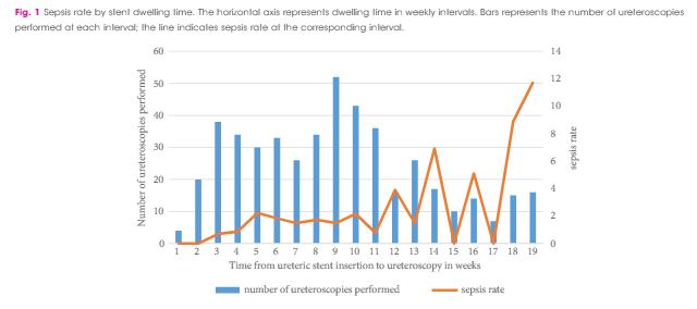
Between October 2010 and April 2016, 1 256 patients underwent ureteroscopy for stone extraction. Risk factors for sepsis included prior stent placement, female gender and Charlson comorbidity index. A total of 601 patients had a ureteric stent inserted before the operation and were included in the study cohort, in which the median age was 56 years, 90 patients were women (30%), and 97 patients were treated for positive preoperative urine cultures (16.1%). Postoperative sepsis, <48 h after surgery, occurred in eight (1.2%) non-stented patients and in 28 patients (4.7%) with prior stent insertion. Sepsis rates after stent dwelling times of 1, 2, 3 and >3 months were 1, 4.9, 5.5 and 9.2%, respectively. On multivariate analysis, stent dwelling time, stent insertion because of sepsis, and female gender were significantly associated with post-ureteroscopy sepsis in patients with prior stent placement.

Conclusions

Patients who undergo ureteroscopy after ureteric stent insertion have a higher risk of postoperative sepsis. Prolonged stent dwelling time, sepsis as an indication for stent insertion, and female gender are independent risk factors. Stent placement should be considered cautiously, and if inserted, ureteroscopy should be performed within 1 month.

Editorial: Pre-stenting and the risk of postoperative sepsis: a shorter dwell time is better

In this edition of the BJUI, Nevo et al. [1] report their retrospective review of 1256 patients who underwent ureteroscopy (URS)/flexible ureterorenoscopy (FURS) for stone disease and identified an overall sepsis rate of 2.8% within 48 h of surgery. About half of the cohort had a previously placed JJ stent, and the key finding of the study was the association between this and postoperative sepsis. In particular, the risk of sepsis in unstented patients was 1.2%, compared with 4.7% in those with a stent, such that overall, 80% of the patients who developed sepsis had a prior JJ stent in situ. Furthermore, this risk increased cumulatively with longer stent dwell-time before definitive surgery, increasing the risk of sepsis to 9.2% in patients who had a stent in situ for >3 months before the treatment of their stone.

Pre-stenting before ureteroscopic stone treatment can be for ‘absolute’ reasons (e.g. achieving ureteric drainage following presentation with an obstructed/infected kidney, or for an inaccessible ureter during the initial attempt at ureteroscopic stone treatment) or ‘relative’ reasons (e.g. emergency pain relief from ureteric colic if stone clearance cannot be offered immediately, or strategically inserted to allow passive ureteric dilatation to facilitate a subsequent definitive procedure). Data from the Clinical Research Office of the Endourological Society (CROES) URS Global Study showed that pre-stenting occurred in 36.4% of patients with rena l stones, and was associated with an increased stone-free rate (SFR), with a small but significant decrease in intraoperative complications. Pre-stenting was less common in ureteric stone management (11.9% of 8189 patients) and showed no difference in the SFR or complications, but was associated with a shorter length of stay [2]. Similarly, Jessen et al. [3] have reported that pre-stenting conferred a significant improvement in SFR for renal stones (83% vs 60%) but not for ureteric stones (94% vs 90%), although complications were reduced for pre-stented vs unstented patients in both renal stones (8.7% vs 19.4%) and ureteric stones (3.1% vs 10.7%) in that study. The advantage of pre-stenting appears to be greatest for larger stones: as a consequence of ureteric dilatation, and therefore improved renal access, in patients with stones >1 cm in whom a multi-phased approach was anticipated, Chu et al. [4] found that pre-stenting significantly reduced the operative time of the first URS, as well as the total operative time to stone clearance, including the need to re-operate at all in some patients.

The study in this edition of the BJUI [1] has shown that these advantages must be balanced against the increased risk of postoperative sepsis in patients with a pre-placed JJ stent, particularly in cases where the stent has been in situ for >1 month. The ability to identify patients who are at greater risk of post-URS/FURS infections is clearly useful: female gender, diabetes mellitus, ischaemic heart disease, an American Society of Anesthesiologists (ASA) score of ≥II, a large-volume stone burden, and same-session bilateral URS, have already been established as significant risk factors for postoperative infection [5, 6]. In addition, preoperative infections, either as a positive midstream specimen of urine (MSU) or previous sepsis also increase the risk of postoperative infectious complications. Specifically, Blackmur et al[6]reported that patients with a positive preoperative MSU were about five-times more likely to have postoperative urosepsis, even if they had been treated with an appropriate course of antibiotics before their stone surgery. Consistent with this, Youssef et al. [7] reported that the complication rate in patients undergoing URS after previous sepsis increased to 20% compared to 7% in matched non-septic controls, with an associated increased length of stay and duration of postoperative antibiotics.

In the present article [1], the overall sepsis rate was low at 2.8%, (and was comparable to recent CROES data that reported an overall prevalence of postoperative fever/UTI after URS or FURS of ≤2.2% [5]), but patients who had stents inserted for sepsis in the presence of an obstructing stone had a four-times higher postoperative infection rate when their stones were eventually treated than pre-stented patients without prior sepsis [1].

Taken together, these studies suggest that pre-stenting may have value in improving SFR, total operation time and reducing complication rates for large renal stones (i.e. >1 cm), but offers less advantage to stone clearance rates or complications in ureteric stones. Given the findings of increased risk of sepsis with stent-dwell time (increasing from 2.2% at 30 days, to 4.9% at 60 days, 5.5% at 90 days and 9.2% for >90 days) the authors recommendation ‘to keep stent dwelling time as short as possible’, is both practically beneficial to the patient and evidence-based. Furthermore, in addition to an awareness of the recognised risk factors for postoperative sepsis mentioned above, patients who have had stents inserted for prior sepsis, patients with positive preoperative MSU (even if treated), and patients with prolonged stent duration before definitive treatment should be counselled of the greater risk of postoperative sepsis, and watched cautiously in the early postoperative period.

In this study [1], patients with a stent in situ for <1 month had a similar risk of UTI to unstented patients. It would therefore seem reasonable to conclude that patients who have a stent inserted, especially for more ‘relative’ reasons to facilitate future surgery, should be scheduled for their definitive procedure within a month to achieve the benefits of pre-stenting, whilst minimising the potential for postoperative septic complications that Nevo et al. [1] have highlighted.

Daron Smith


Institute of Urology, University College Hospital, London, UK


References


Article of the Month: Identification of non-invasive biomarkers of UCPPS: findings from the MAPP Research Network

Every Month the Editor-in-Chief selects an Article of the Month from the current issue of BJUI. The abstract is reproduced below and you can click on the button to read the full article, which is freely available to all readers for at least 30 days from the time of this post.

In addition to the article itself, there is an accompanying editorial written by a prominent member of the urological community. This blog is intended to provoke comment and discussion and we invite you to use the comment tools at the bottom of each post to join the conversation.

If you only have time to read one article this week, it should be this one.

Identification of novel non-invasive biomarkers of urinary chronic pelvic pain syndrome: findings from the Multidisciplinary Approach to the Study of Chronic Pelvic Pain (MAPP) Research Network

Adelle Dagher*, Adam Curatolo*, Monisha Sachdev*, Alisa J. Stephens, Chris Mullins§, J. Richard Landis, Adrie van Bokhoven, Andrew El-Hayek*, John W. Froehlich**, Andrew C. Briscoe**, Roopali Roy*,††, Jiang Yang*,††, Michel A. Pontari‡‡David Zurakowski††§§, Richard S. Lee**††, Marsha A. Moses*,†† for the MAPP Research Network 
*Vascular Biology Program, Department of Surgery, Boston Childrens Hospital, Boston, MA,


Department of Biostatistics and Epidemiology, Perelman School of Medicine at the University of Pennsylvania, Philadelphia, PA, §National Institute of Diabetes and Digestive and Kidney Diseases, National Institutes of Health, Bethesda, MD, Department of Pathology, University of Colorado Anschutz Medical Campus, Aurora, CO, **Department of Urology, Boston Childrens Hospital, Boston, MA, ††Department of Surgery, Harvard Medical School, Boston, MA, ‡‡Lewis Katz School of Medicine at Temple University, Philadelphia, PA, and §§Department of Anesthesia, Boston Childrens Hospital, Boston, MA, USA


Read the full article

How to Cite

Dagher, A., Curatolo, A., Sachdev, M., Stephens, A. J., Mullins, C., Landis, J. R., van Bokhoven, A., El-Hayek, A., Froehlich, J. W., Briscoe, A. C., Roy, R., Yang, J., Pontari, M. A., Zurakowski, D., Lee, R. S., Moses, M. A. and the MAPP Research Network (2017), Identification of novel non-invasive biomarkers of urinary chronic pelvic pain syndrome: findings from the Multidisciplinary Approach to the Study of Chronic Pelvic Pain (MAPP) Research Network. BJU International, 120: 130–142. doi: 10.1111/bju.13832

Abstract

Objective

To examine a series of candidate markers for urological chronic pelvic pain syndrome (UCPPS), selected based on their proposed involvement in underlying biological processes so as to provide new insights into pathophysiology and suggest targets for expanded clinical and mechanistic studies.

Methods

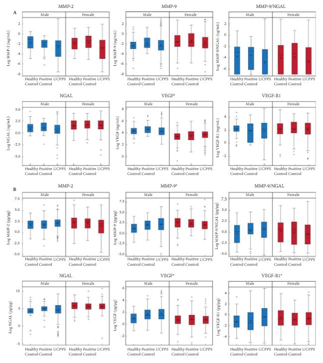
Baseline urine samples from Multidisciplinary Approach to the Study of Chronic Pelvic Pain (MAPP) Research Network study participants with UCPPS (n = 259), positive controls (PCs; chronic pain without pelvic pain, n = 107) and healthy controls (HCs, n = 125) were analysed for the presence of proteins that are suggested in the literature to be associated with UCPPS. Matrix metalloproteinase (MMP)-2, MMP-9, MMP-9/neutrophil gelatinase-associated lipocalin (NGAL) complex (also known as Lipocalin 2), vascular endothelial growth factor (VEGF), VEGF receptor 1 (VEGF-R1) and NGAL were assayed and quantitated using mono-specific enzyme-linked immunosorbent assays for each protein. Log-transformed concentration (pg/mL or ng/mL) and concentration normalized to total protein (pg/μg) values were compared among the UCPPS, PC and HC groups within sex using the Student’s t-test, with P values adjusted for multiple comparisons. Multivariable logistic regression and receiver-operating characteristic curves assessed the utility of the biomarkers in distinguishing participants with UCPPS and control participants. Associations of protein with symptom severity were assessed by linear regression.

jul-aotw-1

Results

Significantly higher normalized concentrations (pg/μg) of VEGF, VEGF-R1 and MMP-9 in men and VEGF concentration (pg/mL) in women were associated with UCPPS vs HC. These proteins provided only marginal discrimination between UCPPS participants and HCs. In men with UCCPS, pain severity was significantly positively associated with concentrations of MMP-9 and MMP-9/NGAL complex, and urinary severity was significantly positively associated with MMP-9, MMP-9/NGAL complex and VEGF-R1. In women with UCPPS, pain and urinary symptom severity were associated with increased normalized concentrations of MMP-9/NGAL complex, while pain severity alone was associated with increased normalized concentrations of VEGF, and urinary severity alone was associated with increased normalized concentrations of MMP-2. Pain severity in women with UCPPS was significantly positively associated with concentrations of all biomarkers except NGAL, and urinary severity with all concentrations except VEGF-R1.

Conclusion

Altered levels of MMP-9, MMP-9/NGAL complex and VEGF-R1 in men, and all biomarkers in women, were associated with clinical symptoms of UCPPS. None of the evaluated candidate markers usefully discriminated UCPPS patients from controls. Elevated VEGF, MMP-9 and VEGF-R1 levels in men and VEGF levels in women may provide potential new insights into the pathophysiology of UCPPS.

Article of the Week: Detection and oncological effect of CTC in patients with variant UCB histology treated with RC

Every week the Editor-in-Chief selects an Article of the Week from the current issue of BJUI. The abstract is reproduced below and you can click on the button to read the full article, which is freely available to all readers for at least 30 days from the time of this post.

In addition to the article itself, there is an accompanying editorial written by a prominent member of the urological community. This blog is intended to provoke comment and discussion and we invite you to use the comment tools at the bottom of each post to join the conversation.

If you only have time to read one article this week, it should be this one.

Detection and oncological effect of circulating tumour cells in patients with variant urothelial carcinoma histology treated with radical cystectomy

Armin Soave*, Sabine Riethdorf, Roland Dahlem*, Sarah Minner, Lars Weisbach*, Oliver Engel*, Margit Fisch*, Klaus Pantel† and Michael Rink*


*Department of Urology, Institute of Tumor Biology, and Department of Pathology, University Medical Center Hamburg-Eppendorf, Hamburg, Germany


Read the full article

How to Cite

Soave, A., Riethdorf, S., Dahlem, R., Minner, S., Weisbach, L., Engel, O., Fisch, M., Pantel, K. and Rink, M. (2017), Detection and oncological effect of circulating tumour cells in patients with variant urothelial carcinoma histology treated with radical cystectomy. BJU International, 119: 854–861. doi: 10.1111/bju.13782

Abstract

Objectives

To investigate for the presence of circulating tumour cells (CTC) in patients with variant urothelial carcinoma of the bladder (UCB) histology treated with radical cystectomy (RC), and to determine their impact on oncological outcomes.

Patients and methods

We prospectively collected data of 188 patients with UCB treated with RC without neoadjuvant chemotherapy. Pathological specimens were meticulously reviewed for pure and variant UCB histology. Preoperatively collected blood samples (7.5 mL) were analysed for CTC using the CellSearch® system (Janssen, Raritan, NJ, USA).

aotw-results-4

Results

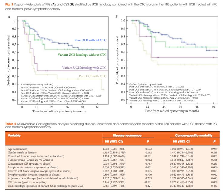
Variant UCB histology was found in 47 patients (25.0%), most frequently of squamous cell differentiation (16.5%). CTC were present in 30 patients (21.3%) and 12 patients (25.5%) with pure and variant UCB histology, respectively. At a median follow-up of 25 months, the presence of CTC and non-squamous cell differentiation were associated with reduced recurrence-free survival (RFS) and cancer-specific survival (pairwise P ≤ 0.016). Patients without CTC had better RFS, independent of UCB histology, than patients with CTC with any UCB histology (pairwise P < 0.05). In multivariable analyses, the presence of CTC, but not variant UCB histology, was an independent predictor for disease recurrence [hazard ratio (HR) 3.45; P < 0.001] and cancer-specific mortality (HR 2.62; P = 0.002).

Conclusion

CTC are detectable in about a quarter of patients with pure or variant UCB histology before RC, and represent an independent predictor for outcomes, when adjusting for histological subtype. In addition, our prospective data confirm the unfavourable influence of non-squamous cell-differentiated UCB on outcomes.

Editorial: Biomarkers for UCPPS: is there light at the end of the tunnel?

Management of urological disease begins with an accurate diagnosis. For this purpose, urologists have PSA and biopsy for prostate cancer, bacterial culture for UTI, CT scan for urolithiasis, cytology, cystoscopy and biopsies for bladder cancer, semen analysis for infertility and the list goes on. We have been able to use these accurate diagnostic markers to kill, maim, remove, fragment or burn our way to therapeutic success. Unfortunately, this has not been true in the case of urological chronic pelvic pain syndrome (UCPPS), where specific diagnoses and therapeutic strategies continue to elude us. Urologists managing patients with UCPPS need something concrete and quantifiable to move the field and improve patient care. A validated and reliable biomarker might fit the bill.

As Dagher et al. [1] clearly show in their study, this is not going to be as easy as we had hoped. The research group analysed urine samples for candidate biological markers from both patients with UCPPS and healthy controls recruited for the Multidisciplinary Approach to the Study of Chronic Pelvic Pain (MAPP) Research Network. The National Institutes of Health/National Institute of Diabetes and Digestive and Kidney Diseases (NIH/NIDDK) developed the MAPP Research Network to provide a comprehensive, multidisciplinary research approach for UCPPS [2, 3]. A major goal of this novel initiative is to better understand UCPPS pathophysiology allowing improved diagnoses and patient stratification that informs identification of therapeutic targets and ultimately improves clinical management.

In the present study, candidate biomarkers were initially selected based on their proposed involvement in underlying processes implicated in UCPPS. The authors showed marginal biomarker discrimination between patients with UCPPS and healthy control subjects. Furthermore, they observed provocative biomarker patterns correlating with pain and urinary symptom severity. None of these observations were conclusive enough, however, to allow a clinically applicable differentiation between patients with UCPPS and control subjects with no UCPPS symptoms. The patterns associated with pain and urinary severity were also not sufficiently robust to provide clinical direction. The underlying reason for these results is probably related to our recent understanding that UCPPS is not a clearly defined disease, but rather a complicated syndrome consisting of a confusing myriad of clinical conditions involving the bladder, prostate and/or pelvic floor and probably systemic contributions from other systems, including the nervous system. Indeed, the chronic pelvic pain and urinary symptoms appear as a result of interrelated, but variable, pathophysiology incorporating inflammatory, microbial, endocrine, neuromuscular, peripheral and CNS and even psychological pathways. The systemic characterization of UCPPS in the MAPP Network supports the idea that each patient with a UCPPS diagnosis represents an individual clinical picture and symptom profile. The promise of well-powered and carefully controlled biomarker research, such as described in the present study, will probably be realized when fully integrated into a systemic clinical profile using phenotypic insights (biological and symptom) still evolving from the MAPP Network and other studies. This is expected to yield a better understanding of specific mechanisms and symptom profiles operative in individual patients or patient subgroups. Identification of biomarker patterns that inform the clinical picture has the promise to allow us, in future, to make individualized diagnoses leading to individualized mechanistically (not only phenotypically) directed prognostic and therapeutic strategies. Success in such efforts will lead to UCPPS becoming less of a urological mystery. Then this enigmatic condition might join other urological diseases for which we have accurate diagnostic algorithms and successful therapeutic interventions.

The ongoing MAPP-2 Research Network’s Symptom Patterns Study is using state-of-the-art approaches to examine closely the interrelationships between the inflammatory, endocrine, microbial, neurological and psychological systems and how these interactions influence patients’ symptoms over a 2–3-year time period. This includes correlations with diverse biological markers over time. It is hoped that MAPP-2 will unravel the tangled web of these mechanistic pathways, allowing better diagnosis and ultimately, better therapy for our patients with UCPPS. That is our promise of the light at the end of a very long urological tunnel.

J. Curtis Nickel *


*Queens University, Kingston, ON, Canada and Urology, Canada Research Chair in Urologic Pain and Inammation, Queens University, Kingston, ON, Canada
 
Read the full article


References


Editorial: Detection and oncological effect of CTC in patients with variant UCB histology treated with RC

I read this article from Hamburg-Eppendorf with great interest [1]. The treatment of invasive urothelial carcinoma has not significantly progressed in the last 30 years, with survivals currently that are little changed since the first introduction of multi-drug platinum-based chemotherapy in the 1980s. Moreover, the broad application of chemotherapy, whether it is in the preoperative or postoperative domains, is associated with significant morbidity in this generally elderly population. As 60–80% of patients are cured by surgery alone, the broad use of chemotherapy in any setting results in unnecessary morbidity and occasionally mortality in some patients unnecessarily. Multiple patients have a permanent reduction in renal function when platinum is used in this setting. The decision to treat preoperatively is limited by inaccurate clinical staging and in the postoperative setting may be compromised by slow or incomplete surgical recovery.

The measurement of preoperative circulating tumour cells (CTC) provides us with a rational approach to more accurately select patients for neoadjuvant chemotherapy and would seem according to this article to independently predict disease recurrence, even when considering aggressive variant histologies. Examining Figure 2, one finds that even with variant histology, 60% of patients will not recur after cystectomy if they are CTC negative. The differences are even more profound in pure urothelial carcinoma, where the presence of detectable CTC decreases survival by 50%. The authors are to be congratulated for providing us with a potential rational methodology to determine the benefit from neoadjuvant chemotherapy in patients with bladder cancer prior to cystectomy. Next we should await the analysis of clinical trials stratified by CTC status.

Read the full article
Michael O. Koch, Chairman and Professor of Urology


Indiana Cancer Pavilion, Indiana University School of Medicine, Indianapolis, IN, USA


Reference


Article of the Week: Evaluation of Sig24, a 24-gene signature

Every week the Editor-in-Chief selects an Article of the Week from the current issue of BJUI. The abstract is reproduced below and you can click on the button to read the full article, which is freely available to all readers for at least 30 days from the time of this post.

In addition to the article itself, there is an accompanying editorial written by a prominent member of the urological community. This blog is intended to provoke comment and discussion and we invite you to use the comment tools at the bottom of each post to join the conversation.

Finally, the third post under the Article of the Week heading on the homepage will consist of additional material or media. This week we feature a video discussing the paper.

If you only have time to read one article this week, it should be this one.

Evaluation of a 24-gene signature for prognosis of metastatic events and prostate cancer-specific mortality

Kathryn L. Pellegrini*, Martin G. Sanda*, Dattatraya Patil*, Qi Long†‡§, MarıSantiago-Jimenez, Mandeep Takhar, Nicholas Erho, Kasra Youse, Elai DavicioniEric A. Klein**, Robert B. Jenkins††, R. Jeffrey Karnes‡‡ and Carlos S. Moreno§§§


*Department of Urology, Emory University School of Medicine, Winship Cancer Institute, Atlanta, GA‡ Department of Biostatistics and Epidemiology and Abramson Cancer Center, University of Pennsylvania Perelman School of Medicine, Philadelphia, PA, §Department of Biomedical Informatics, Emory University School of Medicine, Atlanta, GA, USA, GenomeDx Biosciences, Vancouver, BC, Canada, **Glickman Urological and Kidney Institute, Cleveland Clinic, Cleveland, OH, ††Department of Pathology and Laboratory Medicine, ‡‡Department of Urology, Mayo Clinic, Rochester, MN, and §§Department of Pathology and Laboratory Medicine, Emory University School of Medicine, Atlanta, GA, USA
Read the full article

How to Cite

Pellegrini, K. L., Sanda, M. G., Patil, D., Long, Q., Santiago-Jiménez, M., Takhar, M., Erho, N., Yousefi, K., Davicioni, E., Klein, E. A., Jenkins, R. B., Karnes, R. J. and Moreno, C. S. (2017), Evaluation of a 24-gene signature for prognosis of metastatic events and prostate cancer-specific mortality. BJU International, 119: 961–967. doi: 10.1111/bju.13779

Abstract

Objectives

To determine the prognostic potential of a 24-gene signature, Sig24, for identifying patients with prostate cancer who are at risk of developing metastases or of prostate cancer-specific mortality (PCSM) after radical prostatectomy (RP).

Patients and Methods

Sig24 scores were calculated from previously collected gene expression microarray data from the Cleveland Clinic and Mayo Clinic (I and II). The performance of Sig24 was determined using time-dependent c-index analysis, Cox proportional hazards regression and Kaplan–Meier survival analysis.

aotw-jun-3-results

Results

Higher Sig24 scores were significantly associated with higher pathological Gleason scores in all three cohorts. Analysis of the Mayo Clinic II cohort, which included time-to-event information, indicated that patients with high Sig24 scores also had a higher risk of developing metastasis (hazard ratio [HR] 3.78, 95% confidence interval [CI]: 1.96–7.29; P < 0.001) or of PCSM (HR 6.54, 95% CI: 2.16–19.83; P < 0.001).

Conclusions

The findings of the present study show the applicability of Sig24 for the prognosis of metastasis or PCSM after RP. Future studies investigating the combination of Sig24 with available prognostic tests may provide new approaches to improve risk stratification for patients with prostate cancer.

Editorial: Predicting outcome: role of gene signatures

Pathological assessments, such as Gleason grading, which is a strong clinical predictor of prostate cancer progression [1], have a role to play in predicting the outcome of a patient’s response to therapy. The addition of PSA and TNM staging are further used to inform appropriate treatment strategies in patients who are at low, intermediate and high risk of disease progression. Unfortunately, approximately 30% of men with intermediate-risk prostate cancer will fail to be cured by surgery or radiation therapy approaches. This is not surprising as primary prostate cancer represents a complex heterogeneous disease that is clearly not fully explained by the current clinical prognostic factors, and further molecular characterization is required. There is now significant emerging evidence that the molecular characterization of tumours is important to enable us to stratify prostate cancer patients by their response to primary therapy and to identify the next appropriate steps in their treatment pathway.

Long et al. [2] have identified gene signatures that can define different genomic subtypes of prostate cancer and are predictive of biochemical recurrence. Erho et al. [3] discovered and validated a 22-gene signature, which they termed a genomic classifier for the prediction of early metastasis after radical prostatectomy, while Penny et al. [4] have identified an mRNA expression signature of Gleason grade which is predictive of lethal prostate cancer.

Long et al. [2] identified a 24-gene signature, which they discovered through RNA sequencing analysis of 100 formalin-fixed paraffin-embedded prostatectomy samples. They went on to validate their findings in a publically available independent gene expression microarray dataset of 140 patients. This 24-gene signature forms the basis of the present study by Pellegrini et al. [5], who refer to this gene signature as Sig24. In their study, they firstly undertook to determine if Sig24 was associated with pathological Gleason score as a marker of tumour aggressiveness, and then if it had prognostic value for the identification of patients at risk of metastasis or prostate cancer-specific mortality after radical prostatectomy. The Gleason score association study was carried out using the data from three independent case–control sets, including 182 patients from the Cleveland Clinic and two cohorts (cohort I, n = 545; cohort II, n = 235) from the Mayo Clinic, for which gene expression analysis had previously been conducted by Genome Dx using the Affymetrix Human 1.0 ST Genechip platform. The Sig24 score was calculated for each patient and higher Gleason score was associated with significantly higher Sig24 scores. The association studies for metastatic disease and prostate cancer-specific mortality, however, were only carried out in the Mayo Clinic cohort II. For both clinical endpoints the Sig24 score combined with the clinical model outperformed the clinical model alone of PSA, Gleason score and tumour stage. For metastatic disease the area under the curve for the clinical model alone was 0.69 (0.62–0.77) compared with 0.73 (0.66–0.78) for the clinical model combined with Sig24. For the prostate cancer-specific mortality endpoint, the area under the curve for the clinical model alone was 0.69 (0.67–0.87) compared with 0.74 (0.63–0.85) for the clinical model combined with Sig24.

These gene signatures have significant potential for predicting the progression of disease rather than waiting for PSA relapse, which is currently used to identify disease recurrence, with interval to biochemical failure being the best univariate factor predicting prostate cancer mortality and overall survival [6]. This would allow the initiation of additional therapies before recurrence and a better outcome for the patient. This concept has been demonstrated in a study in which the use of the Decipher gene signature was shown to improve the identification of patients who could benefit from adjuvant radiotherapy and thus only these patients were targeted for therapy [7].

Incorporating these gene signatures in robust clinical assays and integrating them into clinical decision-making is the next essential step in order for these strategies to have an impact on patient outcomes.

Read the full article
Ronald W. Watson
UCD School of Medicine, University College Dublin, Dublin, Ireland

References

1 Gleason DF. Classication of prostatic carcinomas. Cancer Chemother
Rep 1966; 50: 1258


4 Penny KL, Sinnott JA, Fall K et al. mRNA expression signature of Gleason grade predicts lethal prostate cancer. J Clin Oncol 2011; 29:23916


6 Buyyounouski MK, Pickles T, Kestin LL, Allison R, Williams SGValidating the interval to biochemical failure for identication of potentially lethal prostate cancer. J Clin Oncol 2012; 30: 185763


Video: Evaluation of a 24-gene signature for prognosis of metastatic events and PCa-specific mortality

Evaluation of a 24-gene signature for prognosis of metastatic events and prostate cancer-specific mortality

Kathryn L. Pellegrini*, Martin G. Sanda*, Dattatraya Patil*, Qi Long†‡§, MarıSantiago-Jimenez, Mandeep Takhar, Nicholas Erho, Kasra Youse, Elai DavicioniEric A. Klein**, Robert B. Jenkins††, R. Jeffrey Karnes
‡‡ and Carlos S. Moreno§§§


*Department of Urology, Emory University School of Medicine, Winship Cancer Institute, Atlanta, GA‡ Department of Biostatistics and Epidemiology and Abramson Cancer Center, University of Pennsylvania Perelman School of Medicine, Philadelphia, PA, §Department of Biomedical Informatics, Emory University School of Medicine, Atlanta, GA, USA, GenomeDx Biosciences, Vancouver, BC, Canada, **Glickman Urological and Kidney Institute, Cleveland Clinic, Cleveland, OH, ††Department of Pathology and Laboratory Medicine, ‡‡Department of Urology, Mayo Clinic, Rochester, MN, and §§Department of Pathology and Laboratory Medicine, Emory University School of Medicine, Atlanta, GA, USA
Read the full article

How to Cite

Pellegrini, K. L., Sanda, M. G., Patil, D., Long, Q., Santiago-Jiménez, M., Takhar, M., Erho, N., Yousefi, K., Davicioni, E., Klein, E. A., Jenkins, R. B., Karnes, R. J. and Moreno, C. S. (2017), Evaluation of a 24-gene signature for prognosis of metastatic events and prostate cancer-specific mortality. BJU International, 119: 961–967. doi: 10.1111/bju.13779

Abstract

Objectives

To determine the prognostic potential of a 24-gene signature, Sig24, for identifying patients with prostate cancer who are at risk of developing metastases or of prostate cancer-specific mortality (PCSM) after radical prostatectomy (RP).

Patients and Methods

Sig24 scores were calculated from previously collected gene expression microarray data from the Cleveland Clinic and Mayo Clinic (I and II). The performance of Sig24 was determined using time-dependent c-index analysis, Cox proportional hazards regression and Kaplan–Meier survival analysis.

aotw-jun-3-results

Results

Higher Sig24 scores were significantly associated with higher pathological Gleason scores in all three cohorts. Analysis of the Mayo Clinic II cohort, which included time-to-event information, indicated that patients with high Sig24 scores also had a higher risk of developing metastasis (hazard ratio [HR] 3.78, 95% confidence interval [CI]: 1.96–7.29; P < 0.001) or of PCSM (HR 6.54, 95% CI: 2.16–19.83; P < 0.001).

Conclusions

The findings of the present study show the applicability of Sig24 for the prognosis of metastasis or PCSM after RP. Future studies investigating the combination of Sig24 with available prognostic tests may provide new approaches to improve risk stratification for patients with prostate cancer.

Article of the Week: Treatment of ED after RP using nerve grafts and end-to-side somatic-autonomic neurorraphy

Every week the Editor-in-Chief selects an Article of the Week from the current issue of BJUI. The abstract is reproduced below and you can click on the button to read the full article, which is freely available to all readers for at least 30 days from the time of this post.

In addition to the article itself, there is an accompanying editorial written by a prominent member of the urological community. This blog is intended to provoke comment and discussion and we invite you to use the comment tools at the bottom of each post to join the conversation.

If you only have time to read one article this week, it should be this one.

Long-term follow-up of treatment of erectile dysfunction after radical prostatectomy using nerve grafts and end-to-side somatic-autonomic neurorraphy: a new technique

Jose Carlos Souza Trindade*, Fausto Viterbo, Andre Petean TrindadeWagner Josen Favaro§ and Jose Carlos Souza Trindade-Filho*


*Department of Urology, † Divisions of Plastic Surgery,‡ Radiology, Botucatu School of Medicine, State University of Sao Paulo, Sao Paulo, and §Department of Anatomy, Faculty of Medical Sciences, University of Campinas, Campinas, Brazil


Read the full article

Abstract

Objective

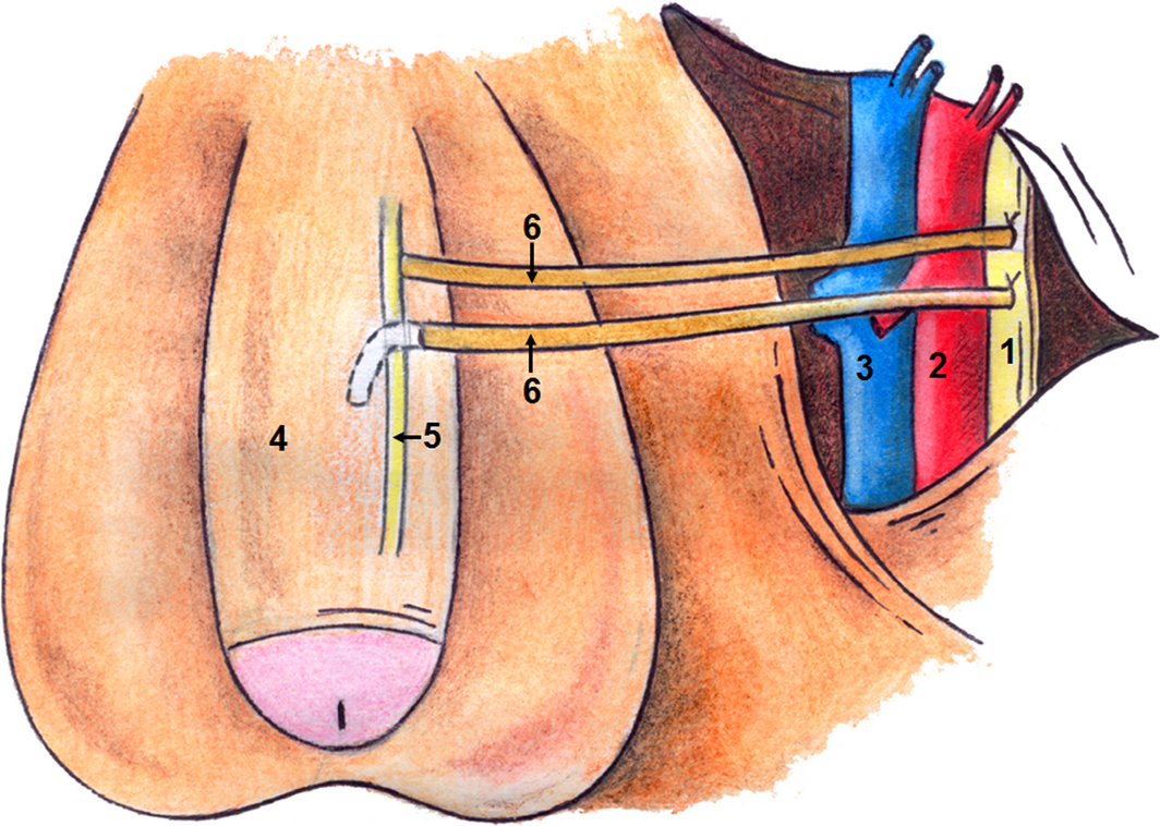
To study a novel penile re innervation technique using four sural nerve grafts and end-to-side neurorraphies connecting bilaterally the femoral nerve and the cavernous corpus and the femoral nerve and the dorsal penile nerves.

Patients and Methods

Ten patients (mean [± sd; range] age 60.3 [± 4.8; 54–68] years), who had undergone radical prostatectomy (RP) at least 2 years previously, underwent penile reinnervation in the present study. Four patients had undergone radiotherapy after RP. All patients reported satisfactory sexual activity prior to RP. The surgery involved bridging of the femoral nerve to the dorsal nerve of the penis and the inner part of the corpus cavernosum with sural nerve grafts and end-to-side neurorraphies. Patients were evaluated using the International Index of Erectile Function (IIEF) questionnaire and pharmaco-penile Doppler ultrasonography (PPDU) preoperatively and at 6, 12 and 18 months postoperatively, and using a Clinical Evolution of Erectile Function (CEEF) questionnaire, administered after 36 months.

Results

The IIEF scores showed improvements with regard to erectile dysfunction (ED), satisfaction with the VitalFlow and general satisfaction. Evaluation of PPDU velocities did not reveal any difference between the right and left sides or among the different time points. The introduction of nerve grafts neither caused fibrosis of the corpus cavernosum, nor reduced penile vascular flow. CEEF results showed that sexual intercourse began after a mean of 13.7 months with frequency of sexual intercourse varying from once daily to once monthly. Acute complications were minimal. The study was limited by the small number of cases.

Conclusions

A total of 60% of patients were able to achieve full penetration, on average, 13 months after reinnervation surgery. Patients previously submitted to radiotherapy had slower return of erectile function. We conclude that penile reinnervation surgery is a viable technique, with effective results, and could offer a new treatment method for ED after RP.

Read more articles of the week
© 2024 BJU International. All Rights Reserved.