Tag Archive for: Article of the Week

Posts

Video: Immortal-Time Bias in Urological Research

Estimating the effect of immortal-time bias in urological research: a case example of testosterone-replacement therapy


Abstract

Objective

To quantify the effect of immortal-time bias in an observational study examining the effect of cumulative testosterone exposure on mortality.

Patients and Methods

We used a population-based matched cohort study of men aged ≥66 years, newly treated with testosterone-replacement therapy (TRT), and matched-controls from 2007 to 2012 in Ontario, Canada to quantify the effects of immortal-time bias. We used generalised estimating equations to determine the association between cumulative TRT exposure and mortality. Results produced by models using time-fixed and time-varying exposures were compared. Further, we undertook a systematic review of PubMed to identify studies addressing immortal-time bias or time-varying exposures in the urological literature and qualitatively summated these.

Results

Among 10 311 TRT-exposed men and 28 029 controls, the use of a time-varying exposure resulted in the attenuation of treatment effects compared with an analysis that did not account for immortal-time bias. While both analyses showed a decreased risk of death for patients in the highest tertile of TRT exposure, the effect was overestimated when using a time-fixed analysis (adjusted hazard ratio [aHR] 0.56, 95% confidence interval [CI]: 0.52–0.61) when compared to a time-varying analysis (aHR 0.67, 95% CI: 0.62–0.73). Of the 1 241 studies employing survival analysis identified in the literature, nine manuscripts met criteria for inclusion. Of these, five used a time-varying analytical method. Each of these was a large, population-based retrospective cohort study assessing potential harms of pharmacological agents.

Conclusions

Where exposures vary over time, a time-varying exposure is necessary to draw meaningful conclusions. Failure to use a time-varying analysis will result in overestimation of a beneficial effect. However, time-varying exposures are uncommonly utilised among manuscripts published in prominent urological journals.

Article of the Week: Profiling microRNA from nephrectomy and biopsy specimens

Every Week the Editor-in-Chief selects an Article of the Week from the current issue of BJUI. The abstract is reproduced below and you can click on the button to read the full article, which is freely available to all readers for at least 30 days from the time of this post.

In addition to the article itself, there is an accompanying editorial written by a prominent member of the urological community. This blog is intended to provoke comment and discussion and we invite you to use the comment tools at the bottom of each post to join the conversation.

If you only have time to read one article this week, it should be this one.

Profiling microRNA from nephrectomy and biopsy specimens: predictors of progression and survival in clear cell renal cell carcinoma


Casey G. Kowalik*, Drew A. Palmer*, Travis B. Sullivan, Patrick A. TeebagyJohn M. Dugan, John A. Libertino*, Eric J. Burks, David Canes* and Kimberly M. Rieger-Christ


Departments of *Urology, Translational Research Ian C. Summerhayes Cell and Molecular Biology Laboratory, and Pathology, Lahey Hospital and Medical Center, Burlington, MA, USA


Read the full article

Abstract

Objective

To identify microRNA (miRNA) characteristic of metastatic clear cell renal cell carcinoma (ccRCC) and those indicative of cancer-specific survival (CSS) in nephrectomy and biopsy specimens. We also sought to determine if a miRNA panel could differentiate benign from ccRCC tissue.

Materials and Methods

RNA was isolated from nephrectomy and kidney biopsy specimens (n = 156 and n = 46, respectively). Samples were grouped: benign, non-progressive, and progressive ccRCC. MiRNAs were profiled by microarray and validated by quantitative reverse transcription-polymerase chain reaction. Biomarker signatures were developed to predict cancer status in nephrectomy and biopsy specimens. CSS was examined using Kaplan–Meier and Cox proportional hazards analyses.

Results

Microarray analysis revealed 20 differentially expressed miRNAs comparing non-progressive with progressive tumours. A biomarker signature validated in nephrectomy specimens had a sensitivity of 86.7% and a specificity of 92.9% for differentiating benign and ccRCC specimens. A second signature differentiated non-progressive vs progressive ccRCC with a sensitivity of 93.8% and a specificity of 83.3%. These biomarkers also discriminated cancer status in biopsy specimens. Levels of miR-10a-5p, -10b-5p, and -223-3p were associated with CSS.

Conclusion

This study identified miRNAs differentially expressed in ccRCC samples; as well as those correlating with CSS. Biomarkers identified in this study have the potential to identify patients who are likely to have progressive ccRCC, and although preliminary, these results may aid in differentiating aggressive and indolent ccRCC based on biopsy specimens.

Read more articles of the week

Editorial: The utility of microRNAs as biomarkers in predicting progression and survival in patients with clear-cell renal cell carcinoma

RCC constitutes a diverse group of malignancies, yet the clear-cell subtype comprises ~80% of all diagnosed RCC cases [1]. The widespread use of abdominal imaging and subsequent stage migration has resulted in improved RCC 5-year cancer-specific survival. However, the overall mortality of RCC remains largely unchanged [2] and one-third of the patients have metastatic disease at the time of presentation [3]. Accordingly, the ability to precisely predict patient outcome has become an increasingly significant question in the management of these patients with RCC.

Accruing evidence suggests that changes in various biomarkers and their consequent downstream pathways affect cancer initiation and progression. Therefore, accurate prediction of the outcome and prognosis after treatment is necessary [4]. MicroRNAs (miRNAs) are small non-coding RNA molecules that can have significant functions in tumorigenesis [5]. Because of their ability in post-transcriptional regulation of gene expression, tumour-specific genetic defects in miRNA biogenesis and production correlate with development of human cancers. Thus, the differential expression of specific miRNA signatures in different tumours might become an important tool to help in directing cancer diagnosis and treatment [5].

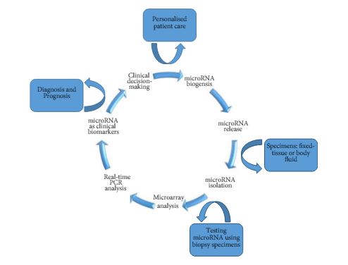
As such, Kowalik et al. [6] report on profiling miRNA to identify biomarker signatures predictive of clear-cell RCC (ccRCC) progression and survival. The authors used 202 formalin-fixed paraffin-embedded samples to isolate RNA from nephrectomy and biopsy specimens (n = 156 and n = 46, respectively) (Fig. 1).

Figure 1. Schematic diagram of miRNA-based biomarkers and potential utility in clinical decision-making approach for targeted therapy and RCC personalised treatment.

The primary analysis of their study [6] focused on the identification of miRNA signatures capable of differentiating between benign and ccRCC, as well as discerning those patients with a non-progressive ccRCC from a progressive clear-cell subtype. The secondary outcome examined the association of miRNA profiles discovered on cancer-specific survival.

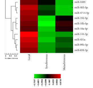
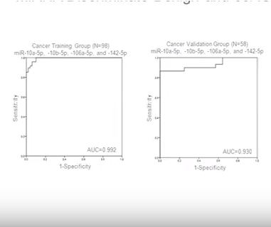
In their initial microarray screening 20 differentially expressed miRNAs, comparing non-progressive with progressive tumours, were identified. The authors found four miRNA panels (10a-5p, 10b-5p, 106a-5p, and 142-5p) as a potential biomarker signature. This model was validated in nephrectomy specimens and resulted in a sensitivity of 86.7%, a specificity of 92.9%, and an area under the curve (AUC) of 0.930 for detecting ccRCC. Further analysis revealed a second signature of two biomarkers (miR-10a-5p and -223-3p) with 93.8% sensitivity, 83.3% specificity, and an AUC of 0.932 when validated for detecting progressive ccRCC. Similarly, the differential expression of these biomarkers could delineate cancer status in biopsy specimens. For correlation of miRNA expression levels with cancer-specific survival, higher expression levels of (miR-10a-5p and miR-10b-5p) and a lower expression level of (miR-223-3p) were significantly associated with survival (P< 0.001), and the median survival times were not reached.

In conclusion, the lack of precise prediction tools has led the authors to explore the potential utility of miRNAs as biomarkers to detect disease presence, biological aggressiveness, and prognosis in ccRCC. However, until future multicentre large prospective studies validate the results of the present work, the transition of miRNA from bench to bedside is emerging on the horizon and has encouraged urologists and scientists to pursue intense translational research in the field. The ability to use miRNAs as biomarkers might be promising for diagnostic and prognostic purposes. These biomarkers may exemplify different aspects of RCC pathogenesis and may potentially have important therapeutic implications to help in a clinical decision-making approach for targeted therapy and RCC personalised treatment.

Firas G. Petrosand Christopher J.D. Wallis†‡
*Department of Urology, The University of Texas MD Anderson Cancer Center, Houston, TX, USADivision of Urology, Department of Surgery, University of Toronto, Toronto, ON, Canada and Division of Urology, Department of Surgery, Sunnybrook Health Sciences Centre, Toronto, ON, Canada


Read the full article


References


1 Reuter VE, Presti JC Jr. Contemporary approach to the classication of renal epithelial tumors. Semin Oncol 2000; 27: 12437


2 Hollingsworth JM, Miller DC, Daignault S, Hollenbeck BK. Rising incidence of small renal masses: a need to reassess treatment effect. J Natl Cancer Inst 2006; 98: 13314


3 Gupta K, Miller JD , Li JZ, Russell MW, Charbonneau C. Epidemiologic and socioeconomic burden of metastatic renal cell carcinoma (mRCC): literature review. Cancer Treat Rev 2008; 34: 193205


5 Esquela-Kerscher A, Slack FJ. Oncomirs microRNAs with a role in cancer. Nat Rev Cancer 2006; 6: 25969


Video: Profiling microRNA from nephrectomy and biopsy specimens

Profiling microRNA from nephrectomy and biopsy specimens: predictors of progression and survival in clear cell renal cell carcinoma


Read the full article

Abstract

Objective

To identify microRNA (miRNA) characteristic of metastatic clear cell renal cell carcinoma (ccRCC) and those indicative of cancer-specific survival (CSS) in nephrectomy and biopsy specimens. We also sought to determine if a miRNA panel could differentiate benign from ccRCC tissue.

Materials and Methods

RNA was isolated from nephrectomy and kidney biopsy specimens (n = 156 and n = 46, respectively). Samples were grouped: benign, non-progressive, and progressive ccRCC. MiRNAs were profiled by microarray and validated by quantitative reverse transcription-polymerase chain reaction. Biomarker signatures were developed to predict cancer status in nephrectomy and biopsy specimens. CSS was examined using Kaplan–Meier and Cox proportional hazards analyses.

Results

Microarray analysis revealed 20 differentially expressed miRNAs comparing non-progressive with progressive tumours. A biomarker signature validated in nephrectomy specimens had a sensitivity of 86.7% and a specificity of 92.9% for differentiating benign and ccRCC specimens. A second signature differentiated non-progressive vs progressive ccRCC with a sensitivity of 93.8% and a specificity of 83.3%. These biomarkers also discriminated cancer status in biopsy specimens. Levels of miR-10a-5p, -10b-5p, and -223-3p were associated with CSS.

Conclusion

This study identified miRNAs differentially expressed in ccRCC samples; as well as those correlating with CSS. Biomarkers identified in this study have the potential to identify patients who are likely to have progressive ccRCC, and although preliminary, these results may aid in differentiating aggressive and indolent ccRCC based on biopsy specimens.

Article of the Week: 11C-acetate PET/CT imaging for detection of recurrent disease after RP or RT in patients with PCa

Every Week the Editor-in-Chief selects an Article of the Week from the current issue of BJUI. The abstract is reproduced below and you can click on the button to read the full article, which is freely available to all readers for at least 30 days from the time of this post.

In addition to the article itself, there is an accompanying editorial written by a prominent member of the urological community. This blog is intended to provoke comment and discussion and we invite you to use the comment tools at the bottom of each post to join the conversation.

If you only have time to read one article this week, it should be this one.

11C-acetate positron-emission tomography/computed tomography imaging for detection of recurrent disease after radical prostatectomy or radiotherapy in patients with prostate cancer

Lukas Hendrik Esch*, Melanie Fahlbusch, Peter Albers‡, Hubertus Hautzel§ and Volker Muller-Mattheis


*Department of Urology, St Antonius Hospital, Gronau, Department of Gynaecology, Bethesda Hospital, Duisburg, Departments of‡ Urology, and §Nuclear Medicine, Medical Faculty, Heinrich-Heine University, Dusseldorf, Germany


Read the full article

Abstract

Objectives

To evaluate, in a prospective study, the effectiveness of computed tomography (CT)-matched 11C-acetate (AC) positron-emission tomography (PET) in patients with prostate cancer (PCa) who had prostate-specific antigen (PSA) relapse after radical prostatectomy (RP) or radiotherapy (RT).

Patients and Methods

In 103 relapsing patients after RP (n = 97) or RT (n = 6) AC-PET images and CT scans were obtained. In patients with AC-PET-positive results with localized PCa recurrence, detected lesions were resected and histologically verified or, after local RT, followed-up by PSA testing. Patients with distant disease on AC-PET were treated with androgen deprivation/chemotherapy.

Results

Of 103 patients, 42 were AC-PET-positive. PSA levels were <1.0, <2.0 and <4.0 ng/mL in six, 16 and 20 patients, respectively. In 25/42 patients AC-PET suggested lymph node metastases: 16/25 patients underwent surgery (10/16 metastasis, 6/16 inflammation); 9/25 patients underwent RT of lymph node metastases, which was followed by decreasing PSA level. In 17/42 patients who had distant disease, systemic treatment was commenced. Combining patients who underwent surgery and those who underwent RT, 19/25 patients were true-positive in terms of AC-PET (positive predictive value 76%). In 5/19 patients, PSA level was <2.0 ng/mL, in 2/19 patients it was <1.0 ng/mL and in 14/19 patients it was 5.4–23.1 ng/mL. In AC-PET-positive patients after surgery or RT (without androgen deprivation), median (range) time to renewed PSA increase was 6 (5–9) months.

Conclusions

Only a minority of patients with relapsing PCa appear to benefit from AC-PET for guiding potential local treatment. False-positive results show that factors other than tumour metabolism induce increased AC uptake. The time free of recurrence after local treatment was shorter than expected.

Read more articles of the week

Editorial: Is choline-based PET imaging still relevant in recurrent prostate cancer?

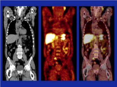
The search for the ideal imaging method to detect small metastatic deposits has largely remained elusive in the field of prostate cancer. However, functional positron emission tomography (PET)/CT is now guiding us in different directions. 11C-acetate PET/CT imaging was one of the first molecular imaging probes showing promise in clinical studies, along with 11C-choline, and more recently challenged by 68Ga-prostate-specific membrane antigen (PSMA) PET/CT [1-3]. So against this background, what does this new study by Esch et al. [4] presented here offer us?

Let us rewind and focus on where molecular imaging may have an impact in prostate cancer. In primary staging, the potential to accurately identify oligometastatic disease or even widespread metastatic disease before primary gland treatment is clearly advantageous. It can allow for wider treatment fields (extended lymphadenectomy or radiation fields), directed treatment of oligometastatic disease, placement into appropriate cytoreductive trials, and in some instances consideration of the use of earlier chemo-hormonal therapy. This study did not address this important clinical scenario [4]. Nor did it focus on primary diagnosis, another ‘holy grail’ for imaging more recently dominated by MRI [5].

The clinical question addressed in this study was whether 11C-acetate PET is able to detect distant metastatic disease in men with PSA relapse after having undergone radical prostatectomy or prostate bed radiotherapy [4]. In other words, if distant metastatic disease is found then prostate bed radiation may be futile, although this assumption has not been subjected to high quality trials, but would seem logical. Also, in the era of oligometastatic disease, treatment (surgery and/or radiation) may be directed at nodal and other deposits to prolong time of systemic therapy such as androgen-deprivation therapy. So what is the ‘sweet spot’? Some would state for men having undergone surgery a PSA level of 0.2 ng/mL places the patient at risk of recurrence, although in clinical practice the level at which investigations are warranted can be as high as 1.0 ng/mL. This study found 11C-acetate PET had few positive results below a PSA level of 1.0 ng/mL. This is in contrast to a recent meta-analysis of 68Ga-PSMA PET/CT reporting positive studies at PSA levels of ≤0.2 ng/mL in 42% of patients [6].

The other group studied the detection of recurrent disease in men having undergone primary radiotherapy, although the numbers were low (six patients). We know this group of men are often undertreated for salvage treatment, and frequently placed on hormonal therapy. Again, early knowledge of locoregional or distant recurrence is required to allow best case selection and thus avoid futile salvage surgery, and also to know who may benefit from salvage lymphadenectomy and treatment of distant oligometastatic disease, if required.

The false-positive rate result for 11C-acetate PET/CT of 24% is notable and concerning. This may lead to unnecessary intervention, or even withholding prostate bed radiation that may have been of benefit. In contrast the false-positive rate for 11C-choline and 68Ga-PSMA PET/CT is far lower, although false-positive PSMA studies may occur in benign conditions and non-prostate cancer tumours.

It should be acknowledged that a strength of this study was comparative histology in a proportion of patients, allowing true sensitivity and specificity to be determined [4]. This is important information, as it guides correct decision-making, and such information is lacking for many other prostate cancer imaging probes, including 68Ga-PSMA, where these data are urgently required.

Overall, this study is important and adds to the rich milieu of available molecular imaging data on staging of possible recurrent or metastatic prostate cancer to date [4]. Current prospective trials exploring 11C-choline, 18F-FACBC (anti-1-amino-3-18F-fluorocyclobutane-1-carboxylic acid), 18F-choline and 68Ga-PSMA with MRI and clinical outcomes should provide further insight into the most appropriate molecular imaging technique for staging prostate cancer.

Nathan Lawrentschuk, BJUI USANZ Editor*,,, Julia M. Coreldand Andrew Scott,§,¶
*Department of Surgery, University of Melbourne and Olivia Newton-John Cancer Research Institute, Austin Hospital , Peter MacCallum Cancer Centre, La Trobe University, Departments of


Medicine, § Molecular Imaging and Therapy
Read the full article


References


1 Afshar-Oromieh A, Zechmann CM, Malcher A et al. Comparison of PET imaging with a (68)Ga-labelled PSMA ligand and (18)F-choline- based PET/CT for the diagnosis of recurrent prostate cancer. Eur J Nucl Med Mol Imaging 2014; 41: 1120


2 Silver DA, Pellicer I, Fair WR, Heston WD, Cordon-Cardo C. Prostate- specic membrane antigen expression in normal and malignant human tissues. Clin Cancer Res 1997; 3: 815


5 Johnson LM, Turkbey B, Figg WD, Choyke PL. Multiparametric MRI in prostate cancer management. Nat Rev Clin Oncol 2014; 11: 34653


Article of the Week: Detecting SNs in patients with BCa intra-operatively

Every Week the Editor-in-Chief selects an Article of the Week from the current issue of BJUI. The abstract is reproduced below and you can click on the button to read the full article, which is freely available to all readers for at least 30 days from the time of this post.

In addition to the article itself, there is an accompanying editorial written by a prominent member of the urological community. This blog is intended to provoke comment and discussion and we invite you to use the comment tools at the bottom of each post to join the conversation.

If you only have time to read one article this week, it should be this one.

Radio-guided sentinel lymph node detection and lymph node mapping in invasive urinary bladder cancer: a prospective clinical study

Firas Aljabery1,2,*, Ivan Shabo2,3,4, Hans Olsson2,5, Oliver Gimm2,6 and Staffan Jahnson1,2

1 Department of Urology, Region Östergötland, Linköping University Hospital, Linköping, Sweden, 2 Department of Clinical and Experimental Medicine, Faculty of Health Sciences, Linköping University, Linköping, sweden 3 Endocrine and Sarcoma Surgery Unit, Department of Molecular Medicine and Surgery, Karolinska Institute, Stockholm, Sweden 4 Department of Breast and Endocrine Surgery, Karolinska University Hospital, Solna Stockholm, Sweden 5 Department of Pathology, Region Östergötland, Linköping University Hospital, Linköping, Sweden 6 Department of Surgery, Region Östergötland, Linköping University Hospital, Linköping, Sweden

Read the full article

Abstract

Objectives

To investigate the possibility of detecting sentinel lymph nodes (SNs) in patients with urinary bladder cancer (BCa) intra-operatively and whether the histopathological status of the identified SNs reflected that of the lymphatic field.

Patients and Methods

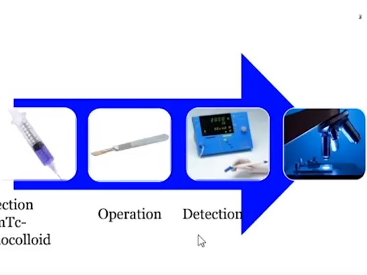
We studied 103 patients with BCa pathological stage T1–T4 who were treated with cystectomy and pelvic lymph node (LN) dissection during 2005–2011 at the Department of Urology, Linköping University Hospital. Radioactive tracer Nanocoll 70 MBq and blue dye were injected into the bladder wall around the primary tumour before surgery. SNs were detected ex vivo during the operation with a handheld Geiger probe (Gamma Detection System; Neoprobe Corp., Dublin, OH, USA). All LNs were formalin-fixed, sectioned three times, mounted on slides and stained with haematoxylin and eosin. An experienced uropathologist evaluated the slides.

Results

The mean age of the patients was 69 years, and 80 (77%) were male. Pathological staging was T1–12 (12%), T2–20 (19%), T3–48 (47%) and T4–23 (22%). A mean (range) number of 31 (7–68) nodes per patient were examined, totalling 3 253 nodes. LN metastases were found in 41 patients (40%). SNs were detected in 83 of the 103 patients (80%). Sensitivity and specificity for detecting metastatic disease by SN biopsy (SNB) varied between LN stations, with average values of 67% and 90%, respectively. LN metastatic density (LNMD) had a significant prognostic impact; a value of ≥8% was significantly related to shorter survival. Lymphovascular invasion (LVI) occurred in 65% of patients (n = 67) and was significantly associated with shorter cancer-specific survival (P < 0.001).

Conclusion

We conclude that SNB is not a reliable technique for peri-operative localization of LN metastases during cystectomy for BCa; however, LNMD has a significant prognostic value in BCa and may be useful in the clinical context and in BCa oncological and surgical research. LVI was also found to be a prognostic factor.

Read more articles of the week

Editorial: Positive messages for bladder cancer management in negative sentinel lymph node study

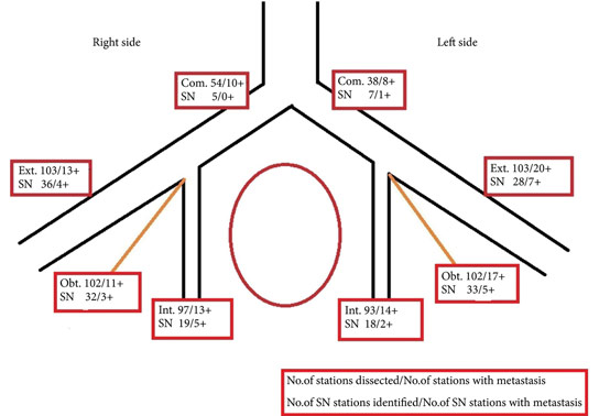
I encourage you to read the study by Aljabery et al. [1] in this edition of BJUI. Their findings are based on some very solid methodology and I think provide a robust answer to their question, which often in science means a ‘negative’ result. The principle of sentinel lymph node biopsy (SLNB) needs no introduction. It is primarily intended to detect the principal LN draining a tumour, allowing its removal and pathological determination of LN metastasis status in that individual [2].The avoidance of an unnecessary LN dissection (LND) and its associated risks is at the heart of any SLNB strategy. On the other hand, particularly for bladder cancer, there is a recognition that a higher number of LNs removed at the time of surgery confers a survival advantage to patients through more accurate staging [3]. With greater numbers of LNs removed pN0 patients are more likely to be truly N0 and pN1 patients with limited metastases have a greater chance that all disease has been completely excised. Thus, when considering SLNB in bladder cancer there is the usual conflict between maximising oncological benefit and minimising surgical harm.Aljabery et al. [1] present an excellent series of cystectomies with a 100% negative margin rate and mean LN count of 30. The 40% rate of LN involvement, is perhaps partly due to the meticulous triple sectioning of each excised LN. Their SLN technique involved four cystoscopic injections to the bladder wall surrounding the tumour and focused on the biggest lesion in multiple tumour cases. The LNs were removed in their packets and studied after removal from the patient. While this is likely to be a more precise method for determining the site of the SLN, it clearly differs from the approach one would take if trying to avoid LND in negative-SLN cases. Furthermore, examination of LNs was performed after formalin fixation. Typically when using SLN techniques frozen sections are also used to guide surgeons during surgery.The results clearly show that SLNB using radiolabelled nanocolloid does not allow accurate identification of pathologically LN-negative patients who could then avoid a complete LN dissection. Sensitivity of the technique in the detection of positive LNs ranged from 67% to 90% at the various LN stations. Overall, of patients with an identifiable SLN that was negative, 19% of patients had positive LNs elsewhere (81% negative predictive value). Effectively one in five patients who might be reassured by a negative SLN result would in fact have undetected positive LNs left behind if this technique were employed. Furthermore, this estimation does not consider errors likely to be introduced with in situ SLN identification and the use of frozen-section analysis rather than non-time-critical analysis of formalin-fixed sections.In such a dangerous disease such inaccuracy is not tolerable and so I totally agree with the authors’ [1] findings that SLNB of pelvic LNs at the time of radical cystectomy for bladder cancer is not a reliable technique for identifying LN metastasis.The positive messages from this study [1] are worth noting by those learning and undertaking cystectomy. The authors’ meticulous approach to surgery is evident from the methodology described and the accumulation of such a well-characterised series. This must be a contributing factor in achieving a 100% negative surgical margin rate and such consistently high LN yields. This should certainly be the aim of all cystectomists. The appropriate time, skill and patience should be given to this step and it should not be compromised upon, particularly when developing robot-assisted or laparoscopic cystectomy services.The findings that T-stage, N-stage and lymphovascular invasion are linked to survival are not that surprising. However, the use of LN metastatic density as a prognostic marker is interesting, as it is not usually discussed in our multidisciplinary meetings. This measure incorporates nodal tumour burden and the extent of LND. The finding of better outcomes in those with a LN metastatic density of <8% reinforces the message that even in those with LN metastases, removing greater numbers of LNs may improve prognosis. Furthermore, the finding that 30% of unilateral LN-positive tumours also had contralateral LNs settles any arguments for unilateral LN dissections.In a recent systematic review of SLNB in bladder cancer [4], the negative predictive value was found to be 92% compared to 81% in the Aljabery et al. [1] study. The authors of the systematic review suggested that SLNB is a promising technique; perhaps in view of technology advances they reviewed that might improve future outcomes of SLNB. While improvements may be possible, current evidence would not encourage me to consider SLNB using radiolabelled nanocolloid for fear of impairing cancer outcomes.

Congratulations to Aljabery et al. [1] on their work. I hope you find reading their paper as constructive as I did.

Tim Dudderidge
Department of Urology, University Hospital Southampton,
Southampton, Hampshire, UK

Read the full article

References

1 Aljabery F, Shabo I, Olson H, Gimm O, Jahnson S. Radio-guided sentinel lymph node detection and lymph node mapping in invasive urinary bladder cancer: a prospective clinical study. BJU Int 2017; 120: 329–36

2 Gould EA, Winship T, Philbin PH, Kerr HH. Observations on a “sentinel node” in cancer of the parotid. Cancer 1960; 13: 77–8

3 Koppie TM, Vickers AJ, Vora K, Dalbagni G, Bochner BH. Standardization of pelvic lymphadenectomy performed at radical cystectomy. Cancer 2006; 107: 2368–74

4 Liss M, Noguchi J, Lee H, Vera D, Kader AK. Sentinel lymph node biopsy in bladder cancer: systematic review and technology update. Indian J Urol 2015; 31: 170–5


Video: Detecting SNs in patients with BCa intra-operatively

Radio-guided sentinel lymph node detection and lymph node mapping in invasive urinary bladder cancer: a prospective clinical study

Read the full article

Abstract

Objectives

To investigate the possibility of detecting sentinel lymph nodes (SNs) in patients with urinary bladder cancer (BCa) intra-operatively and whether the histopathological status of the identified SNs reflected that of the lymphatic field.

Patients and Methods

We studied 103 patients with BCa pathological stage T1–T4 who were treated with cystectomy and pelvic lymph node (LN) dissection during 2005–2011 at the Department of Urology, Linköping University Hospital. Radioactive tracer Nanocoll 70 MBq and blue dye were injected into the bladder wall around the primary tumour before surgery. SNs were detected ex vivo during the operation with a handheld Geiger probe (Gamma Detection System; Neoprobe Corp., Dublin, OH, USA). All LNs were formalin-fixed, sectioned three times, mounted on slides and stained with haematoxylin and eosin. An experienced uropathologist evaluated the slides.

Results

The mean age of the patients was 69 years, and 80 (77%) were male. Pathological staging was T1–12 (12%), T2–20 (19%), T3–48 (47%) and T4–23 (22%). A mean (range) number of 31 (7–68) nodes per patient were examined, totalling 3 253 nodes. LN metastases were found in 41 patients (40%). SNs were detected in 83 of the 103 patients (80%). Sensitivity and specificity for detecting metastatic disease by SN biopsy (SNB) varied between LN stations, with average values of 67% and 90%, respectively. LN metastatic density (LNMD) had a significant prognostic impact; a value of ≥8% was significantly related to shorter survival. Lymphovascular invasion (LVI) occurred in 65% of patients (n = 67) and was significantly associated with shorter cancer-specific survival (P < 0.001).

Conclusion

We conclude that SNB is not a reliable technique for peri-operative localization of LN metastases during cystectomy for BCa; however, LNMD has a significant prognostic value in BCa and may be useful in the clinical context and in BCa oncological and surgical research. LVI was also found to be a prognostic factor.

Article of the Week: sRPLND+PLND for ‘node-only’ recurrent PCa

Every Week the Editor-in-Chief selects an Article of the Week from the current issue of BJUI. The abstract is reproduced below and you can click on the button to read the full article, which is freely available to all readers for at least 30 days from the time of this post.

In addition to the article itself, there is an accompanying editorial written by a prominent member of the urological community. This blog is intended to provoke comment and discussion and we invite you to use the comment tools at the bottom of each post to join the conversation.

If you only have time to read one article this week, it should be this one.

Robotic salvage retroperitoneal and pelvic lymph node dissection for ‘node-only’ recurrent prostate cancer: technique and initial series

Andre Abreu*, Carlos Fay*, Daniel Park*, David Quinn*, Tanya Dorff*,John Carpten*, Peter Kuhn*, Parkash Gill*, Fabio Almeida* and Inderbir Gill*


*University of Southern California (USC) Institute of Urology, Catherine & Joseph Aresty Department of Urology, Keck School of Medicine, USC, Los Angeles, CA, USA, and Pontical Catholic University of Rio Grande do Sul, Porto Alegre, Brazil


Read the full article

Abstract

Objectives

To describe the technique of robot-assisted high-extended salvage retroperitoneal and pelvic lymphadenectomy (sRPLND+PLND) for ‘node-only’ recurrent prostate cancer.

Patients and Methods

In all, 10 patients underwent robot-assisted sRPLND+PLND (09/2015–03/2016) for ‘node-only’ recurrent prostate cancer, as identified by 11C-acetate positron emission tomography/computed tomography imaging. Our anatomical template extends from bilateral renal artery/vein cranially up to Cloquet’s node caudally, completely excising lymphatic-fatty tissue from aorto-caval and iliac vascular trees; RPLND precedes PLND. Meticulous node-mapping assessed nodes at four prospectively assigned anatomical zones.

Results

The median operative time was 4.8 h, estimated blood loss 100 mL and hospital stay 1 day. No patient had an intraoperative complication, open conversion or blood transfusion. Three patients had spontaneously resolving Clavien–Dindo grade II postoperative complications. The mean (range) number of nodes excised per patient was 83 (41–132) and mean (range) number of positive nodes per patient was 23 (0–109). Seven patients (70%) had positive nodes on final pathology. Node-positive rates per anatomical level I, II, III and IV were 28%, 32%, 33% and 33%, respectively. In patients with positive nodes, the median PSA level had decreased by 83% at the 2-month follow-up.

Conclusion

The initial series of robot-assisted sRPLND+PLND is presented, wherein we duplicate open surgery with superior nodal counts and decreased morbidity. Robot-assisted technical details for an anatomical LND template up to the renal vessels are presented. Longer follow-up is necessary to assess oncological outcomes.

Read more articles of the week


© 2024 BJU International. All Rights Reserved.