Tag Archive for: Article of the Week

Posts

Video: Surgical outcomes of PCNL and results of stone analysis

Surgical outcomes of percutaneous nephrolithotomy in 3402 patients and results of stone analysis in 1559 patients

Abstract

Objective

To report our experience of a series of percutaneous nephrolithotomy (PCNL) procedures in a single centre over 18 years in terms of patient and stone characteristics, indications, stone clearance and complications, along with the results of chemical analysis of stones in a subgroup.

Patients and Methods

We retrospectively analysed the outcomes of PCNL in 3402 patients, who underwent the procedure between 1997 and 2014, obtained from a prospectively maintained database. Data analysis included patients’ age and sex, laboratory investigations, imaging, punctured calyx, duration of operation, volume of irrigation fluid, radiation exposure time, blood transfusion, complications and stone-free status at 1-month follow-up. For the present analysis, outcomes in relation to complications and success were divided in two eras, 1997–2005 and 2006–2014, to study the differences.

Results

Of the 3402 patients, 2501 (73.5%) were male and 901 (26.5%) were female, giving a male:female ratio of 2.8:1. Staghorn (partial or complete) calculi were found in 27.5% of patients, while 72.5% had non-staghorn calculi. Intracorporeal energy sources used for stone fragmentation included ultrasonography in 917 patients (26.9%), pneumatic lithoclast in 1820 (53.5%), holmium laser in 141 (4.1%) and Lithoclast® master in 524 (15.4%). In the majority of patients (97.4%) a 18–22-F nephrostomy tube was placed after the procedure, while 69 patients (2.03%) underwent tubeless PCNL. The volume of the irrigation fluid used ranged from 7 to 37 L, with a mean of 28.4 L. The stone-free rate after PCNL in the first era studied was 78%, vs 83.2% in the second era, as assessed by combination of ultrasonography and plain abdominal film of the kidney, ureter and bladder. The complication rate in the first era was 21.3% as compared with 10.3% in the second era, and this difference was statistically significant. Stone analysis showed pure stones in 41% and mixed stones in 58% of patients. The majority of stones consisted of calcium oxalate.

Conclusions

This is the largest series of PCNL reported from any single centre in Pakistan, where there is a high prevalence of stone disease associated with infective and obstructive complications, including renal failure. PCNL as a treatment method offers an economic and effective option in the management of renal stone disease with acceptable stone clearance rates in a resource-constrained healthcare system.


Article of the Week: Effect of MetS on serum PSA levels is concealed by enlarged prostate

Every Week the Editor-in-Chief selects an Article of the Week from the current issue of BJUI. The abstract is reproduced below and you can click on the button to read the full article, which is freely available to all readers for at least 30 days from the time of this post.

In addition to the article itself, there is an accompanying editorial written by a prominent member of the urological community. This blog is intended to provoke comment and discussion and we invite you to use the comment tools at the bottom of each post to join the conversation.

Finally, the third post under the Article of the Week heading on the homepage will consist of additional material or media. This week we feature a video discussing the paper.

If you only have time to read one article this week, it should be this one.

Actual lowering effect of metabolic syndrome on serum prostate-specific antigen levels is partly concealed by enlarged prostate: results from a large-scale population-based study

Sicong Zhao*, Ming Xia*, Jianchun Tang† and Yong Yan*


*Department of Urology, and Department of Cardiology, Beijing Shijitan Hospital, Capital Medical University, Beijing, China


Read the full article

Abstract

Objectives

To clarify the lowering effect of metabolic syndrome (MetS) on serum prostate-specific antigen (PSA) levels in a Chinese screened population.

Subjects and Methods

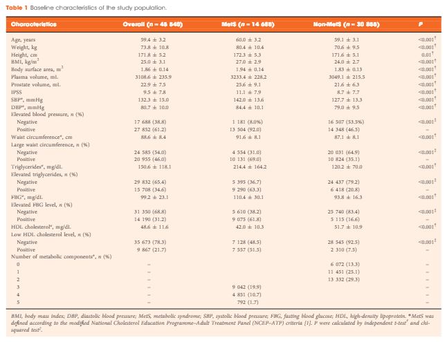
A total of 45 540 ostensibly healthy men aged 55–69 years who underwent routine health check-ups at Beijing Shijitan Hospital between 2008 and 2015 were included in the study. All the men underwent detailed clinical evaluations. PSA mass density was calculated (serum PSA level × plasma volume ÷ prostate volume) for simultaneously adjusting plasma volume and prostate volume. According to the modified National Cholesterol Education Programme–Adult Treatment Panel (NCEP-ATP) III criteria, patients were dichotomized by the presence of MetS, and differences in PSA density and PSA mass density were compared between groups. Linear regression analysis was used to evaluate the effect of MetS on serum PSA levels.

Results

When larger prostate volume in men with MetS was adjusted for, both PSA density and PSA mass density in men with MetS were significantly lower than in men without MetS, and the estimated difference in mean serum PSA level between men with and without MetS was greater than that before adjusting for prostate volume. In the multivariate regression model, the presence of MetS was independently associated with an 11.3% decline in serum PSA levels compared with the absence of MetS. In addition, increasing number of positive MetS components was significantly and linearly associated with decline in serum PSA levels.

Conclusion

The actual lowering effect of MetS on serum PSA levels was partly concealed by the enlarged prostate in men with MetS, and the presence of MetS was independently associated with lower serum PSA levels. Urologists need to be aware of the effect of MetS on serum PSA levels and should discuss this subject with their patients.

Read more articles of the week

Editorial: Anomalous observation with regard to PCa in cancer research

In science, reports showing data deviating from what is expected are called anomalous observations. Metabolic syndrome (MetS) is a promoter of cancer at almost all sites [1]; however, when it comes to prostate cancer (PCa), a series of reports have been published showing an inverse relationship between MetS and its aspects and incident PCa. This lack of coherence in cancer research seriously hampers efforts to fight cancer disorders. It is therefore crucial to find an explanation for this incoherence.

In the search for a reasonable explanation for this anomalous observation, a hypothesis has been formulated, based on the study by Häggström et al. [2], and stating that the PSA-driven diagnostic procedure in PCa, which creates low-stage incident PCa material, is the culprit. The PSA-driven diagnostic procedure introduces several bias mechanisms, which tend to protect men with MetS from being diagnosed with PCa. Thus, men with MetS and its aspects are under-represented in PCa populations generated by PSA-driven diagnostics, thereby creating a distorted incident PCa population. This hypothesis also predicts that high-stage PCa, as well as non-localized and lethal PCa, are not subject to these bias mechanisms, as a minor reduction in the PSA level is of no importance for the PCa diagnosis at these high PSA levels. Finally, the hypothesis predicts that the link between MetS and incident PCa is stage-dependent. A study testing this hypothesis is now in progress.

Several studies have reported that men with MetS had lower PSA levels compared with men without MetS. Zhao et al. [3] address this specific question in this issue of BJUI and confirm that the presence of MetS was independently associated with a lower PSA level and that the enlarged prostate gland, which is an aspect of MetS, partly concealed an even greater PSA level reduction [3]. The findings indicate that a bias mechanism inverses the link between MetS and incident PCa and support the above-mentioned hypothesis.

In short, the following bias mechanisms have been described. MetS is associated with greater body fat with increased aromatase activity, resulting in a reduced testosterone level, which, in turn, is related to a reduced PSA level, as the production of PSA is under androgen control. Another possible bias mechanism, leading to men with MetS being diagnosed less often with PCa, is that these men are more likely to be obese. It is well established that men with a higher BMI also have larger plasma volumes and therefore have greater haemodilution of the PSA production, resulting in a lower PSA level. This means that incident PCa is diagnosed less often in men with MetS, as their PSA level is lower. MetS is also associated with an enlarged prostate gland volume, which means that fewer incident PCas are diagnosed, given the same tumour volume and the same number of biopsies. Another bias mechanism is that a high proportion of men with high socio-economic status undergo PSA testing in the PSA era. It is well established that men with a high socio-economic status have a lower prevalence of MetS and therefore have higher PSA levels, as indicated by the present report in the BJUI [3], and an elevated risk of PCa. Thus, multiple bias mechanisms seem to conceal low-stage PCa in the PSA era.

If it could be confirmed that the negative relationship between MetS and incident PCa is a spurious observation as a result of bias mechanisms, this would open the door for the MetS hypothesis regarding the promotion of multiple cancer disorders. This door has previously been closed by findings in a series of reports of an inverse relationship between MetS and its aspects and incident prostate cancer. Furthermore, this could lead to increased efforts to fight the metabolic aberrations of MetS. It is now well established that MetS and its aspects could be reduced by changes in lifestyle, including physical activity and diet. The most convincing evidence of the effect of diet on MetS comes from studies involving decreased intake of carbohydrates and increased intake of unsaturated fats. Recently, leading authorities in nutrition, endocrinology and metabolism presented a critical review and concluded that carbohydrate restriction is the single most effective intervention to reduce all features of MetS [4]. Another review concluded that carbohydrate restriction is one of the few common interventions that target all features of MetS [5]. This conclusion has recently been confirmed in a meta-analysis by Mansoor et al. [6].

In conclusion, new knowledge challenges the anomalous observation of PCa showing a negative relationship between MetS and PCa. The credibility of the hypothesis that MetS is an important promoting factor for cancer at almost all sites is strengthened. MetS could be treated effectively with a low carbohydrate and high fat diet.

Jan Hammarsten, MD, PhD
Department of Urology, Institute of Clinical SciencesUniversity of Gothenburg, Gothenburg, Sweden


Read the full article


References


1 Esposito K, Chiodini P, Colao AM et al. Metabolic syndrome and risk of cancer. Diabetes Care 2012; 35: 240211 


2Haggstrom C, Stocks T, Ulmert D et al. Prospective study on metabolic factors and risk of prostate cancer. Cancer 2012; 118: 6199206


3 Zhao S, Xia M, Tang J et al. The actual lowering effect of metabolic syndrome on serum prostate-specic antigen levels is partly concealed by enlarged prostate: results from large-scale population-based study. BJU Int 2017; 120: 4829


4 Feinman RD, Pogozelski WK, Astrup A et al. Dietary carbohydrate restriction as the rst approach in diabetes management: critical review and evidence base. Nutrition 2015;31: 113


5 Accurso A, Bernstein RK, Dahlqvist A et al. Dietary carbohydrate restriction in type 2 diabetes mellitus and metabolic syndrome: time for critical appraisal. Nutrition & Metabolism 2008; 5: 9


6 Mansoor N, Vinknes UJ , Veierod MB et al. Effects of low-carbohydrate diets v. low fat diets on body weight and cardiovascular risk factors: meta-analysis of randomized controlled trials. Br J Nutrition 2016; 115: 4667


Video: Effect of MetS on serum PSA levels is concealed by enlarged prostate

Actual lowering effect of metabolic syndrome on serum prostate-specific antigen levels is partly concealed by enlarged prostate: results from a large-scale population-based study

Sicong Zhao*, Ming Xia*, Jianchun Tang† and Yong Yan*


*Department of Urology, and Department of Cardiology, Beijing Shijitan Hospital, Capital Medical University, Beijing, China


Read the full article

Abstract

Objectives

To clarify the lowering effect of metabolic syndrome (MetS) on serum prostate-specific antigen (PSA) levels in a Chinese screened population.

Subjects and Methods

A total of 45 540 ostensibly healthy men aged 55–69 years who underwent routine health check-ups at Beijing Shijitan Hospital between 2008 and 2015 were included in the study. All the men underwent detailed clinical evaluations. PSA mass density was calculated (serum PSA level × plasma volume ÷ prostate volume) for simultaneously adjusting plasma volume and prostate volume. According to the modified National Cholesterol Education Programme–Adult Treatment Panel (NCEP-ATP) III criteria, patients were dichotomized by the presence of MetS, and differences in PSA density and PSA mass density were compared between groups. Linear regression analysis was used to evaluate the effect of MetS on serum PSA levels.

Results

When larger prostate volume in men with MetS was adjusted for, both PSA density and PSA mass density in men with MetS were significantly lower than in men without MetS, and the estimated difference in mean serum PSA level between men with and without MetS was greater than that before adjusting for prostate volume. In the multivariate regression model, the presence of MetS was independently associated with an 11.3% decline in serum PSA levels compared with the absence of MetS. In addition, increasing number of positive MetS components was significantly and linearly associated with decline in serum PSA levels.

Conclusion

The actual lowering effect of MetS on serum PSA levels was partly concealed by the enlarged prostate in men with MetS, and the presence of MetS was independently associated with lower serum PSA levels. Urologists need to be aware of the effect of MetS on serum PSA levels and should discuss this subject with their patients.

Read more articles of the week

Article of the Week: Early surgical outcomes and oncological results of RAPN

Every Week the Editor-in-Chief selects an Article of the Week from the current issue of BJUI. The abstract is reproduced below and you can click on the button to read the full article, which is freely available to all readers for at least 30 days from the time of this post.

In addition to the article itself, there is an accompanying editorial written by a prominent member of the urological community. This blog is intended to provoke comment and discussion and we invite you to use the comment tools at the bottom of each post to join the conversation.

If you only have time to read one article this week, it should be this one.

Early surgical outcomes and oncological results of robot-assisted partial nephrectomy: a multicentre study


Rajan Veeratterapillay*, Sanjai K. Addla, Clare Jelley, John Bailie*, David Rix*,Steve Bromage, Neil Oakley, Robin Weston§ and Naeem A. Soomro*


*Department of Urology, Freeman Hospital, Newcastle Upon Tyne, Department of Urology, Bradford Teaching Hospitals NHS Foundation Trust, Bradford, Department of Urology, Stepping Hill Hospital, Stockport, and §Department of Urology, Royal Liverpool University Hospital, Liverpool, UK


Read the full article

Abstract

Objective

To describe a multicentre experience of robot-assisted partial nephrectomy (RAPN) in northern England, with focus on early surgical outcomes and oncological results.

Patients and Methods

All consecutive patients undergoing RAPN at four tertiary referral centres in northern England in the period 2012–2015 were included for analysis. RAPN was performed via a transperitoneal approach using a standardized technique. Prospective data collection was performed to capture preoperative characteristics (including R.E.N.A.L. nephrometry score), and peri-operative and postoperative data, including renal function. Correlations between warm ischaemia time (WIT), positive surgical margin (PSM) rate, complication rates, R.E.N.A.L. nephrometry scores and learning curve were assessed using univariate and multivariate analyses.

Results

A total of 250 patients (mean age 58.1 ± 13 years, mean ± sd body mass index 27.3 ± 7 kg/m2) were included, with a median (range) follow-up of 12 (3–36) months. The mean ± sd tumour size was 30.6 ± 10 mm, mean R.E.N.A.L. nephrometry score was 6.1 ± 2 and 55% of tumours were left-sided. Mean ± sd operating console time was 141 ± 38 min, WIT 16.7 ± 8 min and estimated blood loss 205 ± 145 mL. There were five conversions (2%) to open/radical nephrectomy. The overall complication rate was 16.4% (Clavien I, 1.6%; Clavien II, 8.8%; Clavien III, 6%; Clavien IV/V; 0%). Pathologically, 82.4% of tumours were malignant and the overall PSM rate was 7.3%. The mean ± sd preoperative and immediate postoperative estimated glomerular filtration rates were 92.8 ± 27 and 80.8 ± 27 mL/min/1.73 m2, respectively (P = 0.001). In all, 66% of patients remained in the same chronic kidney disease category postoperatively, and none of the patients required dialysis during the study period. ‘Trifecta’ (defined as WIT < 25 min, negative surgical margin status and no peri-operative complications) was achieved in 68.4% of patients overall, but improved with surgeon experience. PSM status and long WIT were significantly associated with early learning curve.

Conclusion

This is the largest multicentre RAPN study in the UK. Initial results show that RAPN is safe and can be performed with minimal morbidity. Early oncological outcomes and renal function preservation data are encouraging.

Read more articles of the week

Editorial: From Novick to the NHS – the evolution of minimally-invasive NSS

The publication in this issue of the BJUI by Veeratterapillay et al. [1] of a UK multicentre study in a community setting marks a watershed in the availability and quality of minimally invasive nephron-sparing surgery (NSS) for renal cancer. Such a turning point was predicted almost 17 years ago by Novick [2] when he wrote, ‘minimally invasive modalities of tumour resection or destruction should be reserved for highly select patients and awaits improvements in technology, standardization of technique and long-term outcomes data before they may be completely integrated options’. It appears now that robot-assisted surgery provides such a platform. The present study [1] describes the outcomes of patients treated with robot-assisted partial nephrectomy (RAPN) at four centres in Northern England, and shows very good outcomes within their first 250 cases.

The benefits of NSS have been well described. Indeed, excellent outcomes for PN were described over 20 years ago in carefully selected cases, with benefits including reduced incidence of renal insufficiency compared to radical nephrectomy, which until that time had been viewed as the ‘gold-standard’ for patients with RCC [3]. However, the popularity of PN for small renal masses appeared to decline with the advent of laparoscopy. It became apparent that a minimally invasive approach to radical nephrectomy had the advantage of improved recovery, reduced blood loss with equal cancer control to open nephrectomy [4]. Notwithstanding absolute and relative indications for PN, given the choice between an open PN and a laparoscopic radical nephrectomy, the balance for patients with an elective indication for PN was tipped in favour of a minimally invasive yet radical approach [5]. Techniques for PN were in their infancy, and even in the leading high-volume centres outcomes, including warm ischaemia time (WIT) and positive surgical margin (PSM) rate, failed to match those of open surgery [6].

Fast forward to 2017 with the increasing use of robot-assisted urological surgery carrying the advantages of three-dimensional vision, wristed movement and integrated real-time intraoperative imaging, especially beneficial for procedures such as PN where quick and accurate suturing are essential for a successful outcome. Veeratterapillay et al. [1] present a series of 250 patients from centres in the UK, in which each performs <50 RAPN procedures/year, yet the authors present favourable outcomes overall, with a PSM rate of 7.3%, major complications in 6% and trifecta in 68.4%. An impressive learning curve is seen with improving outcomes over the series, such that in the final 50 cases a trifecta (WIT <25 min, negative surgical margin and absence of complications) was achieved in 82% of cases, with a PSM rate of 2% despite increasing complex nephrometry scores, which compares favourably with larger series from internationally renowned centres [6].

So then, with the results of the present study [1], can we say that Novick’s requirements have been met, and that minimally invasive NSS is now a ‘completely integrated option’? Certainly, with the widespread adoption of robot-assisted surgery, high-quality outcomes are within the grasp of centres other than elite academic institutions. As techniques develop and experience grows robot-assisted surgery can be increasingly offered, even for resection of more complex tumours.

To ensure that minimally invasive NSS is delivered to the highest standards, it will be necessary for providers to ensure both quality assurance and quality control in their processes. The learning curve needs to be minimised with structured teaching and mentoring, and the use of adjuncts such as intraoperative ultrasonography or fluorescence should be a routine part of care.

Centres offering this technique should be mindful of the well documented volume–outcome relationship that appears to be ubiquitous among complex surgical procedures. If centres are performing less than an optimum number of cases, they may consider affiliating themselves with other such centres in networks and forming a joint clinical governance programme, as has been described for robot-assisted radical prostatectomy and which has shown demonstrable improvements in outcomes.

Finally, auditing and reporting of outcomes remains the cornerstone of quality assurance as shown by the introduction of the BAUS complex surgery audit, which is intended to drive standards of care forward. Publications such as that of Veeratterapillay et al. [1] greatly assist in documenting the progress of new techniques and emerging technologies. Increasingly, patients expect transparency from healthcare providers, and with the necessary support processes in place, such initiatives, and the data that they produce will help to further improve the delivery of complex surgery to patients from all areas of our practice.

Benjamin W. Lamb* and Daniel A. Moon*


*Division of Cance r Surgery, Peter MacCallum Cancer Centre, Epworth Healthcare, and Department of Surgery, Central Clinical School, Monash University, Melbourne, Vic., Australia
Read the full article


References


1 Veeratterapillay R, Addla SK, Jelley C et al. Early surgical outcomes and oncological results of robot-assisted partial nephrectomy: a multicentre study. BJU Int 2017; 120: 5505


2 Uzzo RG, Novick AC. Nephron sparing surgery for renal tumors: indications, techniques and outcomes. J Urol 2001; 166: 618


3 Polascik TJ, Pound CR, Meng MV, Partin AW, Marshall FF. Partial nephrectomy: technique, complications and pathological ndings. J Urol 1995; 154: 131218


4 Gill IS, Meraney AM, Schweizer DK et al. Laparoscopic radical nephrectomy in 100 patients. Cancer 2001; 92: 184355


5 Novick AC. Laparoscopic and partial nephrectomy. Clin Cancer Res 2004; 10: 6322S7S


Article of the Week: When to Perform Preoperative Chest CT for RCC Staging

Every Week the Editor-in-Chief selects an Article of the Week from the current issue of BJUI. The abstract is reproduced below and you can click on the button to read the full article, which is freely available to all readers for at least 30 days from the time of this post.

In addition to the article itself, there is an accompanying editorial written by a prominent member of the urological community. This blog is intended to provoke comment and discussion and we invite you to use the comment tools at the bottom of each post to join the conversation.

If you only have time to read one article this week, it should be this one.

When to perform preoperative chest computed tomography for renal cancer staging

Alessandro Larcher*, Paolo DellOglio*, Nicola Fossati*, Alessandro Nini*Fabio Muttin*, Nazareno Suardi*, Francesco De Cobelli, Andrea Salonia*Alberto Briganti*, Xu Zhang§, Francesco Montorsi*, Roberto Bertini*† and Umberto Capitanio*


*Division of Experimental Oncology, URI – Urological Research Institute, Unit of Urology, Vita-Salute San Raffaele University, Unit of Radiology, Vita-Salute San Raffaele University, IRCCS San Raffaele Scientific Institute, Milan, Italy, and § Clinical Division of Surgery, Department of Urology, Chinese PLA General Hospital, Beijing, China


Read the full article

Abstract

Objectives

To provide objective criteria for preoperative staging chest computed tomography (CT) in patients diagnosed with renal cell carcinoma (RCC) because, in the absence of established indications, the decision for preoperative chest CT remains subjective.

Patients and Methods

A total of 1 946 patients undergoing surgical treatment of RCC, whose data were collected in a prospective institutional database, were assessed. The outcome of the study was presence of pulmonary metastases at staging chest CT. A multivariable logistic regression model predicting positive chest CT was fitted. Predictors consisted of preoperative clinical tumour (cT) and nodal (cN) stage, presence of systemic symptoms and platelet count (PLT)/haemoglobin (Hb) ratio.

Results

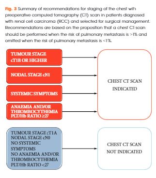
The rate of positive chest CT was 6% (n = 119). At multivariable logistic regression, ≥cT1b, cN1, systemic symptoms and Hb/PLT ratio were all associated with higher risk of positive chest CT (all P < 0.001). After 2000-sample bootstrap validation, the concordance index was found to be 0.88. At decision-curve analysis, the net benefit of the proposed strategy was superior to the select-all and select-none strategies. Accordingly, if chest CT had been performed when the risk of a positive result was >1%, a negative chest CT would have been spared in 37% of the population and a positive chest CT would have been missed in 0.2% of the population only.

Conclusions

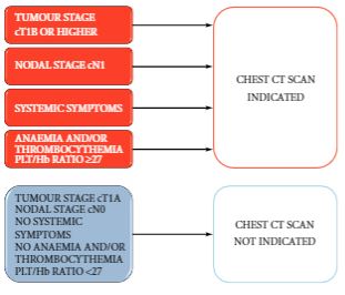
The proposed strategy estimates the risk of positive chest CT at RCC staging with optimum accuracy and the results were statistically and clinically relevant. The findings of the present study support a recommendation for chest CT in patients with ≥cT1b, cN1, systemic symptoms or anaemia and thrombocythemia. Conversely, in patients with cT1a, cN0 without systemic symptoms, anaemia and thrombocythemia, chest CT could be omitted.

Read more articles of the week

Editorial: Do all patients with renal cell carcinoma need a chest computed tomography?

While all patients with RCC need chest imaging for staging evaluation, the answer to the question in the title is ‘No’, and, in fact, many patients would be adequately staged with a chest X-ray, albeit with reduced accuracy. Evidence to support this assertion is provided by Larcher et al. [1] in this issue of BJUI, who retrospectively evaluated 1946 patients with a solitary and sporadic RCC mass. While excluding patients who did not have surgery and those with visceral metastases seen on abdominal imaging, the authors observed pulmonary metastases in 6% (119 patients) of their population. In a multivariable analysis, features associated with a positive chest CT included cT1b+, cN1, systemic symptoms, anaemia, and thrombocytosis. Incorporating these features into a predictive model, the authors report a robust concordance index of 0.88, with the effect of each feature demonstrated in a nomogram. Further, the authors report that if a chest CT is only performed when the risk of a positive result is >1%, 37% of their population could have been spared a chest CT while missing a positive result in only 0.2% (four patients). Patient factors that predict for a <1% risk of a positive chest CT essentially include those with cT1aN0 RCC without systemic symptoms, anaemia, or thrombocytosis. Thus, the authors conclude that in these low-risk patients, a chest CT can be omitted, while any patient that is cT1b+, cN1, or with systemic symptoms, anaemia, or thrombocytosis warrants a dedicated chest CT at diagnosis.

The finding that patients with RCC with smaller tumours (cT1a or ≤4 cm) were unlikely to harbour pulmonary metastases is consistent with prior literature. Observations from the Memorial Sloan-Kettering Cancer Center (MSKCC) [2], and subsequently validated by our group at Mayo Clinic [3], suggested that among surgically treated patients with RCC, risk of M1 disease (at any location) at diagnosis was non-existent for tumours of <2 cm, was <1% for tumours of 2–3 cm, and was only 1–2% for tumours of 3-4 cm in size. Given that it is rare for patients with small renal masses to endorse systemic symptoms or have paraneoplastic symptoms related to the tumour, these prior observations suggest a lack of utility for chest CT for patients with small renal masses supporting the findings from Larcher et al. [1].

In patients with RCC with synchronous metastases, lung is the most common site of spread and guidelines uniformly recommend chest imaging at diagnosis. However, a ‘select-all’ strategy for chest CT in patients with renal masses leads to unnecessary findings in those with a benign primary tumour, increased use of healthcare resources, and relatively frequent findings of indeterminate lesions. In fact, contemporary observations from the MSKCC found that about half of patients with RCC undergoing surgery had indeterminate pulmonary nodules on chest CT that required either additional evaluation or subsequent chest CT to document stability [4]. Further, the presence of indeterminate pulmonary nodules was not associated with distant metastases or death from RCC after surgery unless they were >1 cm, which only represented a small portion (4%) of the entire cohort [4]. Thus, the analysis from Larcher et al. [1] in this issue of BJUI has meaningful clinical relevance; that is, patients with cT1aN0 RCC without symptoms or laboratory abnormalities do not require a chest CT for screening of their lungs.

R. Houston Thompson
Department of Urology, Mayo Clinic, Rochester, MN, USA


Read the full article


References


1 Larcher A, DellOglio P, Fossati N et al. When to perform preoperative chest computed tomography for renal cancer staging. BJU Int 2017; 120: 4906


2 Thompson RH, Hill JR, Babayev Y et al. Metastatic renal cell carcinoma risk according to tumor size. J Urol 2009; 182: 415


3 Umbreit EC, Shimko MS, Childs MA et al. Metastatic potential of renal mass according to original tumour size at presentation. BJU Int 2011; 109: 1904


Article of the Month: Immortal-Time Bias in Urological Research

Every Month the Editor-in-Chief selects an Article of the Month from the current issue of BJUI. The abstract is reproduced below and you can click on the button to read the full article, which is freely available to all readers for at least 30 days from the time of this post.

In addition to the article itself, there is an accompanying editorial written by a prominent member of the urological community. This blog is intended to provoke comment and discussion and we invite you to use the comment tools at the bottom of each post to join the conversation.

Finally, the third post under the Article of the Week heading on the homepage will consist of additional material or media. This week we feature a video discussing the paper.

If you only have time to read one article this week, it should be this one.

Estimating the effect of immortal-time bias in urological research: a case example of testosterone-replacement therapy


Christopher J.D. Wallis*Rek Saskin†‡, Steven A. Narod§, Calvin Law, Girish S. Kulkarni† **, Arun Seth†† and Robert K. Nam*


*Division of Urology, Sunnybrook Health Sciences Centre, Institute for Health Policy, Management and Evaluation, University of Toronto, Institute of Clinical Evaluative Sciences, Sunnybrook Health Sciences Centre, §Department of Public Health Sciences, University of Toronto, Division of General Surgery, Sunnybrook Health Sciences Centre, **Division of Urology, University Health Network, University of Toronto, and ††Department of Anatomic Pathology, Platform Biological Sciences, Sunnybrook Health Sciences Centre, Toronto, ON, Canada


Read the full article

Abstract

Objective

To quantify the effect of immortal-time bias in an observational study examining the effect of cumulative testosterone exposure on mortality.

Patients and Methods

We used a population-based matched cohort study of men aged ≥66 years, newly treated with testosterone-replacement therapy (TRT), and matched-controls from 2007 to 2012 in Ontario, Canada to quantify the effects of immortal-time bias. We used generalised estimating equations to determine the association between cumulative TRT exposure and mortality. Results produced by models using time-fixed and time-varying exposures were compared. Further, we undertook a systematic review of PubMed to identify studies addressing immortal-time bias or time-varying exposures in the urological literature and qualitatively summated these.

Results

Among 10 311 TRT-exposed men and 28 029 controls, the use of a time-varying exposure resulted in the attenuation of treatment effects compared with an analysis that did not account for immortal-time bias. While both analyses showed a decreased risk of death for patients in the highest tertile of TRT exposure, the effect was overestimated when using a time-fixed analysis (adjusted hazard ratio [aHR] 0.56, 95% confidence interval [CI]: 0.52–0.61) when compared to a time-varying analysis (aHR 0.67, 95% CI: 0.62–0.73). Of the 1 241 studies employing survival analysis identified in the literature, nine manuscripts met criteria for inclusion. Of these, five used a time-varying analytical method. Each of these was a large, population-based retrospective cohort study assessing potential harms of pharmacological agents.

Conclusions

Where exposures vary over time, a time-varying exposure is necessary to draw meaningful conclusions. Failure to use a time-varying analysis will result in overestimation of a beneficial effect. However, time-varying exposures are uncommonly utilised among manuscripts published in prominent urological journals.

Read more articles of the week

Editorial: Immortal-Time Bias – A Crucial Yet Overlooked Confounder in Urological Research

The measurement of treatment effect through observational studies has become commonplace in the medical literature. These cohort studies provide valuable data on outcomes that can be difficult to assess in randomized controlled trials, such as long-term mortality. Accurate interpretation of observational data, however, requires accounting for potential confounders of study design, including the immortal-time bias. In this issue of BJUI, Wallis et al. [1] show how accounting for this bias can influence the measured effect of cumulative testosterone exposure on mortality. The implications of their findings extend to several other studies, whose designs may also be subject to immortal-time bias.

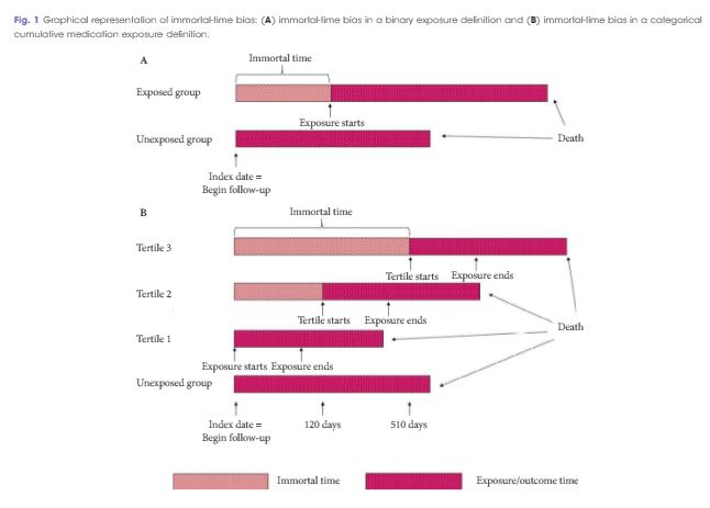
‘Immortal time’ refers to the portion of a follow-up period during which an outcome could not have occurred (e.g. subjects in the ‘exposure group’ cannot die before they receive the exposure); thus, potentially allowing the artificial magnification of an effect on the study outcome [2]. As the authors point out, this concept is not new. It was first identified several decades ago to highlight how a study’s finding of a survival advantage for patients undergoing heart transplant was nullified once immortal time was properly accounted for [3]. Despite its long existence in epidemiological teachings, the authors cite several studies both within and outside of the urological literature that have failed to appropriately account for this bias. Many of these studies employ binary exposure variables, but Wallis et al. delve into relatively uncharted territory by examining the effect of immortal-time bias on multi-level categorical exposures.

The relationship between testosterone replacement therapy (TRT) and mortality, the focus of the accompanying study, is apt because it is a controversial topic that weighs heavily on an accurate assessment of the therapy’s risks and benefits. The authors, using data from their own prior study, show that this delicate balance can be easily tipped when immortal-time bias is not properly accounted for. In their analysis, the overall result was the same regardless of controlling for this bias; men in the lowest tertile of TRT exposure had a higher risk of mortality, and those in the highest tertile had a lower risk of mortality; however, use of a time-fixed as opposed to the more appropriate time-varying analysis led to a substantial magnification of the effect size in each direction. While the overall result may have been the same in this example, the authors cite other instances of high-impact research whose published conclusions were shown to be completely different once accounting for immortal-time bias [4]. One can easily imagine how this type of erroneous data analysis could have deleterious consequences in the clinical setting. Healthcare providers rely on research to make decisions that have far-reaching impacts on patients’ lives. This study highlights the importance of ensuring that such analyses are carried out properly so that patients can receive the high-quality, evidence-based care they deserve.

The authors should be commended for taking the time to deconstruct and evaluate an analytical concept that is pertinent to study designs across several disciplines. Much of the research published today seeks to find answers to important clinical questions, but not nearly enough investigation is devoted to verifying that the analyses to obtain these answers are conducted properly. Urology in particular is a field that is still maturing with respect to the use of secondary data analytical techniques, such as propensity score models and instrumental variables [5]. To sustain our improvement in investigative skills alongside our fellow medical disciplines, we must pay special attention to studies that hold a magnifying glass to commonly used methodologies in the urological literature. In a similar vein, there have been increasing efforts recently to improve the process and transparency of corroborating the results of scientific studies, and these authors’ findings reinforce why these efforts are so crucial. If we expect to continue pushing forward the boundaries of medical research, it is our duty to ensure that our analytical methods are as rigorous and accurate as possible.

Sean A. Fletcher, Philipp Gild and Quoc-Dien Trinh
Division of Urological Surgery and Center for Surgery and Public Health, Harvard Medical School, Brigham and Womens Hospital, Boston, MA, USA


Read the full article


References


2 Suissa S. Immortal time bias in pharmaco-epidemiology. Am J Epidemiol 2008; 167: 4929


3 Gail MH. Does cardiac transplantation prolong life? A reassessment Ann Intern Med 1972; 76: 8157


4 van Walraven C, Davis D, Forster AJ et al. Time-dependent bias was common in survival analyses published in leading clinical journals. J Clin Epidemiol 2004; 57: 67282


5 Cole AP, Trinh QD. Secondary data analysis: techniques for comparing interventions and their limitations. Curr Opin Urol 2017; 27: 3549


© 2024 BJU International. All Rights Reserved.