Tag Archive for: Article of the Week

Posts

Editorial: Is it time for a more ‘proactive’ approach to metastatic prostate cancer?

In this issue of BJU International, Jang et al. [1] investigate the perioperative and oncological outcomes of robot-assisted radical prostatectomy (RARP) in oligometastatic prostate cancer. The authors evaluated a retrospective cohort of 79 patients with oligometastatic prostate cancer, defined as up to five bony metastases on bone scan without visceral metastasis on conventional CT, treated either with RARP (n = 38, 48%) or androgen-deprivation therapy (ADT: n = 41, 52%). They found that cytoreductive RARP was associated with longer progression-free survival (PFS) and cancer-specific survival (CSS) relative to the ADT cohort at a median follow-up of 40 months.

The authors exclusively use the robotic approach for RP in the metastatic prostate cancer setting. Overall, they should be commended for demonstrating the feasibility of RARP in the metastatic setting, with a median operating room time of 147 min and complication rate of ~15%. This is in the same range as the 164 min operative time and 20% complication rate reported in a recent multi-institutional cytoreductive RP (cRP) series, consisting of both open and robotic approaches [2]. However, it is important to note that Jang et al. [1] had a 79% positive margin rate compared to 54% for Sooriakumaran et al. [2]. Furthermore, Jang et al. [1] had a median hospital stay of 5 days compared to 2–3 days in the USA centres [2]. These findings underscore the significant difficulty often encountered in metastatic cases, despite robotic assistance. Thus, we believe the age-old debate of open vs robotic prostatectomy is perhaps less relevant in the cytoreductive setting, where surgeon experience and expertise may be more critical drivers of outcome.

Although there is some evidence in favour of cRP compared to the standard of care, selection bias, limited collection and analysis of much clinical data, and short follow-up often plague most series. Understandably, the current manuscript by Jang et al. [1] also suffers from some of these limitations and leaves some questions unanswered. For instance, did the number of bone metastases, PSA doubling time at diagnosis, and distribution of lymphadenopathy (pelvic vs extra-pelvic) differ between cRP and ADT groups and thus confound the impact of cRP on survival? In addition, the authors may wish to report overall survival, so that their series can be more readily compared to the existing literature. Moreover, the median CSS was only 40 months in the ADT group, whereas it was not even reached in the low-volume arm of the ChemoHormonal Therapy Versus Androgen Ablation Randomized Trial for Extensive Disease in Prostate Cancer (CHAARTED) [3], suggesting that the control group in the current manuscript may have had higher volume metastatic disease. Lastly, there is no evaluation of the impact of adjuvant ADT and radiation therapy on CSS, and further studies may wish to explore if such multi-modal approaches may yield a benefit in the cytoreductive setting.

The existing literature on cRP remains in the nascent stage and is conflicting. The first large studies to suggest a benefit for cRP were retrospective series based on large cancer databases. Subsequently, Heidenreich et al. [4] explored the role of cRP in a case-control study, which found a significantly longer PFS and CSS in a group of 23 men undergoing neoadjuvant ADT + cRP compared to 38 men treated with ADT alone. On the other hand, a prospective investigation comparing 43 men with low-volume bone metastasis treated with cRP to 38 men treated with ADT did not show a benefit for time-to-castration-resistance or overall survival [5]. Moschini et al. [6] also found no survival benefit for cRP with 5-years of follow-up relative to a cohort of ADT patients with CSS more consistent with randomised data from the CHAARTED [3]. The present study by Jang et al. [1] adds to the growing body of retrospective series advocating cRP in select patients.

Whilst a full discussion of the putative biological mechanisms proposed to explain a potential survival benefit of cRP is beyond this editorial, they can be broadly grouped into the following: removal of the primary source of circulating tumour cells, reducing the number of cells that can develop resistant mechanisms for systemic therapy, removal of immunosuppressive cytokines, abscopal effects, and decreasing tumour-growth promoting factors. Ultimately, the ‘proof is in the pudding’, and the results of several randomised trials (Testing radical prostatectomy in men with oligometastatic prostate cancer that has spread to the bone [TRoMbone], NCT01751438, and NCT02454543) are eagerly awaited to determine if cRP can benefit patients.

Matteo Soligo, Vidit Sharma and R. Jeffrey Karnes
Mayo Clinic Urology, Rochester, MN, USA


References


3 Sweeney CJ, Chen YH, Carducci M et al. Chemohormonal therapy in metastatic hormone-sensitive prostate cancer. N Engl J Med 2015; 373: 73746


4 Heidenreich A, Pster D, Porres D. Cytoreductive radical prostatectomy in patients with prostate cancer and low volume skeletal metastases: results of a feasibility and case-control study. J Urol 2015; 193: 8328


6 Moschini M, Morlacco A, Kwon E, Rangel LJ, Karnes RJ. Treatment of M1a/M1b prostate cancer with or without radical prostatectomy at diagnosis. Prostate Cancer Prostatic Dis 2017; 20: 11721


Article of the Week: Comparison of transperineal mpMRI/fusPbx and sysPbx

Every Week the Editor-in-Chief selects an Article of the Week from the current issue of BJUI. The abstract is reproduced below and you can click on the button to read the full article, which is freely available to all readers for at least 30 days from the time of this post.

In addition to the article itself, there is an accompanying editorial written by a prominent member of the urological community. This blog is intended to provoke comment and discussion and we invite you to use the comment tools at the bottom of each post to join the conversation.

If you only have time to read one article this week, it should be this one.

Prospective comparison of transperineal magnetic resonance imaging/ultrasonography fusion biopsy and transrectal systematic biopsy in biopsy-naïve patients


Angelika Borkowetz*, Boris Hadaschik†‡, Ivan Platzek§, Marieta Toma, Georgi TosevTheresa Renner*, Roman Herout*, Martin Baunacke*, Michael Laniado §, Gustavo Baretton, Jan Philipp Radtke, Claudia Kesch, Markus Hohenfellner† , Michael Froehner*, Heinz-Peter Schlemmer**, Manfred Wirth* and Stefan Zastrow*


*Department of Urology, Technische Universitat Dresden, Dresden, Germany, Department of Urology, University Hospital Heidelberg, Heidelberg, Germany, Department of Urology, University Hospital Essen, Essen, Germany, §Department of Radiology and Interventional Radiology, Technische Universitat Dresden, Dresden, Germany, Department of Pathology, Technische Universitat Dresden, Dresden, Germany, and **Department of Radiology, German Cancer Research Centre (DKFZ), Heidelberg, Germany


Read the full article

Abstract

Objectives

To evaluate the value of multiparametric magnetic resonance imaging (mpMRI) in the detection of significant prostate cancer (PCa) and to compare transperineal MRI/ultrasonography fusion biopsy (fusPbx) with conventional transrectal systematic biopsy (sysPbx) in biopsy-naïve patients.

Patients and Methods

This multicentre, prospective trial investigated biopsy-naïve patients with suspicion of PCa undergoing transperineal fusPbx in combination with transrectal sysPbx (comPbx). The primary outcome was the detection of significant PCa, defined as Gleason pattern 4 or 5. We analysed the results after a study period of 2 years.

Results

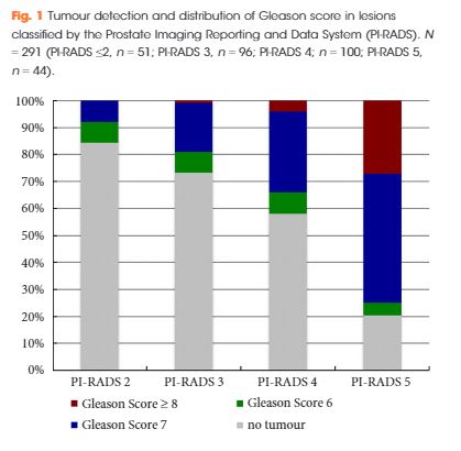
The study included 214 patients. The median (range) number of targeted and systematic cores was 6 (2–15) and 12 (6–18), respectively. The overall PCa detection rate of comPbx was 52%. FusPbx detected more PCa than sysPbx (47% vs 43%; P = 0.15). The detection rate of significant PCa was 38% for fusPbx and 35% for sysPbx (P = 0.296). The rate of missed significant PCa was 14% in fusPbx and 21% in sysPbx. ComPbx detected significantly more significant PCa than fusPbx and sysPbx alone (44% vs 38% vs 35%; P < 0.005). In patients presenting with Prostate Imaging Reporting and Data System (PI-RADS) 4 and 5 lesions there was a higher detection rate of significant PCa than in patients presenting with PI-RADS ≤3 lesions in comPbx (61% vs 14%; P < 0.005).

Conclusions

For biopsy-naïve men with tumour-suspicious lesions in mpMRI, the combined approach outperformed both fusPbx and sysPbx in the detection of overall PCa and significant PCa. Thus, biopsy-naïve patients may benefit from sysPbx in combination with mpMRI targeted fusPbx.

Read more articles of the week

Editorial: The new frontier of prostate biopsy: determining the role of image-guidance in moving the needle

One of the most pressing topics in urological oncology concerns the role of MRI/ultrasonography (US)-fusion guided biopsies in detecting prostate cancer. The literature on this emerging technology is permeated by questions regarding when it should be used, how it should be performed, and which patients stand to benefit. The stakes are high to figure this out, as real patients will continue to suffer from missteps made in diagnosis and treatment of prostate cancer whilst we seek to improve our detection methods.

In this issue of BJUI, Borkowetz et al. [1] compare prostate cancer detection rates between MRI/US-fusion targeted and conventional systematic biopsies. They prospectively enrolled a cohort of biopsy-naïve men who had an elevated PSA level and/or an abnormal DRE. All men received both a transperineal MRI/US-fusion biopsy and a systematic TRUS-guided biopsy. They found that combining both approaches led to improved detection rates for clinically significant and overall prostate cancer compared to either method alone.

A strength of this study [1] is its focus on biopsy-naïve men, for which data on the comparison of biopsy techniques is relatively limited. Siddiqui et al. [2] were instrumental in demonstrating that targeted MRI/US-fusion biopsy, compared to the systematic TRUS biopsy, was associated with improved diagnostic accuracy for higher-risk tumours and decreased detection of low-risk disease. However, the large majority of men in the study had received a prior biopsy, making it difficult to generalise the results to biopsy-naïve men. Focusing on biopsy-naïve men is of great importance – men referred for a biopsy after an elevated PSA level or abnormal DRE increasingly face conflicting opinions about the next best step in diagnostic evaluation, especially given the recent influx of imaging and biomarker tests that aim to guide this decision point.

One group previously compared MRI-guided and systematic TRUS biopsy in a large group of biopsy-naïve of men and found the MRI-guided in-bore technique to be superior in detecting significant cancers and avoiding insignificant cancers [3]. However, that study was limited by both its definition of ‘significant cancer’ (it included Gleason 3 + 3 disease) and the variance in the definition of ‘low-risk’ cancer between MRI-guided and TRUS biopsies. By contrast, the present authors uniformly defined significant cancer as Gleason ≥3 + 4. This is just one example highlighting the widespread disagreement over the parameters used to measure the efficacy of targeted-biopsy techniques in cancer detection. Continued incorporation of standardised scales such as the Prostate Imaging Reporting and Data System (PI-RADS), which was also used in the accompanying article, will help to mitigate some of this disagreement in future studies. Of course, these standardised scales are still subject to inter- and intra-observer variability, which may decrease with further clarification of the grading systems, as well as appropriate reader training [4].

Whilst Borkowetz et al. [1] helped to fill important gaps in the literature, their study was not without limitations. They included PI-RADS 2 scores for targeted biopsies although many urologists would consider these lesions insignificant and not worth targeting. Furthermore, comparing a transperineal MRI/US-fusion with a transrectal systematic approach makes it difficult to separate the true effect of the targeted approach from that of the anatomical approach. Using both methods in each patient also precludes any comparison of complication rates between the biopsy approaches, an important clinical endpoint for both the urologists administering the biopsies and the patients enduring the complications.

Methodology aside, the new frontier of prostate biopsy technique still relies on a basic triad of efficacy: (i) improved accuracy of cancer detection, (ii) reduced complication rates, and (iii) manageable cost and practicality of widespread implementation. For the first tenet, the recently published PROstate MRI Imaging Study (PROMIS) trial cemented the ability of multi-parametric MRI to detect clinically significant prostate cancer with greatly improved sensitivity and negative predictive value over TRUS [5]. The ongoing randomised PRostate Evaluation for Clinically Important Disease: Sampling Using Image-guidance Or Not? (PRECISION) trial will hopefully add to the step taken by Borkowetz et al. [1], by comparing MRI-targeted and systematic TRUS biopsies on the outcomes of accuracy in cancer detection, adverse events, patient health-related quality of life, and cost [6]. Continued investigation in all of these areas will be crucial to guiding how we move the needle in prostate cancer diagnostics.

Sean A. Fletcher, Sebastian Berg and Quoc-Dien Trinh


Division of Urological Surgery, Center for Surgery and Public Health, Brigham and Womens Hospital, Harvard Medical School, Boston, MA, USA


References


1 Borkowetz AHadaschik BPlatzek I et al. Prospective comparison of transperineal magnetic resonance imaging/ultrasonography fusion biopsy and transrectal systematic biopsy in biopsy-naive patients. BJU Int 2018;121: 5360


2 Siddiqui MM, Rais-Bahrami STurkbey B et al. Comparison of MR/ultrasound fusion-guided biopsy with ultrasound-guided biopsy for the diagnosis of prostate cancer. JAMA2015; 313: 3907


3 Pokorny MRde Rooij MDuncan E et al. Prospective study of diagnostic accuracy comparing prostate cancer detection by transrectal ultrasound-guided biopsy versus magnetic resonance (MR) imaging with subsequent MR-guided biopsy in men without previous prostate biopsies. Eur Urol 2014; 66: 229


5 Ahmed HUEl-Shater Bosaily A,Brown LC et al. Diagnostic accuracy of multi-parametric MRI and TRUS biopsy in prostate cancer (PROMIS): a paired validating confirmatory study. Lancet 2017; 389:81522


6 ClinicalTrials.gov. PRostate Evaluation for Clinically Important Disease: Sampling Using Image-guidance or Not? (PRECISION), Identification No. NCT02380027 (2017). Available at: https://clinicaltrials.gov/ct2/show/NCT02380027. Accessed October 2017.


Article of the Week: Impact of warm ischaemia time on postoperative renal function after partial nephrectomy for clinical T1 renal cell carcinoma

Every Week the Editor-in-Chief selects an Article of the Week from the current issue of BJUI. The abstract is reproduced below and you can click on the button to read the full article, which is freely available to all readers for at least 30 days from the time of this post.

In addition to the article itself, there is an accompanying editorial written by a prominent member of the urological community. This blog is intended to provoke comment and discussion and we invite you to use the comment tools at the bottom of each post to join the conversation.

If you only have time to read one article this week, it should be this one.

Impact of warm ischaemia time on postoperative renal function after partial nephrectomy for clinical T1 renal cell carcinoma: a propensity score-matched study

Hakmin Lee*, Byung D. Song*, Seok-Soo Byun*, Sang E. Lee* and Sung K. Hong*
*Department of Urology, Seoul National University Bundang Hospital, Seongnam, and Department of Urology, Seoul National University College of Medicine, Seoul, Korea


Read the full article

Objectives

To analyse the effect of prolonged warm ischaemia time (WIT) on long-term renal function after partial nephrectomy (PN), as controversy still exists as to whether prolonged WIT adversely affects the incidence of chronic kidney disease (CKD) after PN.

Patients and Methods

We reviewed data from 1816 patients who underwent PN for a clinical T1 renal tumour. The propensity scores for prolonged WIT were calculated with the shorter WIT group (<30 min) matched to the longer WIT group (≥30 min) in a 2:1 ratio. Multivariate analysis was used to determine independent predictors for occurrence of postoperative CKD [defined as an estimated glomerular filtration rate (eGFR) of <60 mL/min/1.73 m2] and major renal function deterioration (MRFD; defined as an eGFR decrease of ≥25% postoperatively).

Results

After propensity score matching, there was no significant difference in CKD-free survival between the two WIT groups (P = 0.787). Furthermore, longer WIT did not show any significant associations with postoperative CKD-free survival [hazard ratio (HR) 1.002, 95% confidence interval (CI) 0.989–1.015; P = 0.765) and MRFD-free survival (HR 1.014, 95% CI 1.000–1.028; P = 0.055). From further subgroup analyses using more specific WIT thresholds (≤20, 21–30, 31–40, 41–50, ≥50 min) and status of preoperative CKD, no significant differences were noted in CKD and MRFD-free survival amongst the subgroups (all P > 0.05).

Conclusions

Prolonged WIT was not associated with increased incidence of CKD or MRFD after PN.

Read more articles of the week

Editorial: Impact of warm ischaemia time during partial nephrectomy on renal function – is it really a matter of time?

In the latest edition of the BJUI, Lee et al. [1] have revisited the question of defining the ideal limit of warm ischaemia time (WIT) and its impact on postoperative renal function in patients undergoing partial nephrectomy (PN).

Partial nephrectomy has replaced radical nephrectomy as the preferred treatment for T1 renal masses. This publication challenges the theory that ischaemic nephropathy is inevitable if the renal vessels are clamped beyond 30 min, leading to a long-term decline in renal function.

The authors in this series are to be commended for analysing a prospectively collected database on 1 816 patients in two institutions who underwent PN for clinical T1 renal tumours. Their primary endpoint was to investigate the impact of prolonged WIT on long-term renal function focusing on two clinical endpoints; chronic kidney disease, as estimated by an estimated GFR of <60 mL/min/1.73 m2, and major renal function deterioration defined as an increase in creatinine of >25% of the preoperative value.

Warm ischaemia time using a threshold of 30 min created two comparative groups. Patients were followed for up to 40 months after surgery. In addition to this, patients were further sub-stratified into five subgroups, critiquing the effect of WIT up to 50 min. A key feature of this paper [1] is the use of propensity score matching to adjust for any potential preoperative confounders affecting postoperative renal function, a technique also allowing matching of the two groups.

The authors correctly emphasise the direct relationship between tumour size and duration of WIT, with larger tumours requiring excision of more renal parenchyma and adding to ‘on-clamp’ time. Tumour size and renal function are vital determinants of suitability for PN [2]. This publication [1] clearly demonstrates that although large tumour size equated with prolonged clamp time, this was not the sole determinant of impaired long-term renal function.

The importance of other independent variables such as preoperative renal function, patient age and preserved renal parenchyma have been highlighted here as potentially playing a greater role than was previously appreciated.

The second and possibly more remarkable finding from this paper is that ischaemic time was not an independent predictor of ultimate renal function after PN. This contrasts with most other reports to date. Although not recommending using a WIT of up to 50 min, the results here suggest this may not be relevant to future renal function. It appears that long-term renal function after PN is primarily determined by the quantity and quality of renal parenchyma preserved, although the type and duration of ischaemia remain the most important modifiable factors, and warrant further evaluation [3].

When discussing this topic, it is interesting to refer to the initial bench work on this issue. The current approach to WIT is extrapolated from data derived from the histological changes occurring in nephrons during operative stone cases. From the data presented in this and other studies, it seems more relevant than ever to conduct clinical trials to assess this appropriately. Traditionally the time threshold for WIT is taken as 30 min, an arbitrarily placed time-point based on the above laboratory data. Beyond this value in the setting of room temperature renal ischaemia creates an array of injury centred on cellular adaptations beginning ~20 min after clamping and persisting beyond 60 min. This indicates that the traditional 30-min limit of WIT is a somewhat subjective time point and was not based on clinical outcomes.

Previous evidence suggests a 5% increase in risk for acute renal failure for every additional minute of WIT [4]. It is hard to ignore such data in exchange for this a contemporary study when so much is at stake for patient longevity. Advocators of zero-ischaemia PN have shown that those who benefit most from a zero-ischaemia technique are those with the poorest baseline renal function [5]. Most of these studies have shown that the renal functional outcomes are either equivalent or superior in zero-ischaemia cases involving small renal tumours [6].

On balance, the authors are to be credited with tackling such a controversial matter and highlighting the lack of good quality laboratory data. Clearly, factors other than WIT contribute to postoperative renal function but for now we must conclude that every minute ‘on-clamp’ does count.

Eva M. Bolton and Thomas H. Lynch
St. Jamess Hospital, Dublin, Ireland
Read the full article

References

1 Lee H, Song BD, Byun SS, Lee SE, Hong SK. Impact of warm ischaemia time on postoperative renal function after partial nephrectomy for clinical T1 renal cell carcinoma: a propensity score-matched study. BJU Int 2018; 121: 4652


2 Volpe A, Blute ML, Ficarra V et al. Renal ischemia and function after partial nephrectomy: a collaborative review of the literature. Eur Urol 2015; 68: 6174


3 Lane BR, Russo P, Uzzo RG et al. Comparison of cold and warm ischemia during partial nephrectomy in 660 solitary kidneys reveals predominant role of nonmodiable factors in determining ultimate renal function. J Urol 2011; 185: 4217


4 Thompson RH, Lane BR, Lohse CM et al. Every minute counts when the renal hilum is clamped during partial nephrectomy. Eur Urol 2010;58: 3405


6 Salami SS, George AK, Rais-Bahrami S, Okhunov Z, Waingankar NKavoussi LR. Off-clamp laparoscopic partial nephrectomy for hilar tumors: oncologic and renal functional outcomes. J Endourol 2014; 28: 1915


Article of the Month: NICE Guidance – Routine preoperative tests for elective surgery

Every Month the Editor-in-Chief selects an Article of the Month from the current issue of BJUI. The abstract is reproduced below and you can click on the button to read the full article, which is freely available to all readers for at least 30 days from the time of this post.

In addition to the article itself, there is an accompanying editorial written by a prominent member of the urological community. This blog is intended to provoke comment and discussion and we invite you to use the comment tools at the bottom of each post to join the conversation.

If you only have time to read one article this week, it should be this one.

NICE Guidance – Routine preoperative tests for elective surgery


Read the full article

Overview

This guideline covers routine preoperative tests for people aged over 16 who are having elective surgery. It aims to reduce unnecessary testing by advising which tests to offer people before minor, intermediate and major or complex surgery, taking into account specific comorbidities (cardiovascular, renal and respiratory conditions and diabetes and obesity). It does not cover pregnant women or people having cardiothoracic procedures or neurosurgery.

Who is it for?

  • Healthcare professionals
  • People having elective surgery, their families and carers

This guideline updates and replaces NICE guideline CG3 (published June 2003).

Recommendations

People have the right to be involved in discussions and make informed decisions about their care, as described in your care [https://www.nice.org.uk/about/nice-communities/public-involvement/your-care].

We expect you to take our guidance into account. But you should always base decisions on the person you are working with.

Making decisions using NICE guidelines [https://www.nice.org.uk/about/what-we-do/our-programmes/nice-guidance/nice-guidelines/using-NICE-guidelines-to-make-decisions] explains how we use words to show the strength (or certainty) of our recommendations, and has information about prescribing medicines (including off-label use), professional guidelines, standards and laws (including on consent and mental capacity), and safeguarding.

Guidance on consent for young people aged 16–17 is available from the reference guide to consent for examination or treatment [https://www.gov.uk/government/publications/reference-guide-to-consent-for-examination-or-treatment-second-edition] (Department of Health).

The tests covered by this guideline are:

  • chest X-ray
  • echocardiography (resting)
  • electrocardiography (ECG; resting)
  • full blood count (haemoglobin, white blood cell count and platelet count)
  • glycated haemoglobin (HbA1c) testing
  • haemostasis tests
  • kidney function (estimated glomerular filtration rate, electrolytes, creatinine and sometimes urea levels)
  • lung function tests (spirometry, including peak expiratory flow rate, forced vital capacity and forced expiratory volume) and arterial blood gas analysis
  • polysomnography
  • pregnancy testing
  • sickle cell disease/trait tests
  • urine tests.

The recommendations were developed in relation to the following comorbidities:

  • cardiovascular
  • diabetes
  • obesity
  • renal
  • respiratory.


Read more articles of the week

Editorial: Viewpoint – Rationing and Surgical Care

Limitation in the provision of surgical care has many causes. In a nationalised healthcare system, this often reflects lack of funds, leading to rationing of clinical services. Rationing itself takes a number of forms. Deliberate exclusion of specific operations (usually elective) or specific patient groups (smokers, obese) are the most common examples, but strategic extension of waiting times by the removal of ‘target’ times can also be used as a rationing tool.

Many surgeons are dismayed by these decisions. They feel that the surgical patient is unfairly targeted as the clinical and cost-effectiveness of many planned surgical interventions have been well characterised. Surgeon and institutional outcomes are freely available – unlike the situation in many non-surgical specialties, so how can it be fair to pick on the surgical patient?

The idea that non-urgent elective surgery falls into neat categories where delay has no adverse consequences for the patient mystifies many surgeons. Whilst all would advocate a healthy diet, exercise, weight loss and smoking cessation, decisions to withhold surgery from the obese or those who smoke is rarely evidence-based. Rationing based on such prejudice soon becomes illogical. Why should the obese cancer patient receive an operation when the obese incontinent patient cannot?

In the long term, the absence of a substantial volume of ‘routine’ surgery damages training as exposure to such procedures is limited. Surgery has become the soft target for rationing clinical services. Surgeons should make their patients aware of how this process will affect them. Healthcare planners need to hear a public voice as well as that of the clinicians.

Just occasionally, an apparent limitation can be beneficial. In this issue of the BJUI, the National Institute for Health and Care Excellence (NICE) provides clear guidance on preoperative testing. This is based on sensible recommendations such as: avoiding routine urine dipstick testing, routine chest X-rays, and glycated haemoglobin (HbA1c) in non-diabetic patients. All surgeons irrespective of their specialty would benefit from paying close attention to these important guidelines [1].

Derek Alderson

President of the Royal College of Surgeons of England; Emeritus Professor of Surgery, University of Birmingham; Editor-in-chief of BJS Open.

Read the full article

Reference

1 National Institute of Health and Care Excellence (NICE). Routine preoperative tests for elective surgery: © NICE (2016) Routine preoperative tests for elective surgery. BJU Int 2018; 121: 12–6


Article of the Week: Oncological outcomes and toxicity for LDR prostate brachytherapy

Every Week the Editor-in-Chief selects an Article of the Week from the current issue of BJUI. The abstract is reproduced below and you can click on the button to read the full article, which is freely available to all readers for at least 30 days from the time of this post.

In addition to the article itself, there is an accompanying editorial written by a prominent member of the urological community. This blog is intended to provoke comment and discussion and we invite you to use the comment tools at the bottom of each post to join the conversation.

If you only have time to read one article this week, it should be this one.

Long-term oncological outcomes and toxicity in 597 men aged ≤60 years at time of low-dose-rate brachytherapy for localised prostate cancer

Stephen E. M. Langley, Ricardo Soares, Jennifer Uribe, Santiago Uribe-LewisJulian Money-Kyrle, Carla Perna, Sara Khaksar and Robert Laing


St Lukes Cancer Centre, Guildford, Surrey, UK


Read the full article

Objectives

To report oncological and functional outcomes of men treated with low-dose-rate (LDR) prostate brachytherapy aged ≤60 years at time of treatment.

Patients and Methods

Of 3262 patients treated with LDR brachytherapy at our centre up to June 2016, we retrospectively identified 597 patients aged ≤60 years at treatment with ≥3-years post-implantation follow-up and four prostate-specific antigen (PSA) measurements, of which one was at baseline. Overall survival (OS), prostate cancer-specific survival (PCSS) and relapse free survival (RFS) were analysed together with prospectively collected physician-reported adverse events and patient-reported symptom scores.

Results

The median (range) age was 57 (44-60) years, follow-up was 8.9 (1.5-17.2) years, and PSA follow-up 5.9 (0.8-15) years. Low-, intermediate- and high-risk disease represented 53%, 37% and 10% of the patients, respectively. At 10 years after implantation OS and PCSS were 98% and 99% for low-risk, 99% and 100% for intermediate-risk, and 93% and 95% for high-risk disease, respectively. At 10 years after implantation RFS, using the PSA level nadir plus 2 ng/mL definition, was 95%, 90% and 87% for low-, intermediate-, and high-risk disease, respectively. Urinary stricture was the most common genitourinary adverse event occurring in 19 patients (3.2%). At 5 years after implantation erectile function was preserved in 75% of the patients who were potent before treatment.

Conclusion

LDR brachytherapy is an effective treatment with long-term control of prostate cancer in men aged ≤60 years at time of treatment. It was associated with low rates of treatment-related toxicity and can be considered a first-line treatment for prostate cancer in this patient group.

Read more articles of the week

Editorial: LDR prostate brachytherapy in younger men

Langley et al.1 report on the oncological and functional outcomes of men treated with low dose rate (LDR) prostate brachytherapy in men 60 years old or younger. 597 patients with a median (range) age of 57 (44-60) years had a median follow-up of 8.9 (1.5-17.2) years. The 10- year post-implant relapse free survival using the Phoenix definition for biochemical failure (nadir plus 2 ng/ml) was 95%, 90% and 87% for low, intermediate and high-risk disease, respectively. Potency was preserved in 75% of men potent before treatment. The authors concluded that LDR brachytherapy is an efficacious treatment with excellent long-term control of prostate cancer in men ≤60 years at time of treatment. While the results from this investigation are encouraging, enthusiasm should be tempered given the short follow up, long natural history of prostate cancer and the long-life expectancy for these younger patients.

Although the overall median follow-up was 8.9 years, the calculation of PSA-free failure was derived from a median follow-up of 5.9 years. As this investigation did not identify men who may also be at risk of failure because of a rising PSA and who have not yet reached the Phoenix threshold, I anticipate longer-follow up will further reduce their favorable results. Of the 597 men, 6 (1%) died from prostate cancer. The low incidence of prostate cancer mortality, while impressive, also reflects the short follow-up. Our group has previously reported that PCSM substantially increases between the 10th and 15th year post treatment. The experience of these physicians in prostate brachytherapy is demonstrated in their favorable dosimetry outcomes-the median D90 was 106.4% of the prescription (145 Gy). These results (median D90 154.3 Gy), which were determined and computed on the day of the implant would be 10-15% higher had the CT scans been done on day 30 as most centers do. We and others have reported that patients receiving higher dose implants have improved biochemical and cancer-specific outcomes. While these data help explain their favorable oncological outcomes, they should also serve as a guide to other brachytherapy programs where implant quality should be a primary objective. Erectile function was preserved in 75% of men. These data are consistent with other reports of younger men who were treated for prostate cancer with surgery or radiation. Because this was not a randomized study, it is not possible to make direct comparisons between surgery and brachytherapy. The selection of an IIEF score of > 11 as potent might be challenged as the 12-16 group is considered to have mild to moderate ED. Nonetheless, these data are still encouraging for younger men who are considering treatment for localized prostate cancer where sexual function preservation is important.

Nelson Stone

Mount Sinai Medical Center – Urology 350 E 72nd Street, New York, New York 10021 United States

Read the full article

Reference

  1. Langley SM, Soares R, Uribe J, et al. Long-term oncological outcomes and toxicity in 597 men ≤60 years of age at time of low dose rate brachytherapy for localised prostate cancer. BJU Int 2017


Article of the Week: Prostate Health Index density improves detection of clinically significant prostate cancer

Every Week the Editor-in-Chief selects an Article of the Week from the current issue of BJUI. The abstract is reproduced below and you can click on the button to read the full article, which is freely available to all readers for at least 30 days from the time of this post.

In addition to the article itself, there is an accompanying editorial written by a prominent member of the urological community. This blog is intended to provoke comment and discussion and we invite you to use the comment tools at the bottom of each post to join the conversation.

Finally, the third post under the Article of the Week heading on the homepage will consist of additional material or media. This week we feature a video discussing the paper.

If you only have time to read one article this week, it should be this one.

Prostate Health Index density improves detection of clinically significant prostate cancer

Jeffrey J. Tosoian*, Sasha C. Druskin*, Darian Andreas*, Patrick Mullane*, Meera Chappidi*, Sarah Joo*, Kamyar Ghabili*, Mufaddal Mamawala*, Joseph Agostino*, Herbert B. Carter*, Alan W. Partin*, Lori J. Sokoll*§ and Ashley E. Ross*§


*Department of Urology, James Buchanan Brady Urological Institute, Johns Hopkins University School of Medicine, Baltimore, MD, Virginia Commonwealth University School of Medicine, Richmond, VA, Department of Pathology, and §Sidney Kimmel Comprehensive Cancer Center, Johns Hopkins University School of Medicine, Baltimore, MD, USA
Read the full article

Abstract

Objectives

To explore the utility of Prostate Health Index (PHI) density for the detection of clinically significant prostate cancer (PCa) in a contemporary cohort of men presenting for diagnostic evaluation of PCa.

Patients and Methods

The study cohort included patients with elevated prostate-specific antigen (PSA; >2 ng/mL) and negative digital rectal examination who underwent PHI testing and prostate biopsy at our institution in 2015. Serum markers were prospectively measured per standard clinical pathway. PHI was calculated as ([{−2}proPSA/free PSA] × [PSA]½), and density calculations were performed using prostate volume as determined by transrectal ultrasonography. Logistic regression was used to assess the ability of serum markers to predict clinically significant PCa, defined as any Gleason score ≥7 cancer or Gleason score 6 cancer in >2 cores or >50% of any positive core.

Results

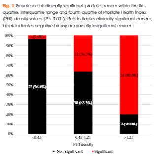
Of 118 men with PHI testing who underwent biopsy, 47 (39.8%) were found to have clinically significant PCa on biopsy. The median (interquartile range [IQR]) PHI density was 0.70 (0.43–1.21), and was 0.53 (0.36–0.75) in men with negative biopsy or clinically insignificant PCa and 1.21 (0.74–1.88) in men with clinically significant PCa (P < 0.001). Clinically significant PCa was detected in 3.6% of men in the first quartile of PHI density (<0.43), 36.7% of men in the IQR of PHI density (0.43–1.21), and 80.0% of men with PHI density >1.21 (P < 0.001). Using a threshold of 0.43, PHI density was 97.9% sensitive and 38.0% specific for clinically significant PCa, and 100% sensitive for Gleason score ≥7 disease. Compared with PSA (area under the curve [AUC] 0.52), PSA density (AUC 0.70), %free PSA (AUC 0.75), the product of %free PSA and prostate volume (AUC 0.79), and PHI (AUC 0.76), PHI density had the highest discriminative ability for clinically significant PCa (AUC 0.84).

Conclusions

Based on the present prospective single-centre experience, PHI density could be used to avoid 38% of unnecessary biopsies, while failing to detect only 2% of clinically significant cancers.

Read more articles of the week
© 2024 BJU International. All Rights Reserved.