Tag Archive for: Article of the Week

Posts

Editorial: Guidelines vs reality of practice (two sides of the same coin) and lifelong learning!

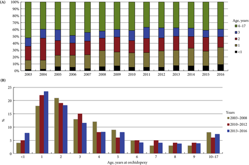
Undescended testis (UDT) is a common paediatric congenital abnormality, with an incidence of 1:100. UDT is managed by paediatricians, paediatric surgeons, paediatric and adult urologists. A consensus document was created to perform this surgery early, at ~6 months of age and definitely before 1 year of age, because of the risk of lower fertility rates and malignancy in the future [1, 2].

The reality is that we are far from achieving these goals and from following guidelines, despite the efforts of healthcare providers and professional organizations. Why is this? Is the following triad not coming together well?

  1. Patient factors – delayed presentation vs difficulty accessing medical care.
  2. Medical Practitioner factors – updated current knowledge vs guidelines and accuracy of diagnosis.
  3. Resources – healthcare costs and availability of expert medical care.

Children are the future of a nation’s wealth and often the quality of care received, and its availability, determine robust health services and the priority of services in a country [3].

In the current issue of BJUI, Boehme et al. [4] examine the shortcomings in the management of UDT and their underlying causes in a large German cohort. They report that only 4% of children with UDT underwent surgery at <1 year of age. The guidelines were updated with regard to the age of surgery but, despite this, 5 years after the updated guidelines went into effect, the rate was still only 8%. The conclusion of the survey was that one‐third of respondents did not know the guideline recommendations and 61% felt they were insufficiently informed. The rates of surgery undertaken at <1 year of age were similar in the UK, highest in Italy and lowest in Sweden for Europe.

Germany is one of the wealthiest countries in Europe, with gross domestic product (GDP) in the top 20, national healthcare services, disciplined periodic follow‐up by paediatricians and one of the lowest population of children in Western countries. Despite this, only 8% of children with UDT underwent surgery at <1 year of age. Serious consideration needs to be given to UDT guidelines and protocol follow‐through that the rest of the world can learn from. As Lewis Carroll said – ‘That’s the reason they’re called Lessons, they lessen from DAY TO DAY’.

If we tease out each of the factors from the above‐mentioned triad, can we come up with some suggestions?

Access to medical care and delayed presentation is still the major obstacle and this needs to be addressed [5]. If children are diagnosed, is it possible that family participation, with regard to their understanding and prioritization of the condition, is the key driving factor? Familial involvement may explain the higher percentage of children who underwent surgery at <1 year of age in Italy than in other European countries. Boehme et al.[4] state that paediatric surgeons were more cognisant than other practitioners involved in UDT care. Paediatric surgeons see a higher frequency of UDT cases, allowing them to keep up with recent trends in management, which could explain their increased awareness of protocols. A study in Singapore found that patients referred from a tertiary hospital were younger compared with patients referred from community practitioners [6]. Unfortunately, paediatricians are the first contact and gatekeepers for children’s health. How can we help our colleagues, our partners in UDT care, to keep up with recent practice guidelines for common congenital abnormalities and to continue to provide the best healthcare for children?

Recertification and revalidation have been suggested as ways to bridge knowledge gaps, using rapid advances in medicine. Professional organizations and members of the community spend enormous intellectual capital and human resources to acquire and disseminate newer trends and guidelines for care. Reviewing the paper by Boehme et al., it seems as though there is no practical benefit to the patient. Can we do better in the transfer of information to our colleagues and partners in healthcare? Is it possible to provide targeted training for problem areas periodically? Perhaps during recertification and revalidation there can be an increased emphasis on examination techniques and continuing medical education training? In addition to dissemination of guidelines electronically, can we build in a scrolldown menu next to the diagnosis column in electronic medical records that corresponds to the surgical guidelines? Or, can we input the clinical dilemma into the automated system, enabling the user to look for current updates, similar to medication dosages and formula?

Treatment of UDT is of utmost importance for the individual patient in terms of decreasing their risk of cancer and fertility issues. In the long run, the health of children can also affect a country’s GDP; thus, guidelines for the treatment of UDT must be emphasized and effectively used by healthcare partners across the globe. Visit the https://www.northraleighpediatrics.com/our-providers/ website to get all the details.

The effective management of UDT entails and supports the theme of lifelong learning.

Mohan S. Gundeti

Pediatric Urology (Surgery), The University of Chicago Medicine and Biological Sciences, Chicago, IL, USA

References

  1. Ritzén EM, Bergh A, Bjerknes R et al. Nordic consensus on treatment of undescended testes. Acta Paediatr 2007; 96: 638–43
  2. British Association of Pediatric Urologists. The BAPU Consensus Statement on the Management of Undescended Testis 2013. https://www.bapu.org.uk/udt-consensus-statement/
  3. Bruijnen CJ, Vogels HD, Beasley SW. Age at orchidopexy as an indicator of the quality of regional child health services. J Paediatr Child Health 2012; 48: 556–9
  4. Boehme P, Geis B, Doerner J. Shortcomings in the management of undescended testis: guideline intention vs. reality and the underlying causes – insights from the biggest German cohort. BJU Int 2018; 122: 644–53
  5. Yiee JH, Saigal CS, Lai J. Timing of orchiopexy in the United States: a quality‐of‐care indicator. Urology 2012; 80: 1121–6
  6. Nah SA, Yeo CS, How GY et al. Undescended testis: 513 patients’ characteristics, age at orchidopexy and patterns of referral. Arch Dis Child 2014; 99: 401–6

 

Residents’ podcast: Urinary continence recovery after radical prostatectomy

Maria Uloko is a Urology Resident at the University of Minnesota Hospital and Giulia Lane is a Female Pelvic Medicine and Reconstructive Surgery Fellow at the University of Michigan.

In this podcast they discuss the BJUI Article of the Week ‘Investigating the mechanism underlying urinary continence recovery after radical prostatectomy: effectiveness of a longer urethral stump to prevent urinary incontinence‘.


Investigating the mechanism underlying urinary continence recovery after radical prostatectomy: effectiveness of a longer urethral stump to prevent urinary incontinence


Yoshifumi Kadono*, Takahiro Nohara*, Shohei Kawaguchi*, Renato Naito*, Satoko Urata*, Kazufumi Nakashima*, Masashi Iijima*, Kazuyoshi Shigehara*, Kouji Izumi*, Toshifumi Gabata† and Atsushi Mizokami*

*Department of Integrative Cancer Therapy and Urology, Kanazawa University Graduate School of Medical Science, Kanazawa, Japan; †Department of Radiology, Kanazawa University School of Medicine, 13‐1 Takara‐machi, Kanazawa, Ishikawa 920‐8640, Japan

Read the full article

Abstract

Objective

To assess the chronological changes in urinary incontinence and urethral function before and after radical prostatectomy (RP), and to compare the findings of pelvic magnetic resonance imaging (MRI) before and after RP to evaluate the anatomical changes.

Patients and Methods

In total, 185 patients were evaluated with regard to the position of the distal end of the membranous urethra (DMU) on a mid‐sagittal MRI slice and urethral sphincter function using the urethral pressure profilometry. The patients also underwent an abdominal leak point pressure test before RP and at 10 days and 12 months after RP. The results were then compared with the chronological changes in urinary incontinence.

Fig. 1 Intraoperative view of the apex of the prostate transection line between the urethra and prostate at the normal (straight line) and long urethral stump (dashed line) positions.

Results

The MRI results showed that the DMU shifted proximally to an average distance of 4 mm at 10 days after RP and returned to the preoperative position at 12 months after RP. Urethral sphincter function also worsened 10 days after RP, with recovery after 12 months. The residual length of the urethral stump and urinary incontinence were significantly associated with the migration length of the DMU at 10 days after RP. The residual length of the urethral stump was a significant predictor of urinary incontinence after RP.

Conclusion

This is the first study to elucidate that the slight vertical repositioning of the membranous urethra after RP causes chronological changes in urinary incontinence. A long urethral residual stump reduces urinary incontinence after RP.


Read more articles of the week

BJUI Podcasts now available on iTunes, subscribe here https://itunes.apple.com/gb/podcast/bju-international/id1309570262


Editorial: Towards an individualized approach for predicting post‐prostatectomy urinary incontinence: the role of nerve preservation and urethral stump length

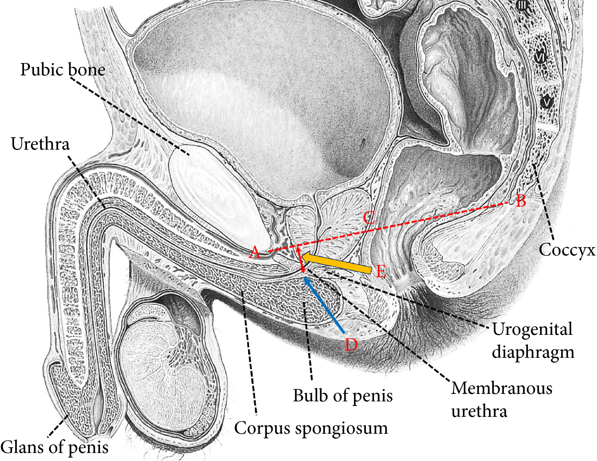
Traditionally, MRI of the prostate has been mainly applied in the diagnosis and staging of prostate cancer. Kadono et al. [1] used pre‐ and postoperative pelvic MRI to assess the repositioning of the urethra 10 days and 12 months after prostatectomy, hypothesizing that these alterations could correlate with urinary incontinence and urethral function. Recent MRI measurements of anatomical structures of the pelvic floor, such as membranous urethral length and inner levator distance, were found to be independent predictors of early continence recovery at 12 months after prostatectomy [2] A meta‐analysis has also shown a strong correlation between membranous urethral length and continence recovery at 3‐, 6‐ and 12‐month follow‐up [3] Kadono et al. [1] add another metric to the pelvic floor dimensions that may help predict continence. Cranial migration of the lower end of the membranous urethra early after prostatectomy was associated with urinary incontinence and urinary sphincter function, as objectively assessed by urethral pressure profile. Interestingly, return of the membranous urethra to the more distal preoperative position after 12 months was associated with improvement in continence. In a multivariate model, urethral stump length was a strong predictor of continence outcome at 10 days as well as 12 months after prostatectomy. This observation suggests that urethral length may partly improve post-prostatectomy continence through better compression of the membranous urethra in the pelvic floor membrane rather than through transfer of the intra‐abdominal pressure onto the intra‐abdominally located urethra. If confirmed, this observation may imply that more cranial fixation of the bladder neck in a more intra‐abdominal position may not necessarily improve continence after prostatectomy, in line with data from randomized controlled studies comparing median fibrous raphe reconstruction with standard anastomosis that failed to show a benefit [4,5].

Besides anatomical location, innervation of the proximal urethra is important for post-prostatectomy continence [6]. Kadono et al. found that nerve preservation was an independent predictor of early and long‐term continence outcome, with a b value similar to that of urethral stump length at 12‐month follow‐up. To improve post-prostatectomy continence outcome, proper patient selection seems crucial. In the era of personalized medicine, MRI could be a valuable tool to assess preoperatively the risks of postoperative urinary incontinence and counsel patients accordingly. Avoiding prostatectomy in men with short preoperative membranous urethral length may be an important approach for improving outcome, in particular in light of the fact that many attempts to surgically correct anatomical alignment of the pelvic floor have not clearly improved continence outcome. If surgery is considered, nerve preservation should be performed where possible to improve continence.

Henk G. van der Poel and Nikos Grivas

Department of Urology, Netherlands Cancer Institute, Amsterdam, The Netherlands

References

  1. Kadono Y, Nohara T, Kawaguchi S et al. Investigating the mechanism underlying urinary continence recovery after radical prostatectomy: effectiveness of a longer urethral stump to prevent urinary incontinence. BJU Int 2018. 37: 463–9
  2. Grivas N, van der Roest R, Schouten D et al. Quantitative assessment of fascia preservation improves the prediction of membranous urethral length and inner levator distance on continence outcome after robot-assisted radical prostatectomy. Neurourol Urodyn 2018; 37: 417–25
  3. Mungovan SF, Sandhu JS , Akin O, Smart NA, Graham PL, Patel MI. Preoperative membranous urethral length measurement and continence recovery  following radical prostatectomy: a systematic review and meta-analysis. Eur Urol 2017; 71: 368–78
  4. Joshi N, de Blok W, van Muilekom E, van der Poel H. Impact of posterior musculofascial reconstruction on early continence after robot-assisted laparoscopic radical prostatectomy: results of a prospective parallel group trial. Eur Urol 2010; 58: 84–9
  5. Menon M, Muhletaler F, Campos M, Peabody JO. Assessment of early continence after reconstruction of the periprostatic tissues in patients undergoing computer assisted (robotic) prostatectomy: results of a 2 group parallel randomized controlled trial. J Urol 2008; 180: 1018–23
  6. van der Poel HG, de Blok W, Joshi N, van Muilekom E. Preservation of lateral prostatic fascia is associated with urine continence after robotic-assisted prostatectomy. Eur Urol 2009; 55: 892–900Dearnaley DP, Jovic G, Syndikus I et al. The. Lancet Oncol 2014; 15:464–73


Video: Urinary continence recovery after radical prostatectomy

Investigating the mechanism underlying urinary continence recovery after radical prostatectomy: effectiveness of a longer urethral stump to prevent urinary incontinence

Read the full article

Abstract

Objective

To assess the chronological changes in urinary incontinence and urethral function before and after radical prostatectomy (RP), and to compare the findings of pelvic magnetic resonance imaging (MRI) before and after RP to evaluate the anatomical changes.

Patients and Methods

In total, 185 patients were evaluated with regard to the position of the distal end of the membranous urethra (DMU) on a mid‐sagittal MRI slice and urethral sphincter function using the urethral pressure profilometry. The patients also underwent an abdominal leak point pressure test before RP and at 10 days and 12 months after RP. The results were then compared with the chronological changes in urinary incontinence.

Results

The MRI results showed that the DMU shifted proximally to an average distance of 4 mm at 10 days after RP and returned to the preoperative position at 12 months after RP. Urethral sphincter function also worsened 10 days after RP, with recovery after 12 months. The residual length of the urethral stump and urinary incontinence were significantly associated with the migration length of the DMU at 10 days after RP. The residual length of the urethral stump was a significant predictor of urinary incontinence after RP.

Conclusion

This is the first study to elucidate that the slight vertical repositioning of the membranous urethra after RP causes chronological changes in urinary incontinence. A long urethral residual stump reduces urinary incontinence after RP.


View more videos

Article of the week: Effectiveness of a longer urethral stump to prevent urinary incontinence after radical prostatectomy

Every Week, the Editor-in-Chief selects an Article of the Week from the current issue of BJUI. The abstract is reproduced below and you can click on the button to read the full article, which is freely available to all readers for at least 30 days from the time of this post.

In addition to the article itself, there is an accompanying editorial written by a prominent member of the urological community. This blog is intended to provoke comment and discussion and we invite you to use the comment tools at the bottom of each post to join the conversation.

Finally, the third post under the Article of the Week heading on the homepage will consist of additional material or media. This week we feature both a video and a podcast discussing the paper.

If you only have time to read one article this week, it should be this one.

Investigating the mechanism underlying urinary continence recovery after radical prostatectomy: effectiveness of a longer urethral stump to prevent urinary incontinence

Yoshifumi Kadono*, Takahiro Nohara*, Shohei Kawaguchi*, Renato Naito*, Satoko Urata*, Kazufumi Nakashima*, Masashi Iijima*, Kazuyoshi Shigehara*, Kouji Izumi*, Toshifumi Gabata† and Atsushi Mizokami*

*Department of Integrative Cancer Therapy and Urology, Kanazawa University Graduate School of Medical Science, Kanazawa, Japan; †Department of Radiology, Kanazawa University School of Medicine, 13‐1 Takara‐machi, Kanazawa, Ishikawa 920‐8640, Japan

Read the full article

Abstract

Objective

To assess the chronological changes in urinary incontinence and urethral function before and after radical prostatectomy (RP), and to compare the findings of pelvic magnetic resonance imaging (MRI) before and after RP to evaluate the anatomical changes.

Patients and Methods

In total, 185 patients were evaluated with regard to the position of the distal end of the membranous urethra (DMU) on a mid‐sagittal MRI slice and urethral sphincter function using the urethral pressure profilometry. The patients also underwent an abdominal leak point pressure test before RP and at 10 days and 12 months after RP. The results were then compared with the chronological changes in urinary incontinence.

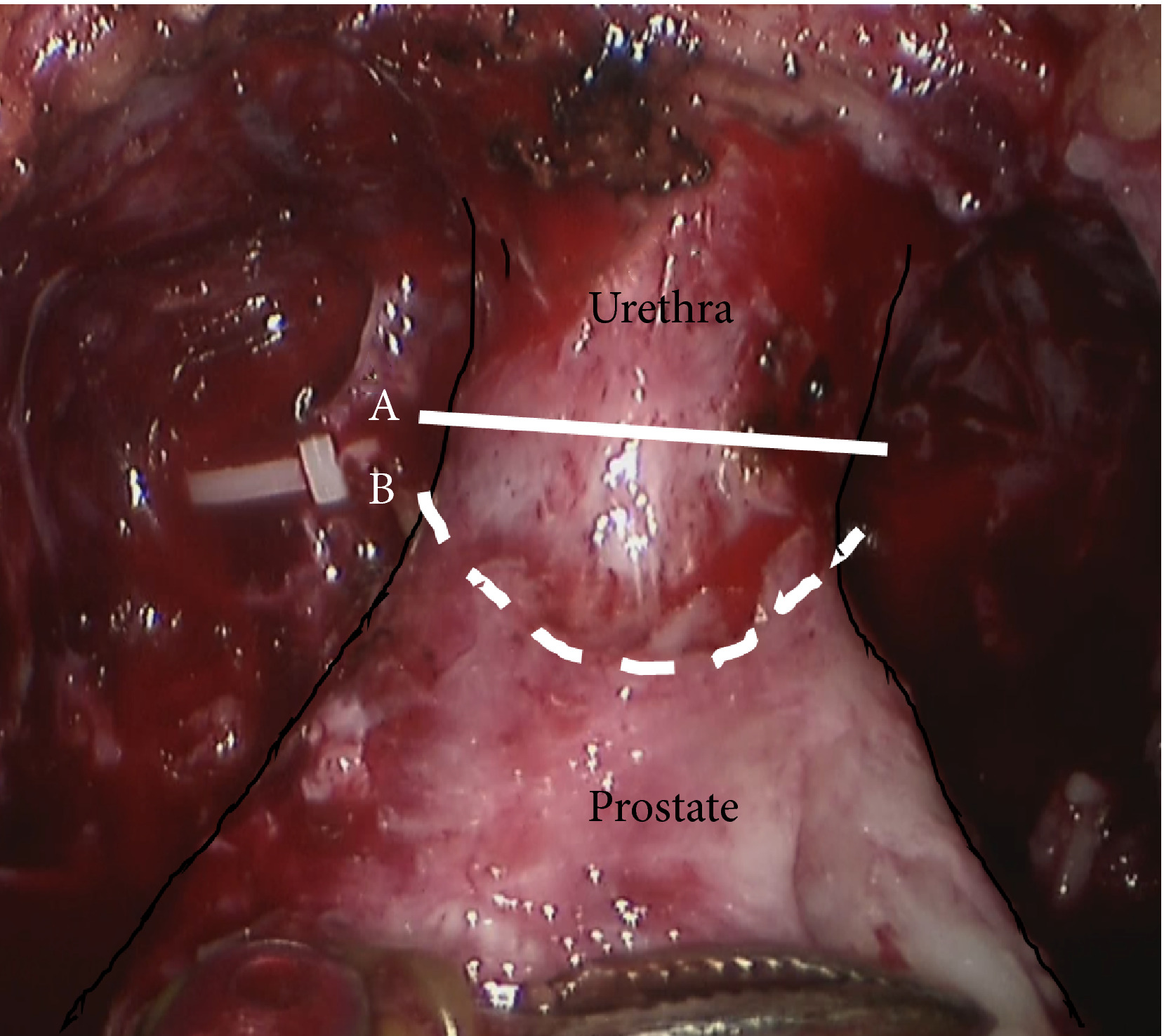
Fig. 1 Intraoperative view of the apex of the prostate transection line between the urethra and prostate at (A) the normal and (B) long urethral stump positions.

Results

The MRI results showed that the DMU shifted proximally to an average distance of 4 mm at 10 days after RP and returned to the preoperative position at 12 months after RP. Urethral sphincter function also worsened 10 days after RP, with recovery after 12 months. The residual length of the urethral stump and urinary incontinence were significantly associated with the migration length of the DMU at 10 days after RP. The residual length of the urethral stump was a significant predictor of urinary incontinence after RP.

Conclusion

This is the first study to elucidate that the slight vertical repositioning of the membranous urethra after RP causes chronological changes in urinary incontinence. A long urethral residual stump reduces urinary incontinence after RP.


Read more articles of the week

Editorial: Prognostic and predictive models in hormone-sensitive PCa

The article by Abdel‐Rahmen and Cheung [1] in the current issue of BJUI takes the Prostascore model [2] developed from epidemiological Surveillance, Epidemiology and End Results (SEER) data and applies it to the prospective randomised trial data from the ChemoHormonal Therapy Versus Androgen Ablation Randomized Trial for Extensive Disease in Prostate Cancer (CHAARTED) [3]. This sort of modelling is invaluable, given the inevitable limitations of epidemiological data (retrospective, no data cleaning, key items such as performance status missing, etc.). For models to be useful, they need to be able to discern prognostic categories reliably and with sufficient difference in outcome that they can usefully contribute to clinical decision‐making. They also need to perform better than existing oncological models, such as TNM stage, as outcome predictors. To be truly useful, they also need to identify subgroups of patients who may differentially benefit from different treatments (e.g. human epidermal growth factor receptor 2 [HER2] status in breast cancer therapy with trastuzumab). It is thus important to differentiate prognostic models (which predict outcome but do not help select treatment) from predictive models (which predict outcome and also help select who may benefit from treatment). A common mistake in this setting is to assume that patients with worse outcomes may benefit from more aggressive treatment, whilst those with better outcomes may require less treatment. This is a frequent confounding bias in retrospective data such as SEER. The key test is to look at good‐ and poor‐prognosis patients and examine whether the benefit from (say) chemotherapy is proportionately the same or different in both groups. If the proportional benefit is the same, the model is purely prognostic, if the proportional benefit is different (e.g., a bigger benefit in worse patients), the model is, in addition, predictive. A randomised trial such as CHAARTED is thus ideal for separating these two factors. It is also worth noting that just because a model predicts a relatively better outcome in one subgroup vs another, it does not mean that all patients do not benefit. For example, programmed death 1 (PD‐1) pathway molecule staining is generally prognostic in advanced bladder cancer and partly predictive of better response to PD‐1 pathway drugs such as atezolizumab [4] and pembrolizumab [5], but all patients derive benefit and hence PD‐1 pathway staining is not useful in selecting for treatment.

How does the Prostascore model fare against these various tests? As a prognostic model it clearly separates patients into groups with plausible differences in outcome. However, there exist many systems already that do this and in the clinic, these are probably not useful – it is well understood that men with extensive disease and visceral metastases will do worse than those with less disease – treatment for all will include androgen‐deprivation therapy (ADT). More recently, trial data have emerged showing survival benefit from the first‐line addition of either docetaxel [3, 6] or abiraterone [7, 8] to ADT. There is controversy about whether this benefit is confined to men with high‐volume disease as claimed by some [9], or applies more generally, as per Systemic Therapy in Advancing or Metastatic Prostate Cancer: Evaluation of Drug Efficacy (STAMPEDE) [6] and the Systemic Treatment Options for Cancer of the Prostate (STOpCaP) meta‐analysis [10]. Can Prostascore help to resolve this problem? Sadly the answer is no. The analysis presented here [1] does not offer convincing evidence of a useful predictive value with respect to selection for first‐line docetaxel. Firstly, the sample size is small (702 men), with only 118 in the lowest risk category (who of course also have the lowest event rate, hence proportionately even less power). In the higher‐risk categories, there seems to be clear evidence of benefit from docetaxel (larger numbers of men and higher event rates). No pooled analysis is presented, but it seems likely that this would show a benefit from docetaxel in the whole sample set, exactly as reported in CHAARTED [3]. Data from underpowered, post hoc, subgroup analysis should not be over‐interpreted [11].

How should we move forward from here? The STAMPEDE group will be presenting data from both the docetaxel and abiraterone parts of the study, classified both by the high‐/low‐volume split in CHAARTED [3] and the high‐/low‐risk split in the LATITUDE trial (multinational, randomised placebo‐controlled phase III clinical trial of men with newly diagnosed, high‐risk metastatic prostate cancer who had not previously received ADT. All patients had at least two of three risk factors: Gleason score of ≥8, ≥3 bone metastases, or ≥3 visceral metastases) [7]. If evidence of a differential response to these agents by these prognostic tools exists, it may be worth applying the Prostascore tool to the STAMPEDE dataset. If there is no evidence of these existing prognostic classifiers also being predictive of best therapy choice, there is probably only a limited role for Prostascore as a prognostic tool in an already crowded space. risk factors and risk of developing cancer.

Nicholas D. James

Institute of Cancer and Genomic Sciences, University of Birmingham,
Queen Elizabeth Hospital, Birmingham, UK

References
  1. Abdel‐Rahman O, Cheung WY. External validation of the prostascore model in patients with metastatic hormone‐sensitive prostate cancer recruited to the CHAARTED study. BJU Int 2018; 122: 394–400
  2. Abdel‐Rahman O. Prostascore: a simplified tool for predicting outcomes among patients with treatment‐naive advanced prostate cancer. Clin Oncol (R Coll Radiol) 2017; 29: 732–8
  3. Sweeney CJ, Chen YH, Carducci M et al. Chemohormonal therapy in metastatic hormone‐sensitive prostate cancer. N Engl J Med 2015; 373: 737–46
  4. Balar AV, Galsky MD, Rosenberg JE et al. Atezolizumab as first‐line treatment in cisplatin‐ineligible patients with locally advanced and metastatic urothelial carcinoma: a single‐arm, multicentre, phase 2 trial. Lancet 2017; 389: 67–76
  5.  Bellmunt J, de Wit R, Vaughn DJ et al. Pembrolizumab as second‐line therapy for advanced urothelial carcinoma. N Engl J Med 2017; 376: 1015–26
  6. James ND, Sydes MR, Clarke NW et al. Addition of docetaxel, zoledronic acid, or both to first‐line long‐term hormone therapy in prostate cancer (STAMPEDE): survival results from an adaptive, multiarm, multistage, platform randomised controlled trial. Lancet 2016; 387: 1163–77
  7. Fizazi K, Tran N, Fein L et al. Abiraterone plus prednisone in metastatic, castration‐sensitive prostate cancer. N Engl J Med 2017; 377: 352–60
  8. James ND, de Bono JS, Spears MR et al. Abiraterone for prostate cancer not previously treated with hormone therapy. N Engl J Med 2017; 377: 338–51
  9. Gravis G, Boher JM, Chen YH et al. Burden of metastatic castrate naive prostate cancer patients, to identify men more likely to benefit from early docetaxel: further analyses of CHAARTED and GETUG‐AFU15 Studies. Eur Urol 2018; 73: 847–55
  10. 10 Vale CL, Burdett S, Rydzewska LH et al. Addition of docetaxel or bisphosphonates to standard of care in men with localised or metastatic, hormone‐sensitive prostate cancer: a systematic review and meta‐analyses of aggregate data. Lancet Oncol 2016; 17: 243–56
  11. Spears MR, James ND, Sydes MR. Thursday’s child has far to go’ – interpreting subgroups and the STAMPEDE trial. Ann Oncol 2017; 28: 2327–30


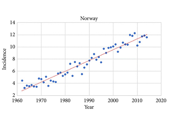
Editorial: Occupational exposure and risk of testicular cancer: what can an ecological study in the Nordic countries tell us?

Examining the association between occupational exposure and incidence rates of testicular cancer over time in several countries may provide useful insights into the relative importance of lifestyle and environmental risk factors. The study by Ylonen et al. [1] assessed an ecological, rather than biological, effect of occupational exposure in order to understand differences in testicular cancer rates among populations; therefore, the authors could not draw causal inferences about the effect of occupational exposure on testicular cancer at the individual level [2]. Ecological studies are, however, a way of performing hypothesis‐generating population‐based research. They take advantage of the natural experiment following the changes in occupational exposure across countries [3].

Keeping the strengths and limitations of an ecological study design in mind, we should consider what can we learn from the study by Ylonen et al. [1]. Firstly, it is interesting to note that the authors themselves state in their discussion that ‘occupational exposure is probably not relevant’ for testicular cancer because the disease ‘is mainly diagnosed in young adults and the duration of occupational exposure before cancer diagnosis is short’. This highlights the fact that occupational exposure should probably be considered here as a proxy variable for other risk factors of testicular cancer: environmental exposure, physical activity, education, etc. This large study based on linkages of high‐quality data registers therefore merely aims to generate hypotheses. No clear patterns were observed, however, and future studies may benefit from similar ecological approaches using risk factors with a better rationale in the context of the aetiology of testicular cancer, such as socio‐economic statuts, diet or body mass index.

Secondly, the authors also note that this lack of unambiguous risk determinants and underlying mechanisms of testicular cancer makes it difficult to explain the geographic and temporal variations observed [1]. As the study did not have a hypothesis a priori, we find ourselves in a situation where occupational exposure may not be the best risk factor to examine in relation to risk of testicular cancer in an ecological study. The rationale for choosing occupational exposure as the risk factor is weak, and perhaps the authors would have been able to observe more clear patterns if they had conducted the study to assess bladder cancer, for which occupation has been a much more established risk factor and time to exposure has been found to be more relevant.

Thirdly, a clinical understanding of testicular cancer detection may inform changes in incidence over time and between countries. No additional information was provided by the authors, but changes in raising awareness of potential symptoms may have resulted in an increased incidence in a specific age group and/or country. Moreover, it would be of interest to know about differences among countries in terms of occupational exposure groups as this may also explain some of the patterns observed. A further assessment of the characteristics of different categories of occupational exposure could inform the patterns observed in this study and may shed light on the aetiology of testicular cancer.

In conclusion, ecological studies force us to think carefully about patterns of cancer incidence over time and among countries. Unfortunately, the findings cannot always lead to further hypotheses and careful consideration about potential risk factors needs to occur before conducting analyses. Moreover, detailed (clinical) knowledge is required about changes in diagnostic activity over time as well as potential changes in risk factor exposure. Future ecological studies using highly valuable resources, such as those used by Ylonen et al. [1] can help us understand cancer aetiology and prevention in more detail. They can be considered as a natural experiment to fill the gap in our understanding of the link between potential risk factors and risk of developing cancer.

Mieke Van Hemelrijck
Translational Oncology & Urology Research, Kings College
London, London, UK


Read the full article
References

1 Ylonen O, Jyrkkio S, Pukkala E, Syvanen K, Bostrom P. Time trends and occupational variation in the incidence of testicular cancer in the Nordic countries. BJU Int 2018; 122: 384–93

2 Morgenstern H. Ecologic studies. In Rothman K, Greenland S eds, Modern Epidemiology, 2nd edn, Philadelphia, PA: Lippincott Williams &Wilkins, 1998: 511–31

3 Sedgwick P. Ecological studies: advantages and disadvantages. BMJ 2014; 348: g2979


Article of the Week: Analysis of hydrogel spacer for PCa RT

Every Week, the Editor-in-Chief selects an Article of the Week from the current issue of BJUI. The abstract is reproduced below and you can click on the button to read the full article, which is freely available to all readers for at least 30 days from the time of this post.

In addition to the article itself, there is an accompanying editorial written by a prominent member of the urological community. This blog is intended to provoke comment and discussion and we invite you to use the comment tools at the bottom of each post to join the conversation.

Finally, the third post under the Article of the Week heading on the homepage will consist of additional material or media. This week we feature a video discussing the paper.

If you only have time to read one article this week, it should be this one.

Prospective analysis of hydrogel spacer for patients with prostate cancer undergoing radiotherapy

Michael Chao*†, Huong Ho† , Yee Chan*‡, Alwin Tan§, Trung Pham¶, Damien Bolton*‡, Andrew Troy*, Catherine Temelcos**, Shomik Sengupta*†† , Kevin McMillan‡, Chee Wee Cham§, Madalena Liu‡, Wei Ding†, Brindha Subramanian†, Jason Wasiak*‡‡, Daryl Lim Joon*†, Sandra Spencer† and Nathan Lawrentschuk*

*The Austin Hospital, Heidelberg, Vic., Australia, †Genesis Cancer Care Victoria, Ringwood East, Vic., Australia, ‡Ringwood Private Hospital, Ringwood East, Vic., Australia, §The Bays Hospital, Mornington, Vic., Australia, ¶ The Valley Private Hospital, Mulgrave, Vic., Australia, **St Vincent’s Hospital, Fitzroy, Vic., Australia, ††Melbourne University; Eastern Health Clinical School, Monash University, Clayton, Vic., Australia, and ‡‡University of Melbourne, Melbourne, Vic., Australia

Read the full article

Abstract

Objective

To report on the dosimetric benefits and late toxicity outcomes after injection of hydrogel spacer (HS) between the prostate and rectum for patients treated with prostate radiotherapy (RT).

Patients and Methods

In all, 76 patients with a clinical stage of T1–T3a prostate cancer underwent general anaesthesia for fiducial marker insertion plus injection of the HS into the perirectal space before intensity‐modulated RT (IMRT) or volumetric‐modulated arc RT (VMAT). HS safety, dosimetric benefits, and the immediate‐ to long‐term effects of gastrointestinal (GI) toxicity were assessed.

Results

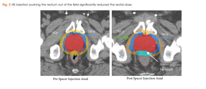
There were no postoperative complications reported. The mean (range) prostate size was 66.0 (25.0–187.0) mm. Rectal dose volume parameters were observed and the volume of rectum receiving 70 Gy (rV70), 75 Gy (rV75) and 78 Gy (rV78) was 7.8%, 3.6% and 0.4%, respectively. In all, 21% of patients (16/76) developed acute Grade 1 GI toxicities, but all were resolved completely by 3 months after treatment; whilst, 3% of patients (2/76) developed late Grade 1 GI toxicities. No patients had acute or late Grade ≥2 GI toxicities.

Conclusion

Injection of HS resulted in a reduction of irradiated rectal dose volumes along with minimal GI toxicities, irrespective of prostate size.

Read more articles of the week

Editorial: 2018 – A Spacer Odyssey

The optimisation of radical external-beam radiotherapy treatment is ultimately a compromise. The aim is to deliver a clinically effective radiation dose to the tumour target, whilst limiting the irradiation of surrounding normal tissue to minimise acute and late side effects. Modern image-guided intensity-modulated radiotherapy (IG-IMRT) allows very accurate treatment delivery, which improves this therapeutic ratio. However, there are limitations in its ability to reduce toxicity, due to the constraints of regional anatomy and the physical properties of photon beams. A variety of additional organ-specific techniques exist to further minimise the impact of radiotherapy on adjacent healthy tissue. These range in complexity from ovarian auto-transplantation in selected patients awaiting pelvic radiotherapy for gynaecological malignancies, to delivering radiotherapy for left-sided breast cancers at inspiratory breath-hold to maximise the distance of the chest wall from the left anterior descending coronary artery.

For prostate cancer radiotherapy, the normal structures that determine the optimal safely deliverable dose include the rectum (anterior rectal wall), bladder, femoral heads and penile bulb. IG-IMRT has facilitated dose-escalation, and the treatment of patients previously considered to be ineligible for a radical treatment; both with acceptable toxicity. Higher doses result in improved prostate-cancer biochemical (PSA) control rates, and metastasisfree survival, but as yet no overall survival benefit has been demonstrated in individual trials, despite 10-year follow-up [1]. This may due to the long natural history of localised prostate cancer, the impact of competing comorbidities [2], and the ever-increasing efficacy of treatments for metastatic disease on relapse.

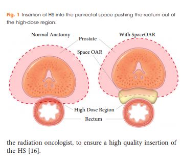
Rectal spacers are a further refinement to the delivery of radical IG-IMRT to the prostate gland. By inserting a biodegradable substance / inflatable biodegradable balloon into the anterior perirectal space, the distance between the prostate and the anterior rectal wall can be increased by approximately 1cm. This reduces the volume of rectum lying within the high-dose radiation field, thereby reducing late bowel toxicity. A recently reported randomised Phase III trial found a reduction the 3-year incidence of ≥ Grade 1 and ≥ Grade 2 rectal toxicity (9.2% v 2.0%; p=0.28; and 5.7% v 0%; p=0.12 respectively) with the addition of a rectal spacer [3]. This was in a very select group of patients: PSA ≤20, cT1-2, International Society of Urological Pathology (ISUP) Prostate Cancer Grade 1-3 (<50% of cores involved), prostate volume <80cc, no use of androgen deprivation therapy, and the use of MRI-CT fusion to aid prostate delineation during the planning process.

In this issue of the BJUI, Chao et al. [4] report their prospective single-centre experience of using a rectal spacer device. This is not a randomised study, and no control arm exists to assist in quantifying the clinical impact of inserting a spacer. However, the patient population studied closely reflects that undergoing radical radiotherapy in most oncology departments world-wide. There were no limitations placed on prostate size (45% were 50- 100cc; 17% >100cc; maximum 187cc), PSA (10% had a PSA >20; maximum 117), or ISUP histological grade (27% were Grade 4 and 5); and 27% of patients were cT3a on imaging. They demonstrated that spacer insertion was safe across a diverse population of men with localised prostate cancer. Further, acceptable prostate-rectal wall separation and rectal dosimetry could be achieved irrespective of prostate size – which was reflected by the low rates of cumulative late rectal toxicity when assessed at a median follow-up of 14 months.

As with any new technique, additional clarification is required to determine exactly where rectal spacers fit into the radiotherapy armoury. In the study by Chao et. al. the Radiation Oncologists did not have access to CT-MRI fusion at the planning stage. As the electron densities for the prostate, rectal-spacer and rectal wall are similar, it can be challenging to determine the anatomical boundaries on CT alone. However, if growing clinical experience now permits accurate delineation by merely adjusting the window levels on the Planning CT, this could facilitate more widespread introduction of the technique, outside the specialist centres with MR-fusion capabilities.

Patient selection is also key to the introduction of this technique. Patient series have reported the safety of the spacer insertion technique across a range of prostate sizes, and prostate cancer risk groups. However, prospective randomised data is lacking on whether treatment efficacy is affected by using a spacer in high-risk / locally advanced disease – the population most likely to benefit from dose-escalated radical radiotherapy [5]. Further, in the only randomised study to date, >90% of patients in the control arm experienced no late rectal toxicity, and >94% experienced no ≥Grade 2 rectal toxicity. Based on long-term follow-up data from the RT01 study, this was an appropriate time-point at which to assess late bowel toxicity, which peaks at 12-36 months before declining [6]. Therefore, the majority of patients do not experience clinically significant late toxicity even without a spacer.

Rectal spacers have the potential to make a valuable contribution to radical IG-IMRT treatment for localised prostate cancer. However, predictive factors are required to identify which patients are likely to benefit from the technique. For many patients, with access to the latest radiotherapy planning and delivery systems, spacers may represent an additional costly procedure with limited benefit.

S.R.Hughes
Oncology Department, Guy’s & St. Thomas’ NHS Trust, London, UK

Read the full article

References

1. Dearnaley DP, Jovic G, Syndikus I, et. al. The Lancet Oncol. 2014; 15(4): 464-473

2. Lu-Yao GL, Albertsen PC, Moore DF, Lin Y, DiPaola RS, Yao SL. Fifteen-year outcomes following conservative management among men aged 65 years or older with localised prostate cancer. Eur Urol. 2015; 68(5): 805-11

3. Hamstra DA, Mariados N, Sylvester J et. al. Continued Benefit to Rectal Separation for Prostate Radiation Therapy: Final Results of a Phase III Trial. Int J Radiat Oncol Biol Phys 2017; 97(5): 976-985

4. Chao M, Ho H, Chan Y et. al. Prospective Analysis of Hydrogel Spacer for Prostate Cancer Patients Undergoing Radiotherapy. BJUI 2018

5. Kalbasi A, Li J, Berman AT. Dose-Escalated Irradiation and Overall Survival in Men with Non-metastatic Prostate Cancer. JAMA Oncol. 2015; 1(7): 897-906

6. Syndikus I, Morgan RC, Sydes MR, Graham JD, Dearnaley DP. Late Gastrointestinal Toxicity After Dose-Escalated Conformal Radiotherapy For Early Prostate Cancer:


Video: Hydrogel spacer for PCa RT

Prospective analysis of hydrogel spacer for patients with prostate cancer undergoing radiotherapy

Read the full article

Abstract

Objective

To report on the dosimetric benefits and late toxicity outcomes after injection of hydrogel spacer (HS) between the prostate and rectum for patients treated with prostate radiotherapy (RT).

Patients and Methods

In all, 76 patients with a clinical stage of T1–T3a prostate cancer underwent general anaesthesia for fiducial marker insertion plus injection of the HS into the perirectal space before intensity‐modulated RT (IMRT) or volumetric‐modulated arc RT (VMAT). HS safety, dosimetric benefits, and the immediate‐ to long‐term effects of gastrointestinal (GI) toxicity were assessed.

Results

There were no postoperative complications reported. The mean (range) prostate size was 66.0 (25.0–187.0) mm. Rectal dose volume parameters were observed and the volume of rectum receiving 70 Gy (rV70), 75 Gy (rV75) and 78 Gy (rV78) was 7.8%, 3.6% and 0.4%, respectively. In all, 21% of patients (16/76) developed acute Grade 1 GI toxicities, but all were resolved completely by 3 months after treatment; whilst, 3% of patients (2/76) developed late Grade 1 GI toxicities. No patients had acute or late Grade ≥2 GI toxicities.

Conclusion

Injection of HS resulted in a reduction of irradiated rectal dose volumes along with minimal GI toxicities, irrespective of prostate size.

View more videos
© 2024 BJU International. All Rights Reserved.